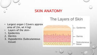
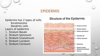
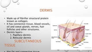
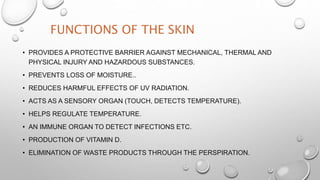
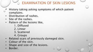
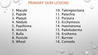
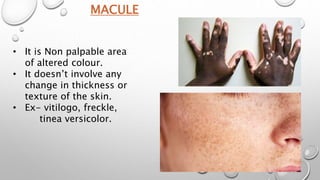
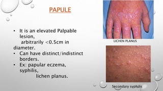
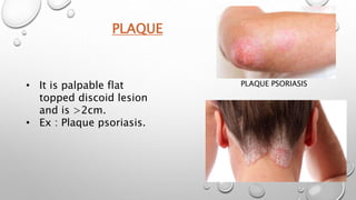
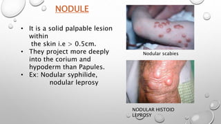
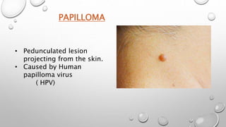
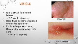
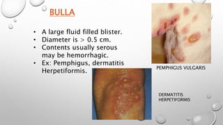
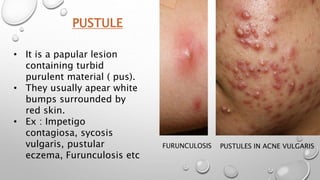
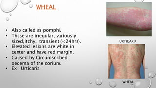
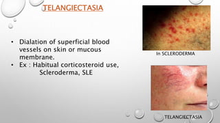
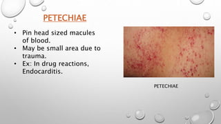
DEPARTMENTAL SAMINAR ON PRIMARY SKIN
LESIONS
BY
DR SAEEDA M MAKANDAR
1ST YEAR PG SCHOLAR DEPT OF PANCHAKARMA
GUIDED BY
DEPT OF PG STUDIES IN PANCHAKARMA
S V M AYURVEDIC MEDICAL COLLEGE AND
RESEARCH CENTER, ILKAL
SKIN AND PRIMARY SKIN
LESIONS
SKIN ANATOMY
• Largest organ ( Covers approx
area of 2m, wt 4 kg)
• Layers of the skin-
1. Epidermis
2. Dermis
3. Hypodermis (Subcutaneous
tissue)
EPIDERMIS
Epidermis has 2 types of cells
Keratinocytes
Dendritic cells
Layers of epidermis
1. Stratum Basale
2. Stratum Spinosum
3. Stratum Granulosum
4. Stratum Lucidum
5. Stratum Corneum
DERMIS
• Made up of fibrillar structural protein
known as collagen.
• It has connective tissue, blood vessels,
oil and sweat glands, nerves, hair
follicles and other structures.
• Dermis layers-
1. Papillary dermis
2. Reticular dermis
SUBCUTANEOUS
TISSUE
FUNCTIONS OF THE SKIN
• PROVIDES A PROTECTIVE BARRIER AGAINST MECHANICAL, THERMAL AND
PHYSICAL INJURY AND HAZARDOUS SUBSTANCES.
• PREVENTS LOSS OF MOISTURE..
• REDUCES HARMFUL EFFECTS OF UV RADIATION.
• ACTS AS A SENSORY ORGAN (TOUCH, DETECTS TEMPERATURE).
• HELPS REGULATE TEMPERATURE.
• AN IMMUNE ORGAN TO DETECT INFECTIONS ETC.
• PRODUCTION OF VITAMIN D.
• ELIMINATION OF WASTE PRODUCTS THROUGH THE PERSPIRATION.
EXAMINATION OF SKIN LESIONS
• History taking asking symptoms of which patient
complains.
• Distribution of rashes.
• Site of the rashes.
• Pattern of the lesions like,
1. Diffused
2. Linear
3. Scattered
4. Groups
• Related scars of previously dameged skin.
• Colour of the skin.
• Shape and size of the lesions.
• Border.
Types of skin lesions, are
1. Primary skin lesions
2. Secondary skin lesions
PRIMARY SKIN LESIONS
1. Macule
2. Papule
3. Plaque
4. Nodule
5. Papilloma
6. Vesicle
7. Bulla
8. Pustule
9. Wheal
10. Talengiectasia
11. Patechia
12. Purpura
13. Ecchymosis
14. Haematoma
15. Paikiloderma
16. Erythema
17. Burrow
18. Comedo
1. Scale
2. Crust
3. Excoriation
4. Lichenification
5. Fissure
6. Errosion
7. Ulcer
8. Sinus
9. Scar
10. Keloid
scar
11. Atrophy
12. Stria
SECONDARY SKIN LESIONS
MACULE
• It is Non palpable area
of altered colour.
• It doesn’t involve any
change in thickness or
texture of the skin.
• Ex- vitilogo, freckle,
tinea versicolor.
PAPULE
LICHEN PLANUS
Secondary syphilis
• It is an elevated Palpable
lesion,
arbitrarily <0.5cm in
diameter.
• Can have distinct/indistinct
borders.
• Ex: papular eczema,
syphilis,
lichen planus.
PLAQUE
PLAQUE PSORIASIS
• It is palpable flat
topped discoid lesion
and is >2cm.
• Ex : Plaque psoriasis.
NODULE
• It is a solid palpable lesion
within
the skin i.e > 0.5cm.
• They project more deeply
into the corium and
hypoderm than Papules.
• Ex: Nodular syphilide,
nodular leprosy
Nodular scabies
NODULAR HISTOID
LEPROSY
PAPILLOMA
• Pedunculated lesion
projecting from the skin.
• Caused by Human
papilloma virus
( HPV)
VESICLE
• It is a small fluid filled
blister.
• < 0.5 cm In diameter.
• Here fluid becomes trapped
under the epidermis
• Ex: In Allergic reactions,
Dermatitis, poison ivy, cold
sore
( herpis simplex)
DERMATITIS
HERPIS SIMPLEX
BULLA
• A large fluid filled blister.
• Diameter is > 0.5 cm.
• Contents usually serous
may be hemorrhagic.
• Ex: Pemphigus, dermatitis
Herpetiformis.
PEMPHIGUS VULGARIS
DERMATITIS
HERPETIFORMIS
PUSTULE
• It is a papular lesion
containing turbid
purulent material ( pus).
• They usually apear white
bumps surrounded by
red skin.
• Ex : Impetigo
contagiosa, sycosis
vulgaris, pustular
eczema, Furunculosis etc
PUSTULES IN ACNE VULGARIS
FURUNCULOSIS
WHEAL
• Also called as pomphi.
• These are irregular, variously
sized,itchy, transient (<24hrs).
• Elevated lesions are white in
center and have red margin.
• Caused by Circumscribed
oedema of the corium.
• Ex : Urticaria
URTICARIA
WHEAL
TELANGIECTASIA
• Dialation of superficial blood
vessels on skin or mucous
membrane.
• Ex : Habitual corticosteroid use,
Scleroderma, SLE
In SCLERODERMA
TELANGIECTASIA
PETECHIAE
PETECHIAE
• Pin head sized macules
of blood.
• May be small area due to
trauma.
• Ex: In drug reactions,
Endocarditis.
PURPURA
• Larger petechiae which do
not blanch on pressure.
• Reddish or purplish spots of
various sizes and shapes.
• Ex : Vasciculitis, in clotting
defects.
VASCICULITIS
ECCHYMOSIS
TRAUMA
• These are larger petechiae
which do not blanch on
pressure.
• Reddish or purplish spots
of various size and shape.
• Ex : Trauma,
Thrombocytopnia.
HAEMATOMA
• Swelling due to gross
bleeding out side the blood
vessels.
• In case of injury.
POIKILODERMA
• It is the combination of atrophy,
Hyperpigmentation and
telangiectasia.
• Has cappilary haemorhages
accompanied by general pruritus.
• Site- All over body Including
scalp.
• Ex: Mycosis fungoides,
Epitheliomatosis
MYCOSIS
FUNGOIDES
ERYTHEMA
• Redness of skin that
blanches on pressure.
• It may localized or
widespread.
BURROW
• Linear or carved track left by the
burrowing scabies mite.
• Intense itching.
• Ex: Scabies
COMEDO
• Commonly known as blackheads.
• Dark horny keratin and
sebaceous plugs within
Pilosebaceous opening.
• Ex : Acne vulgaris
Primary skin lesions

Primary skin lesions

  • 1.
    DEPARTMENTAL SAMINAR ONPRIMARY SKIN LESIONS BY DR SAEEDA M MAKANDAR 1ST YEAR PG SCHOLAR DEPT OF PANCHAKARMA GUIDED BY DEPT OF PG STUDIES IN PANCHAKARMA S V M AYURVEDIC MEDICAL COLLEGE AND RESEARCH CENTER, ILKAL
  • 2.
    SKIN AND PRIMARYSKIN LESIONS
  • 3.
    SKIN ANATOMY • Largestorgan ( Covers approx area of 2m, wt 4 kg) • Layers of the skin- 1. Epidermis 2. Dermis 3. Hypodermis (Subcutaneous tissue)
  • 4.
    EPIDERMIS Epidermis has 2types of cells Keratinocytes Dendritic cells Layers of epidermis 1. Stratum Basale 2. Stratum Spinosum 3. Stratum Granulosum 4. Stratum Lucidum 5. Stratum Corneum
  • 5.
    DERMIS • Made upof fibrillar structural protein known as collagen. • It has connective tissue, blood vessels, oil and sweat glands, nerves, hair follicles and other structures. • Dermis layers- 1. Papillary dermis 2. Reticular dermis SUBCUTANEOUS TISSUE
  • 6.
    FUNCTIONS OF THESKIN • PROVIDES A PROTECTIVE BARRIER AGAINST MECHANICAL, THERMAL AND PHYSICAL INJURY AND HAZARDOUS SUBSTANCES. • PREVENTS LOSS OF MOISTURE.. • REDUCES HARMFUL EFFECTS OF UV RADIATION. • ACTS AS A SENSORY ORGAN (TOUCH, DETECTS TEMPERATURE). • HELPS REGULATE TEMPERATURE. • AN IMMUNE ORGAN TO DETECT INFECTIONS ETC. • PRODUCTION OF VITAMIN D. • ELIMINATION OF WASTE PRODUCTS THROUGH THE PERSPIRATION.
  • 7.
    EXAMINATION OF SKINLESIONS • History taking asking symptoms of which patient complains. • Distribution of rashes. • Site of the rashes. • Pattern of the lesions like, 1. Diffused 2. Linear 3. Scattered 4. Groups • Related scars of previously dameged skin. • Colour of the skin. • Shape and size of the lesions. • Border.
  • 8.
    Types of skinlesions, are 1. Primary skin lesions 2. Secondary skin lesions
  • 9.
    PRIMARY SKIN LESIONS 1.Macule 2. Papule 3. Plaque 4. Nodule 5. Papilloma 6. Vesicle 7. Bulla 8. Pustule 9. Wheal 10. Talengiectasia 11. Patechia 12. Purpura 13. Ecchymosis 14. Haematoma 15. Paikiloderma 16. Erythema 17. Burrow 18. Comedo
  • 10.
    1. Scale 2. Crust 3.Excoriation 4. Lichenification 5. Fissure 6. Errosion 7. Ulcer 8. Sinus 9. Scar 10. Keloid scar 11. Atrophy 12. Stria SECONDARY SKIN LESIONS
  • 11.
    MACULE • It isNon palpable area of altered colour. • It doesn’t involve any change in thickness or texture of the skin. • Ex- vitilogo, freckle, tinea versicolor.
  • 12.
    PAPULE LICHEN PLANUS Secondary syphilis •It is an elevated Palpable lesion, arbitrarily <0.5cm in diameter. • Can have distinct/indistinct borders. • Ex: papular eczema, syphilis, lichen planus.
  • 13.
    PLAQUE PLAQUE PSORIASIS • Itis palpable flat topped discoid lesion and is >2cm. • Ex : Plaque psoriasis.
  • 14.
    NODULE • It isa solid palpable lesion within the skin i.e > 0.5cm. • They project more deeply into the corium and hypoderm than Papules. • Ex: Nodular syphilide, nodular leprosy Nodular scabies NODULAR HISTOID LEPROSY
  • 15.
    PAPILLOMA • Pedunculated lesion projectingfrom the skin. • Caused by Human papilloma virus ( HPV)
  • 16.
    VESICLE • It isa small fluid filled blister. • < 0.5 cm In diameter. • Here fluid becomes trapped under the epidermis • Ex: In Allergic reactions, Dermatitis, poison ivy, cold sore ( herpis simplex) DERMATITIS HERPIS SIMPLEX
  • 17.
    BULLA • A largefluid filled blister. • Diameter is > 0.5 cm. • Contents usually serous may be hemorrhagic. • Ex: Pemphigus, dermatitis Herpetiformis. PEMPHIGUS VULGARIS DERMATITIS HERPETIFORMIS
  • 18.
    PUSTULE • It isa papular lesion containing turbid purulent material ( pus). • They usually apear white bumps surrounded by red skin. • Ex : Impetigo contagiosa, sycosis vulgaris, pustular eczema, Furunculosis etc PUSTULES IN ACNE VULGARIS FURUNCULOSIS
  • 19.
    WHEAL • Also calledas pomphi. • These are irregular, variously sized,itchy, transient (<24hrs). • Elevated lesions are white in center and have red margin. • Caused by Circumscribed oedema of the corium. • Ex : Urticaria URTICARIA WHEAL
  • 20.
    TELANGIECTASIA • Dialation ofsuperficial blood vessels on skin or mucous membrane. • Ex : Habitual corticosteroid use, Scleroderma, SLE In SCLERODERMA TELANGIECTASIA
  • 21.
    PETECHIAE PETECHIAE • Pin headsized macules of blood. • May be small area due to trauma. • Ex: In drug reactions, Endocarditis.
  • 22.
    PURPURA • Larger petechiaewhich do not blanch on pressure. • Reddish or purplish spots of various sizes and shapes. • Ex : Vasciculitis, in clotting defects. VASCICULITIS
  • 23.
    ECCHYMOSIS TRAUMA • These arelarger petechiae which do not blanch on pressure. • Reddish or purplish spots of various size and shape. • Ex : Trauma, Thrombocytopnia.
  • 24.
    HAEMATOMA • Swelling dueto gross bleeding out side the blood vessels. • In case of injury.
  • 25.
    POIKILODERMA • It isthe combination of atrophy, Hyperpigmentation and telangiectasia. • Has cappilary haemorhages accompanied by general pruritus. • Site- All over body Including scalp. • Ex: Mycosis fungoides, Epitheliomatosis MYCOSIS FUNGOIDES
  • 26.
    ERYTHEMA • Redness ofskin that blanches on pressure. • It may localized or widespread.
  • 27.
    BURROW • Linear orcarved track left by the burrowing scabies mite. • Intense itching. • Ex: Scabies
  • 28.
    COMEDO • Commonly knownas blackheads. • Dark horny keratin and sebaceous plugs within Pilosebaceous opening. • Ex : Acne vulgaris