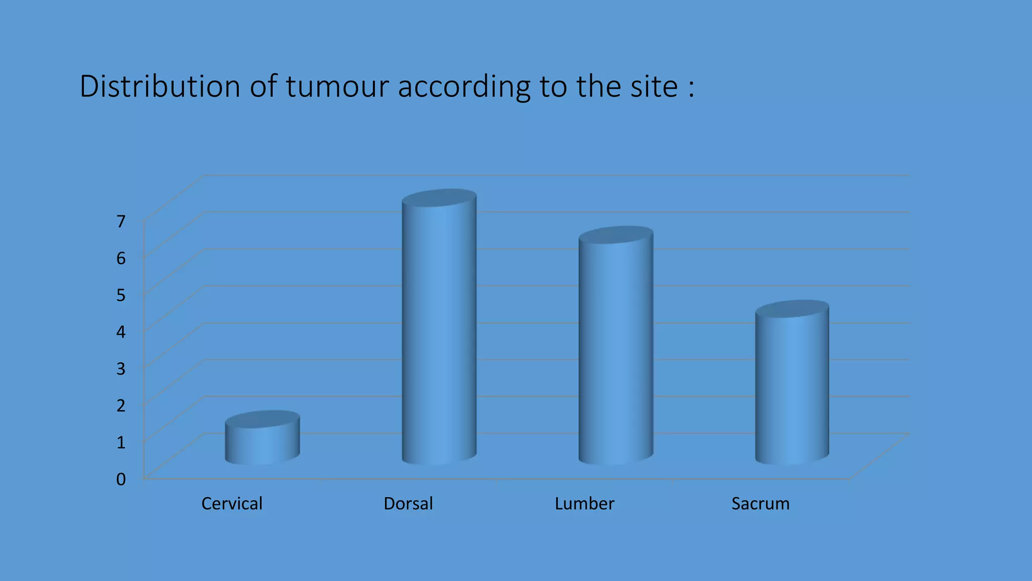
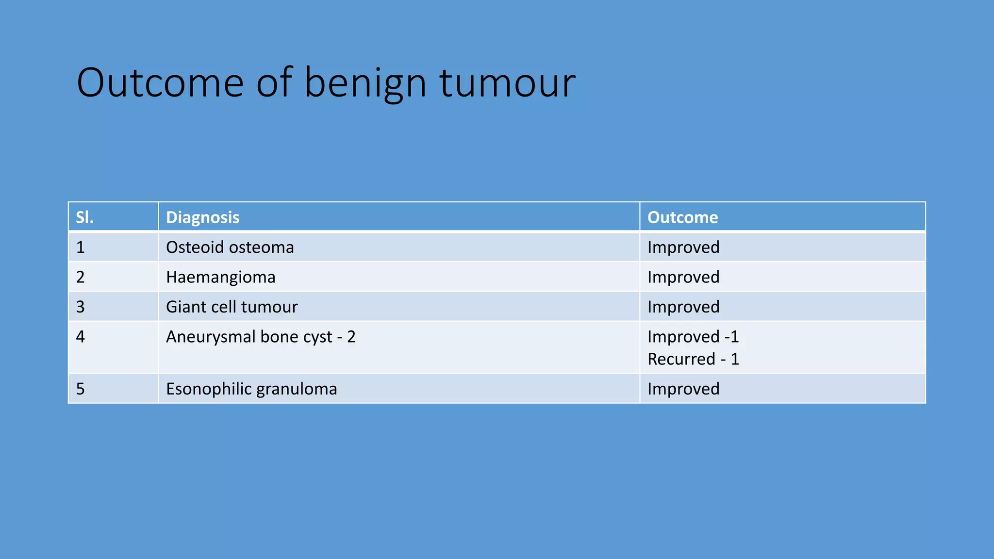
Primary bone tumour of the spine :
Experience of 18 cases.
Dr. Md. Shafiul Alam
Associate Professor
National Institute of Neurosciences & Hospital
Introduction
Primary bone tumour of the spine is a heterogynous group
of conditions that includes both benign and malignant
tumours, with only 6% of these tumours considered
malignant.
The spine is a frequent site of metastasis but primary
bone tumour of the spine are rare and make up only
4.2% of spinal tumour.
Eleven percent (11%) of primary musculoskeletal tumour
occur in the spine with certain tumours having an affinity
for the spine.
Although rare, these tumour offer a considerable
diagnostic dilemma and therapeutic challenge.
Timely diagnosis is crucial, as immediate attention could
affect overall prognosis.
The most common primary tumour of the spine is
metastatic deposits.
A number of both benign and malignant tumours may
arise primarily from the spine.
Benign tomours are
- Osteoid osteoma,
- osteoblastoma,
- Osteochondroma,
- Giant cell tumour (GCT),
- Aneurysmal bone cyst (ABC),
- Eosinophilic granuloma (EG),
- Haemangioma.
Malignant tumours are
- Malignant lymphoma,
- Sarcoma,
- Ewing sarcoma,
- Osteosarcoma,
- Chondrosarcoma,
- Malignant fibrous histiocytoma (MFH),
- Fibrosarcoma,
- Chordoma,
- Plasmacytoma/multiple myeloma
- Peripheral primitive neuroectodermal tumour (pPNET)
Primary bone tumours are more common in the
thoracic and lumbosacral regions than in the cervical
spine.
These tumours occur according to a typical anatomical
distribution within the vertebra. In general, malignant
tumours occur more frequently in the anterior elements
and benign tumours in the posterior elements.
The aims of treatment of these axial neoplasms include
complete resection if feasible, preservation of
neurological function and stabilisation of the spine if
needed.
The aim of our study was to review the primary bone
tumours of the spine treated at our unit regarding the
presentation, the surgical strategy employed and the
outcome.
Materials and methods
This is a retrospective study.
All the primary bone tumours of the spine that received
surgical treatment at the department of Neurosurgery of
Dhaka Medical College Hospital and National Institute of
Neurosciences and Hospital Dhaka, Bangladesh from
2007 to 2016 were included in this study.
We excluded all secondary deposition in the spine.
Materials and Methods
We treated 18 cases during this period that were included
according to these criteria.
A case note and imaging review was done where patient
demographics, details of presentation, clinical information
and the management strategy were analyzed.
All complications and mortalities were recorded.
Results
Between 2007 and 2016, a total 18 patients were diagnosed as primary
bone tumour of the spine.
The median age at presentation was 36 years (range 14 to 49 years)
with eleven(61.12%) male and seven(38.88%) female patients affected.
The median follow-up was 12 months (range 3 to 36 months).
Table 1 : Distribution of patient by age.
Age group Frequency Percentage
0 to 10 years 0 0
11 to 20 years 3 16.67
21 to 30 years 7 38.89
31 to 40 years 4 22.22
41 to 50 years 4 22.22
Total 18 100
Table 2 : Distribution of patient by sex.
Sex Frequency Percentage
Male 11 61.12
Female 7 38.88
Total 18 100
11
7
Sex distribution
Table 3 : Distribution of patient by site of involvement.
Site of involvement Frequency Percentage
Cervical 1 5.55
Dorsal 7 38.89
Lumber 6 33.34
Sacrum 4 22.22
Total 18 100
Distribution of tumour according to the site :
0
1
2
3
4
5
6
7
Cervical Dorsal Lumber Sacrum
Investigation
The most utilized diagnostic tool was MRI and CT scan of
the spine. It helped to diagnose the case as well as
planning of surgery.
X ray is no longer a standard diagnostic tool of spine
tumour, it was used in all cases as a complimentary
diagnostic tool. It also helped to find out the extra spinal
involvement like rib cases in two cases.
Pre-operative CT guided FNAC was done in seven cases.
Post-operative histopathological study was done in all cases.
Primary bone tumour of the spine most commonly
involved the dorsal (38.89%) and the Lumber (33.34%).
It affect the sacral (22.22%) and the cervical spine
(5.55%) less frequently.
Histopathological findings
Benign
-Osteoid osteoma
-Haemangioma
-Giant cell tumour
-Aneurysmal bone cyst
-Esonophilic granuloma
1
1
1
2
1
5.55
5.55
5.55
11.11
5.55
Malignant
-Myxoid Chondrosarcoma
-ewing’s sarcoma
-Chondrosarcoma
-Chordoma
-Multiple myeloma
-pNET
1
2
2
3
2
2
5.55
11.11
22.22
33.33
22.22
22.22
6
12
Type of tumour
Benign
Malignant
Pain was the most common presenting symptom (15 from 18
cases) and four cases presented with pain alone.
Pain was mostly axial pain in the affected region (11 of 18
cases), although three patients presented with chest pain and
one with flank pain.
Other presenting symptoms included neurological symptoms
and odynophagia
Recurance of tumor
Two of the recurrent cases were in malignant tumours
and one in benign aggressive tumour.
In the one recurrent malignant tumour a repeat
attempted marginal resection was performed.
The other two recurrent cases were not re-operated as
resection was not possible with acceptable morbidity.
We had five deaths in the series.
F/35yrs. : Ewing Sarcoma
Fig. : 1
M/27yrs. : Ewing Sarcoma
F/30 yrs. : Chondrosarcoma
F/21 yrs. : Aneurysmal bone cyst
Outcome of benign tumour
Sl. Diagnosis Outcome
1 Osteoid osteoma Improved
2 Haemangioma Improved
3 Giant cell tumour Improved
4 Aneurysmal bone cyst - 2 Improved -1
Recurred - 1
5 Esonophilic granuloma Improved
Outcome of malignat tumour
Sl. Diagnosis Outcome
1 Myxoid chondrosarcoma - 1 Expired
2 Ewing sarcoma - 2 Improved -1
Expired -1
3 Chondrosarcoma -2 Improved -1
Expired -1
4 Chordoma -3 Improved -2
Deteriorate -1
5 Multiple myeloma -2 Improved -2
6 pNET -2 Expied -2
Discussion
The most common presenting complaint of patients with
primary tumour is pain, with roughly 60% complaining of axial
pain and 25% of radicular symptoms.
The presence of a neurological deficit carries a worse
prognosis. Pain at rest or night pain is red flag symptoms that
warrant further investigation. Spinal deformity is obvious
when present, but occurs in less than 10% of patients.
The most common cause of a painful scoliosis in adolescents is
an osteoid osteoma. A delay in diagnosis is common and our
finding is confirmed in other series.
This is due to the presenting symptoms often being vague,
pain often being the only symptom, and the initial radiographs
often only showing subtle or no abnormalities leading to the
symptoms not being properly investigated.
Most patients report a slow, gradual onset of pain in the
involved area. In benign tumours of the spine an average of
26 weeks of symptoms before presentation are reported.
This was confirmed in our series where the average delay in
presentation was 7 months.
Recent advances in chemotherapy have led to improved survival in
malignant primary bone tumour, including in the spine.
Adjuvant radiotherapy is indicated in malignant and some benign
aggressive lesions.
The use of local radiotherapy does increase the risk of major local
complications, including sepsis. We used radio- and chemotherapy
in a number of cases as guided by oncology unit.
.
Limitation of the study
The main limitations of this study are the retrospective
character and the relatively small numbers.
Conclusion
Most primary bone tumour of the spine in our study occurred
in adults and were malignant.
Surgical treatment resulted in pain reduction and neurological
improvement, allowing a rapid adjuvant therapy in good
clinical condition.
Malignant hematopoietic tumors had an intermediate one-
year prognosis ranging between benign and other malignant
PBTS.
Although hematopoietic tumors are characterized by early
systemic spread, spinal surgeons may well manage the ones
presenting with acute primary spinal manifestation.
An experienced multidisciplinary team of tertiary care center
should perform diagnosis and treatment of these extremely
rare tumors.
We conclude that primary bone tumours of the spine are
often associated with a significant delay in diagnosis.
Surgical strategy and adjuvant therapy should be
individualised for each case.
Acceptable results with minimal complications can be
achieved with this approach.
Thank you all.

Primary Bone Tumour of the Spine

  • 1.
    Primary bone tumourof the spine : Experience of 18 cases. Dr. Md. Shafiul Alam Associate Professor National Institute of Neurosciences & Hospital
  • 3.
    Introduction Primary bone tumourof the spine is a heterogynous group of conditions that includes both benign and malignant tumours, with only 6% of these tumours considered malignant.
  • 4.
    The spine isa frequent site of metastasis but primary bone tumour of the spine are rare and make up only 4.2% of spinal tumour. Eleven percent (11%) of primary musculoskeletal tumour occur in the spine with certain tumours having an affinity for the spine.
  • 5.
    Although rare, thesetumour offer a considerable diagnostic dilemma and therapeutic challenge. Timely diagnosis is crucial, as immediate attention could affect overall prognosis.
  • 6.
    The most commonprimary tumour of the spine is metastatic deposits. A number of both benign and malignant tumours may arise primarily from the spine.
  • 7.
    Benign tomours are -Osteoid osteoma, - osteoblastoma, - Osteochondroma, - Giant cell tumour (GCT), - Aneurysmal bone cyst (ABC), - Eosinophilic granuloma (EG), - Haemangioma.
  • 8.
    Malignant tumours are -Malignant lymphoma, - Sarcoma, - Ewing sarcoma, - Osteosarcoma, - Chondrosarcoma, - Malignant fibrous histiocytoma (MFH), - Fibrosarcoma, - Chordoma, - Plasmacytoma/multiple myeloma - Peripheral primitive neuroectodermal tumour (pPNET)
  • 9.
    Primary bone tumoursare more common in the thoracic and lumbosacral regions than in the cervical spine. These tumours occur according to a typical anatomical distribution within the vertebra. In general, malignant tumours occur more frequently in the anterior elements and benign tumours in the posterior elements.
  • 10.
    The aims oftreatment of these axial neoplasms include complete resection if feasible, preservation of neurological function and stabilisation of the spine if needed. The aim of our study was to review the primary bone tumours of the spine treated at our unit regarding the presentation, the surgical strategy employed and the outcome.
  • 11.
    Materials and methods Thisis a retrospective study. All the primary bone tumours of the spine that received surgical treatment at the department of Neurosurgery of Dhaka Medical College Hospital and National Institute of Neurosciences and Hospital Dhaka, Bangladesh from 2007 to 2016 were included in this study. We excluded all secondary deposition in the spine.
  • 12.
    Materials and Methods Wetreated 18 cases during this period that were included according to these criteria. A case note and imaging review was done where patient demographics, details of presentation, clinical information and the management strategy were analyzed. All complications and mortalities were recorded.
  • 13.
    Results Between 2007 and2016, a total 18 patients were diagnosed as primary bone tumour of the spine. The median age at presentation was 36 years (range 14 to 49 years) with eleven(61.12%) male and seven(38.88%) female patients affected. The median follow-up was 12 months (range 3 to 36 months).
  • 14.
    Table 1 :Distribution of patient by age. Age group Frequency Percentage 0 to 10 years 0 0 11 to 20 years 3 16.67 21 to 30 years 7 38.89 31 to 40 years 4 22.22 41 to 50 years 4 22.22 Total 18 100
  • 15.
    Table 2 :Distribution of patient by sex. Sex Frequency Percentage Male 11 61.12 Female 7 38.88 Total 18 100
  • 16.
  • 17.
    Table 3 :Distribution of patient by site of involvement. Site of involvement Frequency Percentage Cervical 1 5.55 Dorsal 7 38.89 Lumber 6 33.34 Sacrum 4 22.22 Total 18 100
  • 18.
    Distribution of tumouraccording to the site : 0 1 2 3 4 5 6 7 Cervical Dorsal Lumber Sacrum
  • 19.
    Investigation The most utilizeddiagnostic tool was MRI and CT scan of the spine. It helped to diagnose the case as well as planning of surgery. X ray is no longer a standard diagnostic tool of spine tumour, it was used in all cases as a complimentary diagnostic tool. It also helped to find out the extra spinal involvement like rib cases in two cases.
  • 20.
    Pre-operative CT guidedFNAC was done in seven cases. Post-operative histopathological study was done in all cases.
  • 21.
    Primary bone tumourof the spine most commonly involved the dorsal (38.89%) and the Lumber (33.34%). It affect the sacral (22.22%) and the cervical spine (5.55%) less frequently.
  • 22.
    Histopathological findings Benign -Osteoid osteoma -Haemangioma -Giantcell tumour -Aneurysmal bone cyst -Esonophilic granuloma 1 1 1 2 1 5.55 5.55 5.55 11.11 5.55
  • 23.
    Malignant -Myxoid Chondrosarcoma -ewing’s sarcoma -Chondrosarcoma -Chordoma -Multiplemyeloma -pNET 1 2 2 3 2 2 5.55 11.11 22.22 33.33 22.22 22.22
  • 24.
  • 25.
    Pain was themost common presenting symptom (15 from 18 cases) and four cases presented with pain alone. Pain was mostly axial pain in the affected region (11 of 18 cases), although three patients presented with chest pain and one with flank pain. Other presenting symptoms included neurological symptoms and odynophagia
  • 26.
    Recurance of tumor Twoof the recurrent cases were in malignant tumours and one in benign aggressive tumour. In the one recurrent malignant tumour a repeat attempted marginal resection was performed. The other two recurrent cases were not re-operated as resection was not possible with acceptable morbidity. We had five deaths in the series.
  • 27.
    F/35yrs. : EwingSarcoma Fig. : 1
  • 28.
  • 29.
    F/30 yrs. :Chondrosarcoma
  • 31.
    F/21 yrs. :Aneurysmal bone cyst
  • 33.
    Outcome of benigntumour Sl. Diagnosis Outcome 1 Osteoid osteoma Improved 2 Haemangioma Improved 3 Giant cell tumour Improved 4 Aneurysmal bone cyst - 2 Improved -1 Recurred - 1 5 Esonophilic granuloma Improved
  • 34.
    Outcome of malignattumour Sl. Diagnosis Outcome 1 Myxoid chondrosarcoma - 1 Expired 2 Ewing sarcoma - 2 Improved -1 Expired -1 3 Chondrosarcoma -2 Improved -1 Expired -1 4 Chordoma -3 Improved -2 Deteriorate -1 5 Multiple myeloma -2 Improved -2 6 pNET -2 Expied -2
  • 35.
    Discussion The most commonpresenting complaint of patients with primary tumour is pain, with roughly 60% complaining of axial pain and 25% of radicular symptoms. The presence of a neurological deficit carries a worse prognosis. Pain at rest or night pain is red flag symptoms that warrant further investigation. Spinal deformity is obvious when present, but occurs in less than 10% of patients.
  • 36.
    The most commoncause of a painful scoliosis in adolescents is an osteoid osteoma. A delay in diagnosis is common and our finding is confirmed in other series. This is due to the presenting symptoms often being vague, pain often being the only symptom, and the initial radiographs often only showing subtle or no abnormalities leading to the symptoms not being properly investigated.
  • 37.
    Most patients reporta slow, gradual onset of pain in the involved area. In benign tumours of the spine an average of 26 weeks of symptoms before presentation are reported. This was confirmed in our series where the average delay in presentation was 7 months.
  • 38.
    Recent advances inchemotherapy have led to improved survival in malignant primary bone tumour, including in the spine. Adjuvant radiotherapy is indicated in malignant and some benign aggressive lesions. The use of local radiotherapy does increase the risk of major local complications, including sepsis. We used radio- and chemotherapy in a number of cases as guided by oncology unit. .
  • 39.
    Limitation of thestudy The main limitations of this study are the retrospective character and the relatively small numbers.
  • 40.
    Conclusion Most primary bonetumour of the spine in our study occurred in adults and were malignant. Surgical treatment resulted in pain reduction and neurological improvement, allowing a rapid adjuvant therapy in good clinical condition.
  • 41.
    Malignant hematopoietic tumorshad an intermediate one- year prognosis ranging between benign and other malignant PBTS. Although hematopoietic tumors are characterized by early systemic spread, spinal surgeons may well manage the ones presenting with acute primary spinal manifestation.
  • 42.
    An experienced multidisciplinaryteam of tertiary care center should perform diagnosis and treatment of these extremely rare tumors.
  • 43.
    We conclude thatprimary bone tumours of the spine are often associated with a significant delay in diagnosis. Surgical strategy and adjuvant therapy should be individualised for each case. Acceptable results with minimal complications can be achieved with this approach.
  • 44.