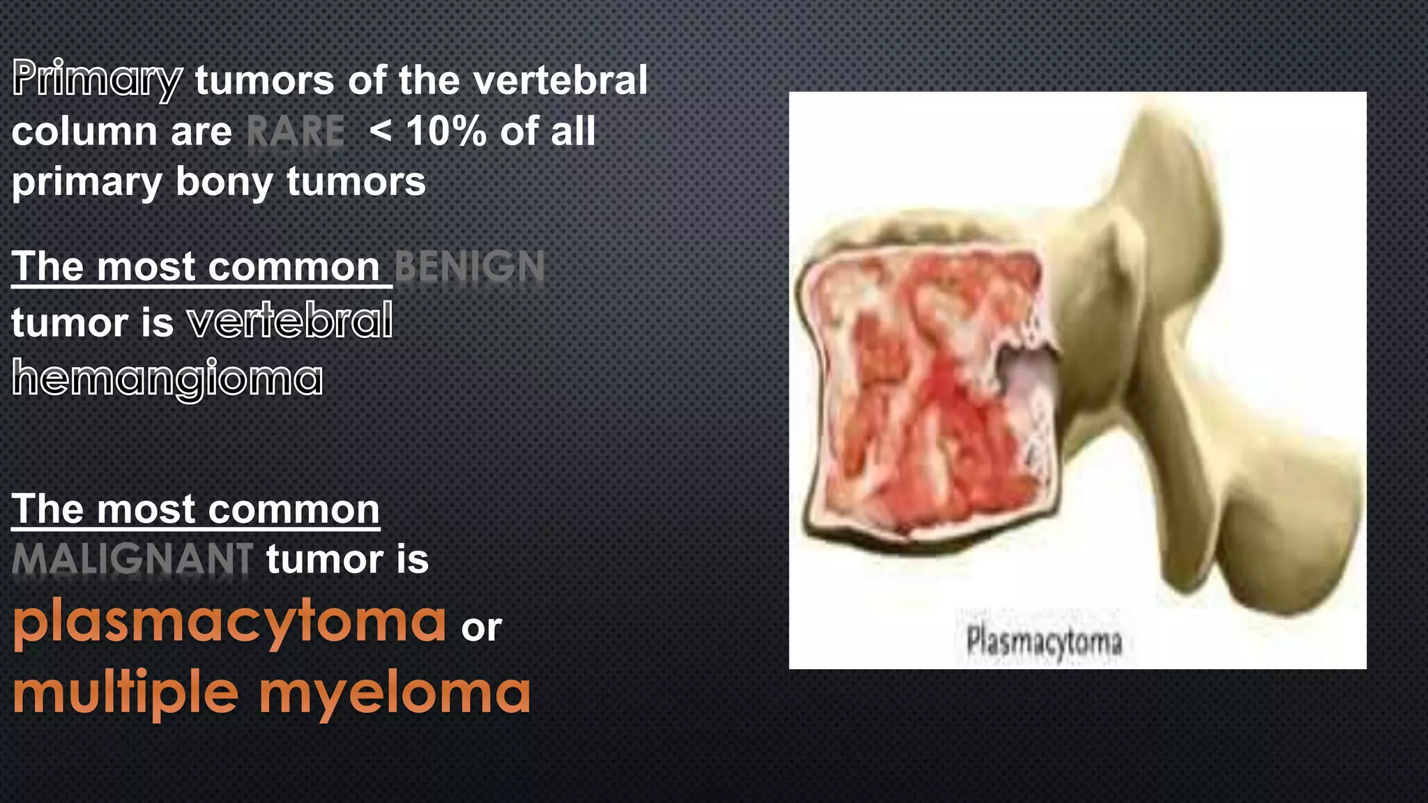
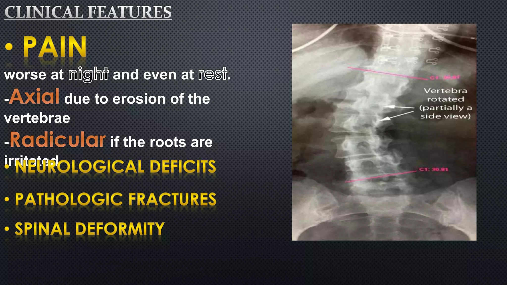
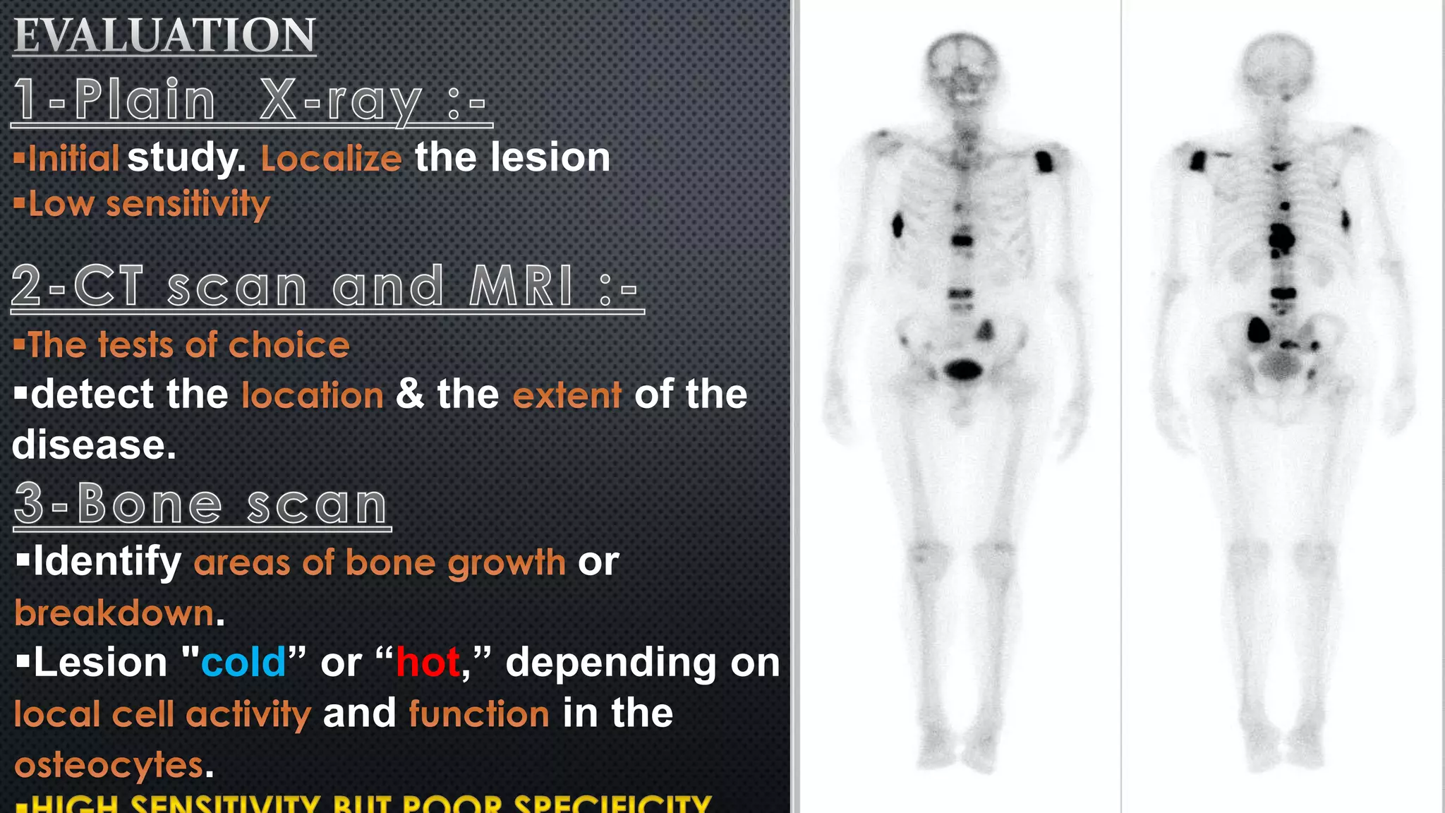
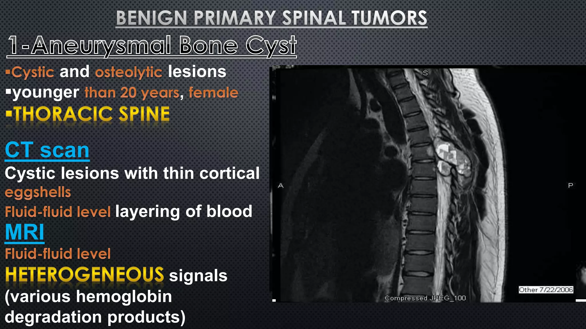
tumors of the vertebral 
column are RARE < 10% of all 
primary bony tumors 
The most common BENIGN 
tumor is 
The most common 
MALIGNANT tumor is 
or
worse at and even at . 
- due to erosion of the 
vertebrae 
- if the roots are 
irritated
study. the lesion 
detect the & the of the 
disease. 
Identify or 
. 
Lesion "cold” or “hot,” depending on 
and in the 
.
Use a 
( ) 
and ( 
) information regarding a 
tumor. 
Determine extent of the 
Preoperative
An accurate is essential in the 
evaluation and 
to or 
therapy 
, economical, , 
and minimally invasive 
procedure. 
of up to
and lesions 
younger , 
CT scan 
Cystic lesions with thin cortical 
layering of blood 
MRI 
signals 
(various hemoglobin 
degradation products)
Histological 
filled with blood 
products separated by 
. associated with 
Treatment 
to decrease 
intraoperative blood loss 
Complete excision (difficult) 
Incomplete (recurrence) 
(adjuvant)
The tumors 
Incidental findings, 
are 
Myelopathy 
Pathologic 
fractures 
Pain 
Radiculopathy 
CT scan 
MRI 
signals on both T1 
Hanids tTo2pathology 
Aggregate of irregularly shaped 
channels of 
with erosion of the bony
thoracic and lumbar spine. 
The is the most 
frequent site of 
Association between 
and the development of 
hemangiomas 
Treatment 
hemangiomas 
percutaneous injection of 
spinal cord compression or 
neurological symptoms
benign, , 
Teenage years, 
Involve the 
worse at 
 with 
CT scan 
Bone scans
Histopathology 
in a background of 
with 
sTurreraoutmndeinngt 
spinal cord compression or 
neurological symptoms 
 
Constant pain that is not relieved 
by salicylates 
is the
Benign, 
of immature 
or 
of 
CT scan 
MRI 
signals on T2 
signals on T1 
Treatment
Benign, cartilage 
radiation-induced 
cervical spine 
, 
CT scan 
MRI
Treatment 
Symptomatic lesions, Complete 
excision 
Rare ,arises from immature cartilage 
Radiographic findings are 
Round chondroblast-like cells and 
multinucleated giant cells in a 
background of cartilaginous 
intercellular matrix and focal 
calcification
3rd - 4th decades, 
is the most common site 
originates from 
Expansile mass with destruction 
of the vertebral bodies 
Histopathology:- 
Treatment 
to 
decrease intraoperative blood loss 
(adjuvant)
arise from remnants 
of the 
Found from the to the 
 5th – 6th decade , 
CT scan 
near the center 
of the vertebral body
MRI 
T1- within the 
lesion. 
T2- (high water 
cHoinstteonpt)athology 
Elongated cords of clear cells 
The cells contain intracytoplasmic 
vTarceuaotlmese wnitth a copious amount of 
(adjuvant) 
is traditionally 
age, , and 
adjuvant
very similar features to 
 5th – 6th decade , 
CT scan 
MRI 
demonstrate the relationship 
between the tumor and 
surrounding soft tissue 
Treatment
2ry to previous 
or . 
CT scan 
Treatment
primary malignant 
tumors of the vertebral column 
Arise within of the 
vertebral column ( ) 
by definition is 
is truly 
C6tTh – s7cthadnecade , 
MRI 
diffuse abnormal marrow 
signals of the vertebral column 
serum and urine
Treatment 
en bloc spondylectomy 
(adjuvant)
Spinal axis tumors

Spinal axis tumors

  • 2.
    tumors of thevertebral column are RARE < 10% of all primary bony tumors The most common BENIGN tumor is The most common MALIGNANT tumor is or
  • 3.
    worse at andeven at . - due to erosion of the vertebrae - if the roots are irritated
  • 4.
    study. the lesion detect the & the of the disease. Identify or . Lesion "cold” or “hot,” depending on and in the .
  • 5.
    Use a () and ( ) information regarding a tumor. Determine extent of the Preoperative
  • 6.
    An accurate isessential in the evaluation and to or therapy , economical, , and minimally invasive procedure. of up to
  • 7.
    and lesions younger, CT scan Cystic lesions with thin cortical layering of blood MRI signals (various hemoglobin degradation products)
  • 8.
    Histological filled withblood products separated by . associated with Treatment to decrease intraoperative blood loss Complete excision (difficult) Incomplete (recurrence) (adjuvant)
  • 9.
    The tumors Incidentalfindings, are Myelopathy Pathologic fractures Pain Radiculopathy CT scan MRI signals on both T1 Hanids tTo2pathology Aggregate of irregularly shaped channels of with erosion of the bony
  • 10.
    thoracic and lumbarspine. The is the most frequent site of Association between and the development of hemangiomas Treatment hemangiomas percutaneous injection of spinal cord compression or neurological symptoms
  • 11.
    benign, , Teenageyears, Involve the worse at  with CT scan Bone scans
  • 12.
    Histopathology in abackground of with sTurreraoutmndeinngt spinal cord compression or neurological symptoms  Constant pain that is not relieved by salicylates is the
  • 13.
    Benign, of immature or of CT scan MRI signals on T2 signals on T1 Treatment
  • 14.
    Benign, cartilage radiation-induced cervical spine , CT scan MRI
  • 15.
    Treatment Symptomatic lesions,Complete excision Rare ,arises from immature cartilage Radiographic findings are Round chondroblast-like cells and multinucleated giant cells in a background of cartilaginous intercellular matrix and focal calcification
  • 16.
    3rd - 4thdecades, is the most common site originates from Expansile mass with destruction of the vertebral bodies Histopathology:- Treatment to decrease intraoperative blood loss (adjuvant)
  • 17.
    arise from remnants of the Found from the to the  5th – 6th decade , CT scan near the center of the vertebral body
  • 18.
    MRI T1- withinthe lesion. T2- (high water cHoinstteonpt)athology Elongated cords of clear cells The cells contain intracytoplasmic vTarceuaotlmese wnitth a copious amount of (adjuvant) is traditionally age, , and adjuvant
  • 19.
    very similar featuresto  5th – 6th decade , CT scan MRI demonstrate the relationship between the tumor and surrounding soft tissue Treatment
  • 20.
    2ry to previous or . CT scan Treatment
  • 21.
    primary malignant tumorsof the vertebral column Arise within of the vertebral column ( ) by definition is is truly C6tTh – s7cthadnecade , MRI diffuse abnormal marrow signals of the vertebral column serum and urine
  • 22.
    Treatment en blocspondylectomy (adjuvant)