Malignant vs. BenignTumors
“Rapid growth, warmth, tenderness,
and ill defined edges are suggestive of
malignancy.”
3.
Classification of malignanttumors of
bone:
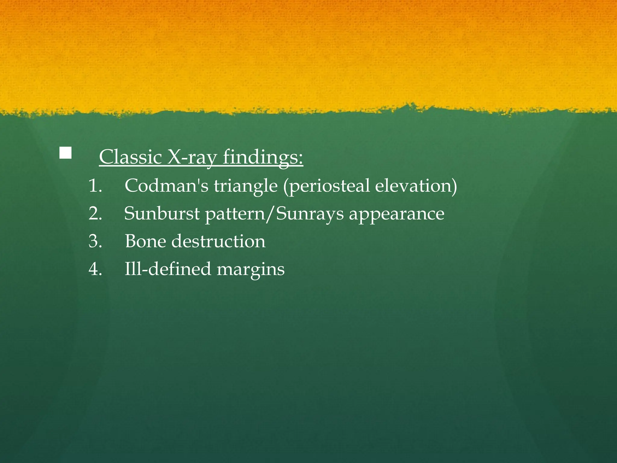
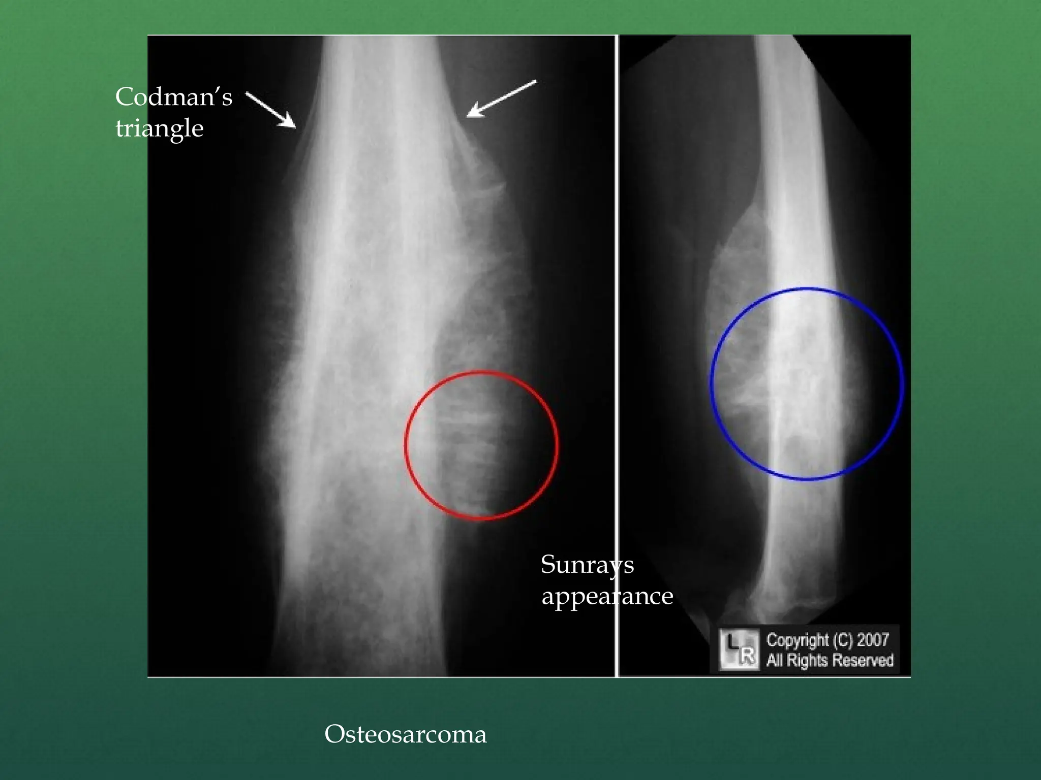
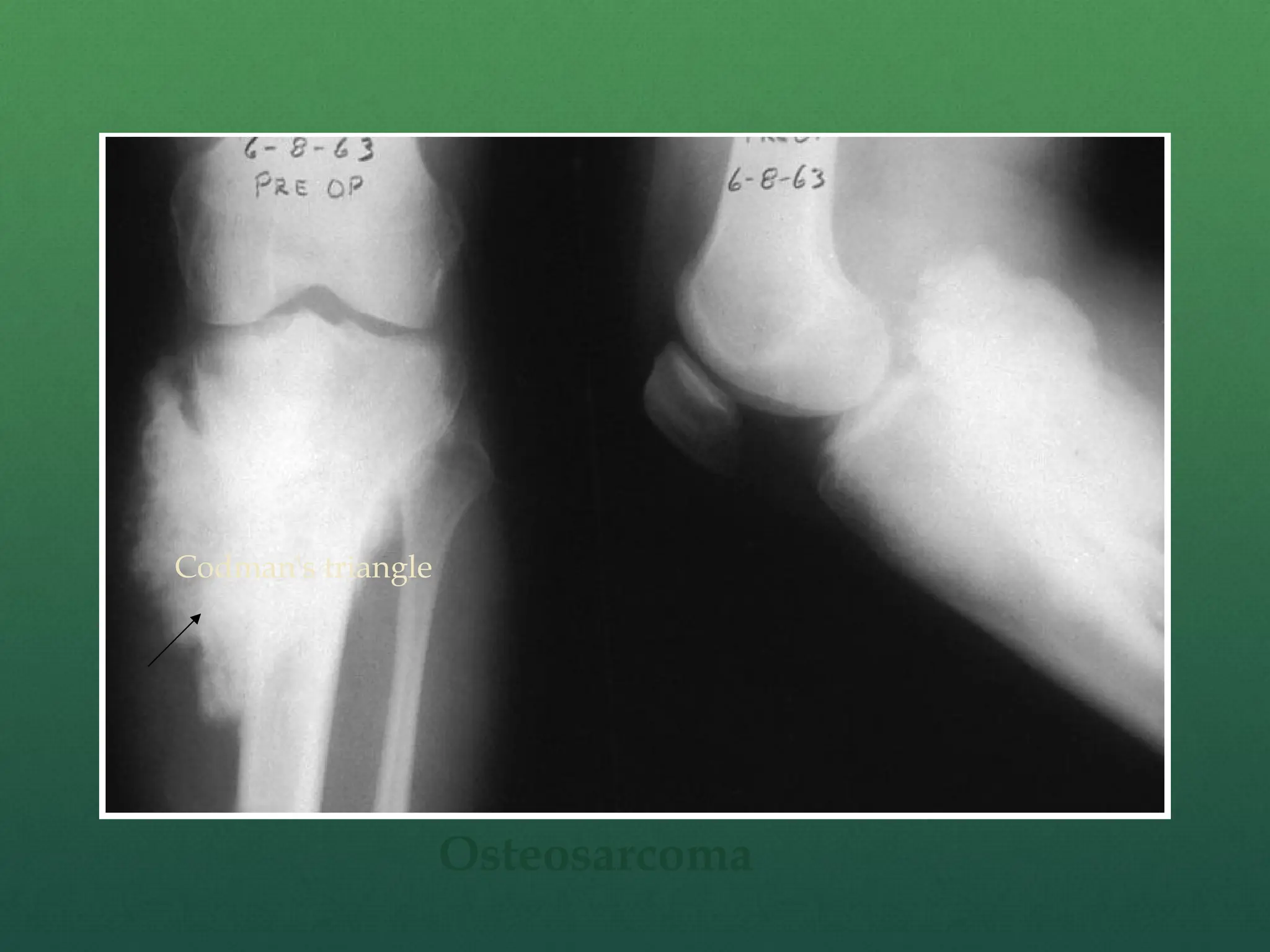
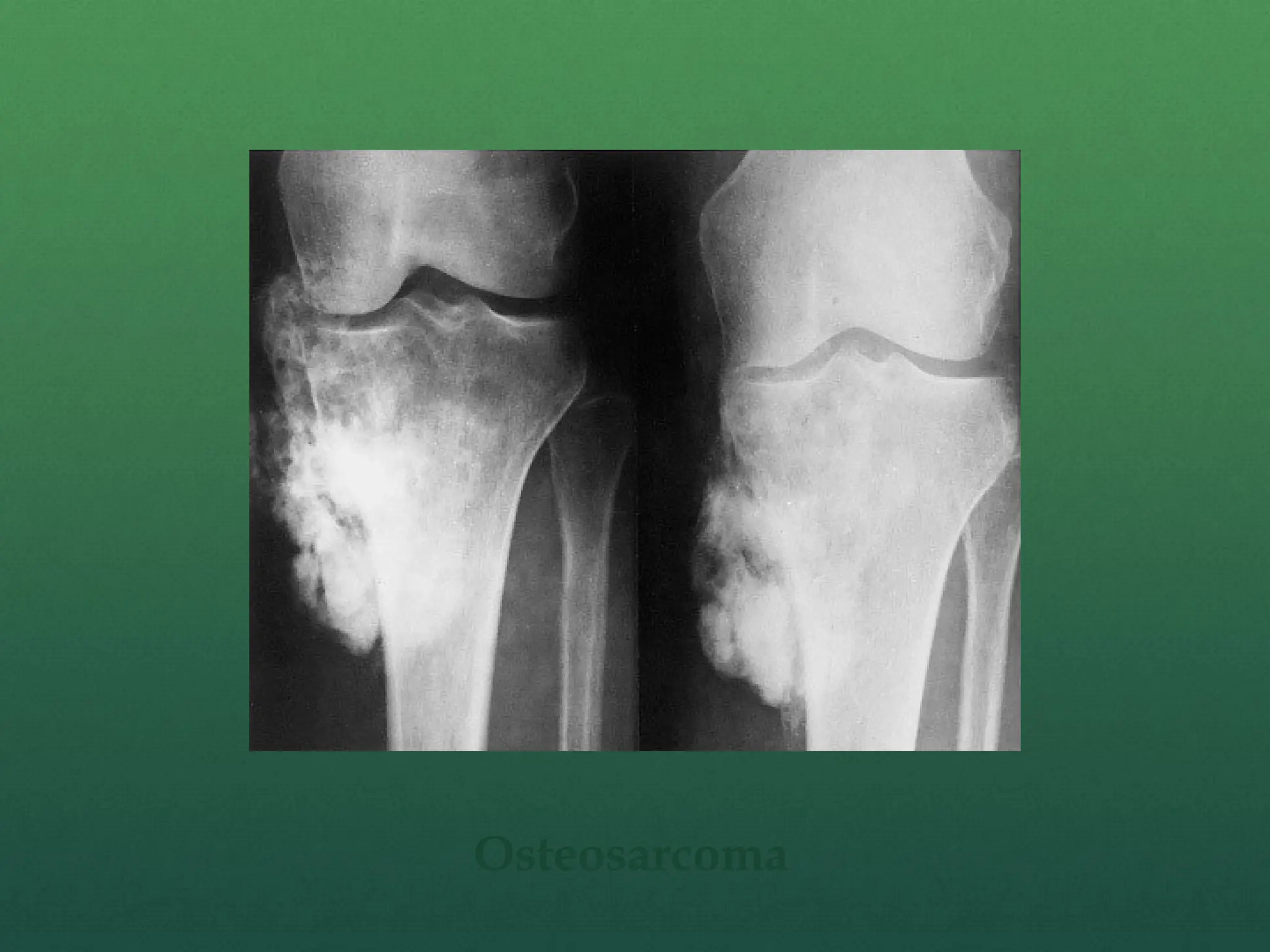
1. Osteosarcoma (Osteogenic sarcoma)
2. Chondrosarcoma
3. Ewing’s sarcoma
4. Multiple myeloma
4.
Osteosarcoma
(Osteogenic sarcoma)
Itis a malignant mesenchymal tumor in which
cancellous cell produce bone matrix.
Most common primary malignant tumor of bone
Occurs in all age group but has bimodal age distribution
75% occur in person younger than 20 years of age
Second peak occur in elderly who have predisposing
condition – Paget disease, bone infarct, prior irradiation
Males> females
5.
Usually arisefrom metaphysis of long bones of
extremities, and almost 50% occur about the knee.
Beyond the age of 25 years incidence in flat bones
and long bones is almost equal.
6.
Pathogenesis
Approx. 70%have acquired genetic abnormalities such as
ploidy changes and chromosomal aberrations, none of
which is specific for this tumor.
Mutation of RB gene (cell cycle regulator) and p53 gene
(gene whose product regulate DNA repair and cellular
metabolism) frequently associated with osteosarcoma.
Germline mutation in RB gene roughly 1000-fold
increase the risk of osteosarcoma
Patient with Li-Fraumeni syndrome (germline p53
mutation) greatly elevate the incidence of osteosarcoma.
7.
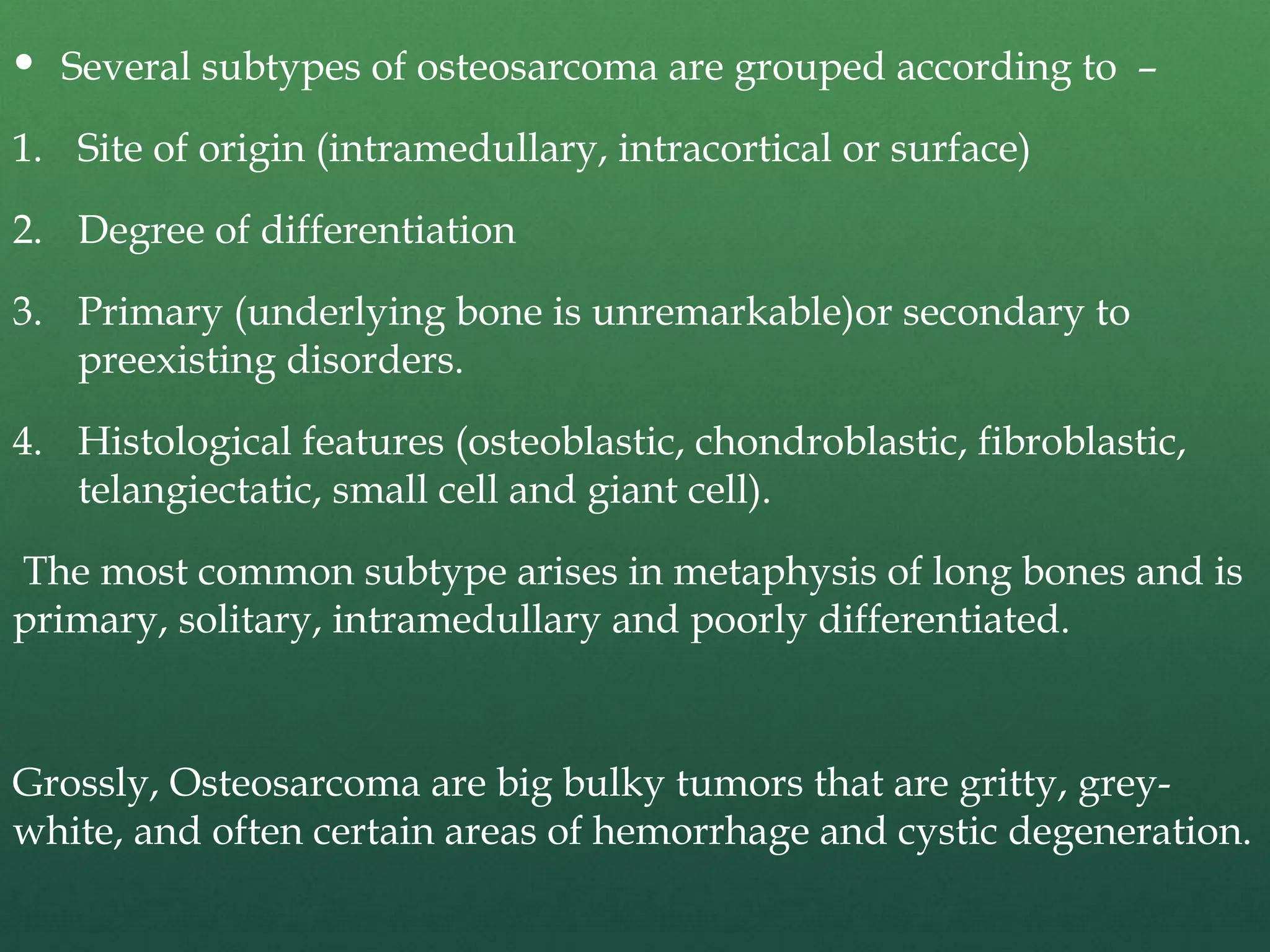
Several subtypesof osteosarcoma are grouped according to –
1. Site of origin (intramedullary, intracortical or surface)
2. Degree of differentiation
3. Primary (underlying bone is unremarkable)or secondary to
preexisting disorders.
4. Histological features (osteoblastic, chondroblastic, fibroblastic,
telangiectatic, small cell and giant cell).
The most common subtype arises in metaphysis of long bones and is
primary, solitary, intramedullary and poorly differentiated.
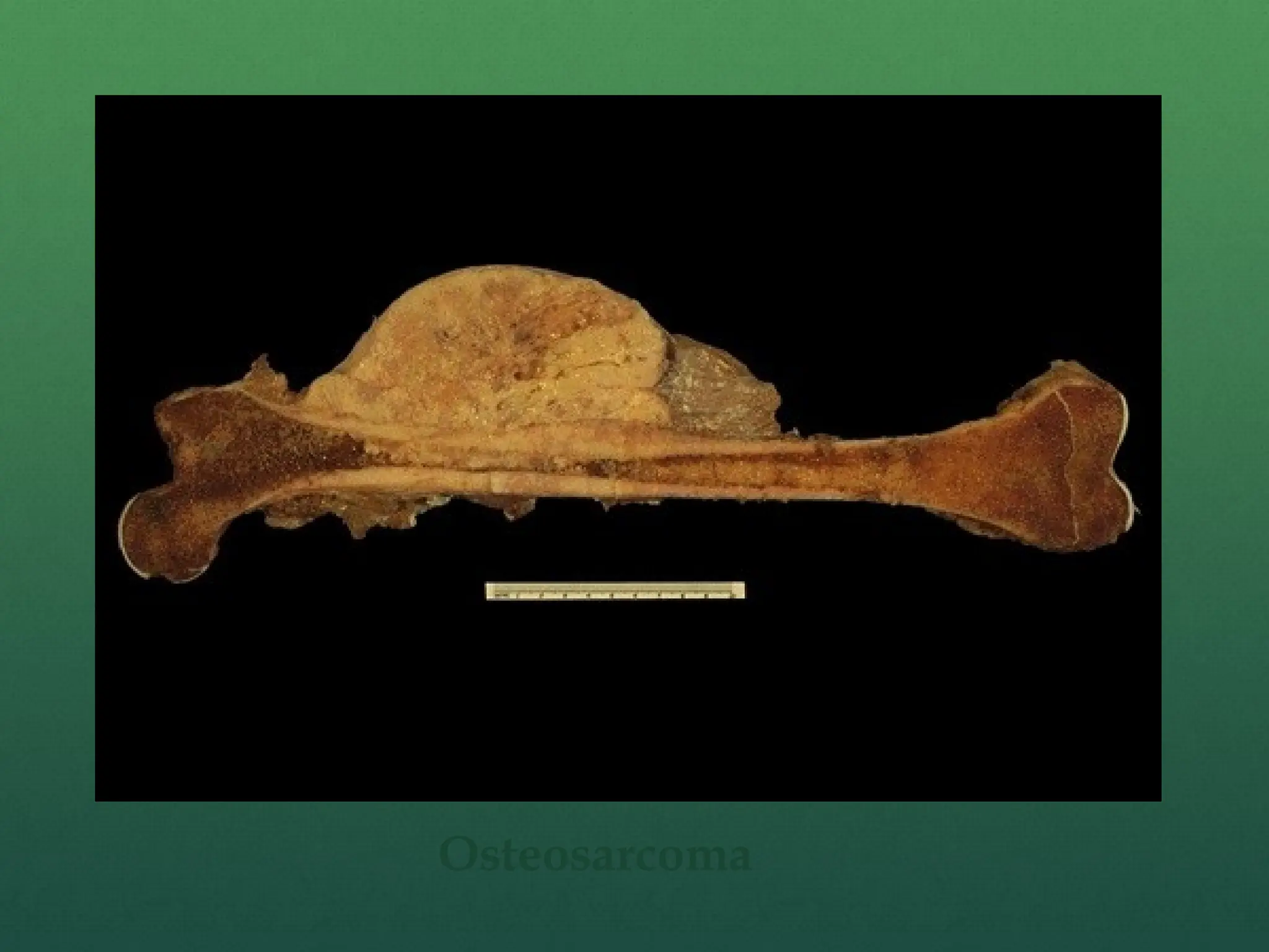
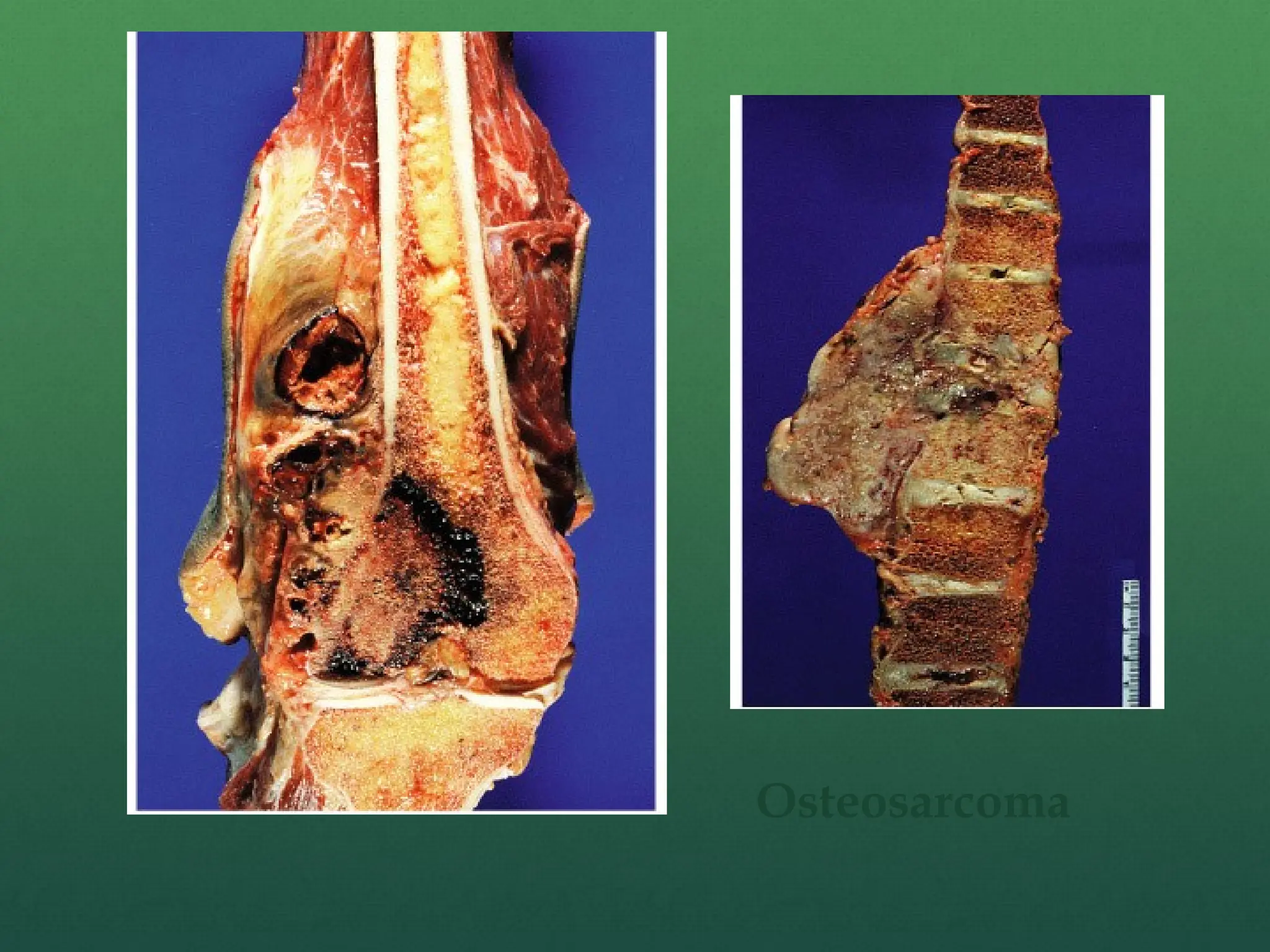
Grossly, Osteosarcoma are big bulky tumors that are gritty, grey-
white, and often certain areas of hemorrhage and cystic degeneration.
Clinical features
Localizedpain and swelling
Fast growing tumor
Progressive weakness and weight loss
Skin over the tumor is shiny and stretched with prominent veins
Warm, tender and ill defined margins.
Pulsatile tumor
Movement of adjacent joint restricted due to mechanical
obstruction and effusion.
Regional lymph node enlarged only in 25-30% cases.
If distal neurovascular deficit present strongly suggest
malignancy.
Lung metastasis occur in 10-12 months if left untreated.
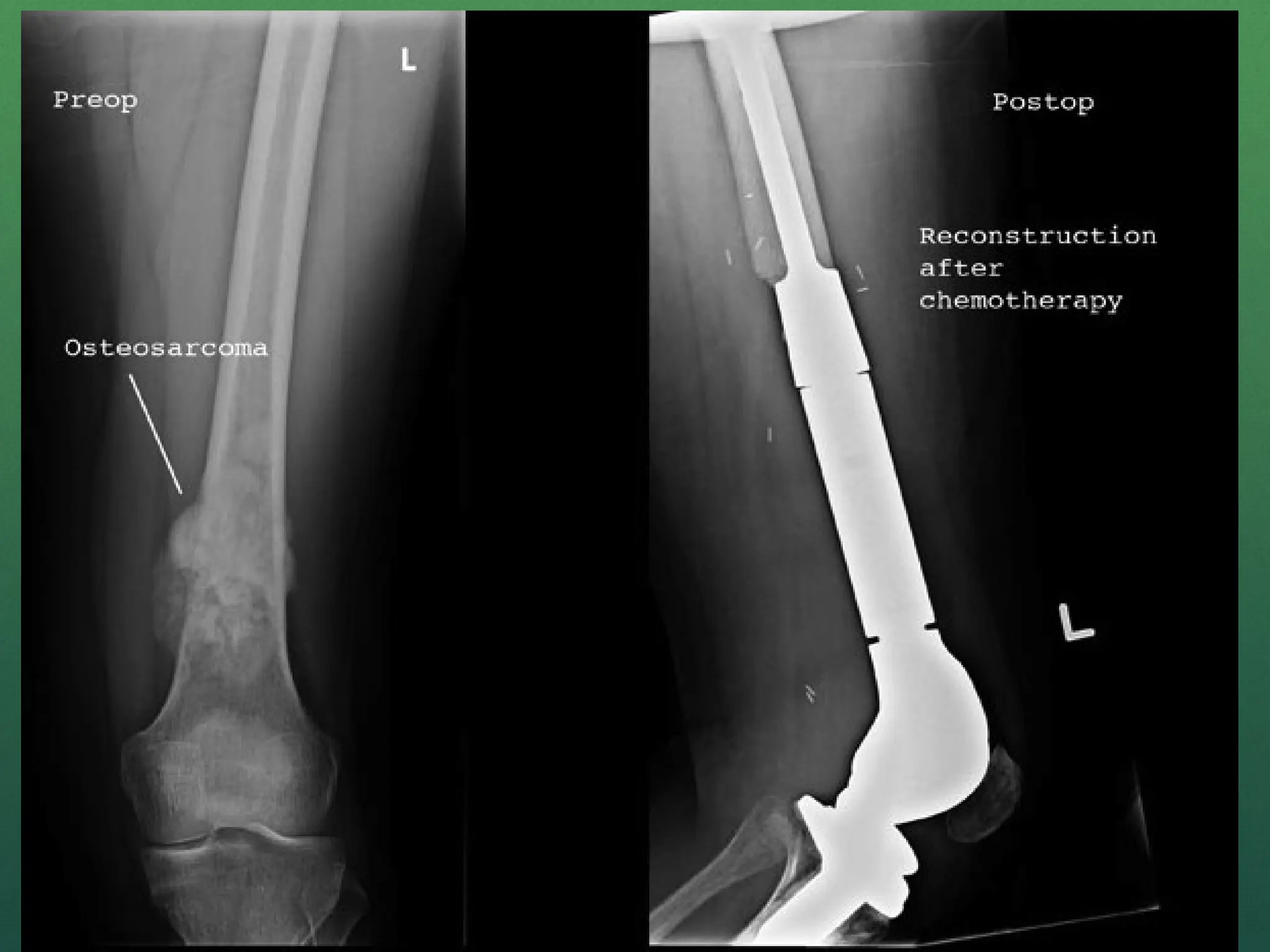
Surgery
Disarticulation
Amputation
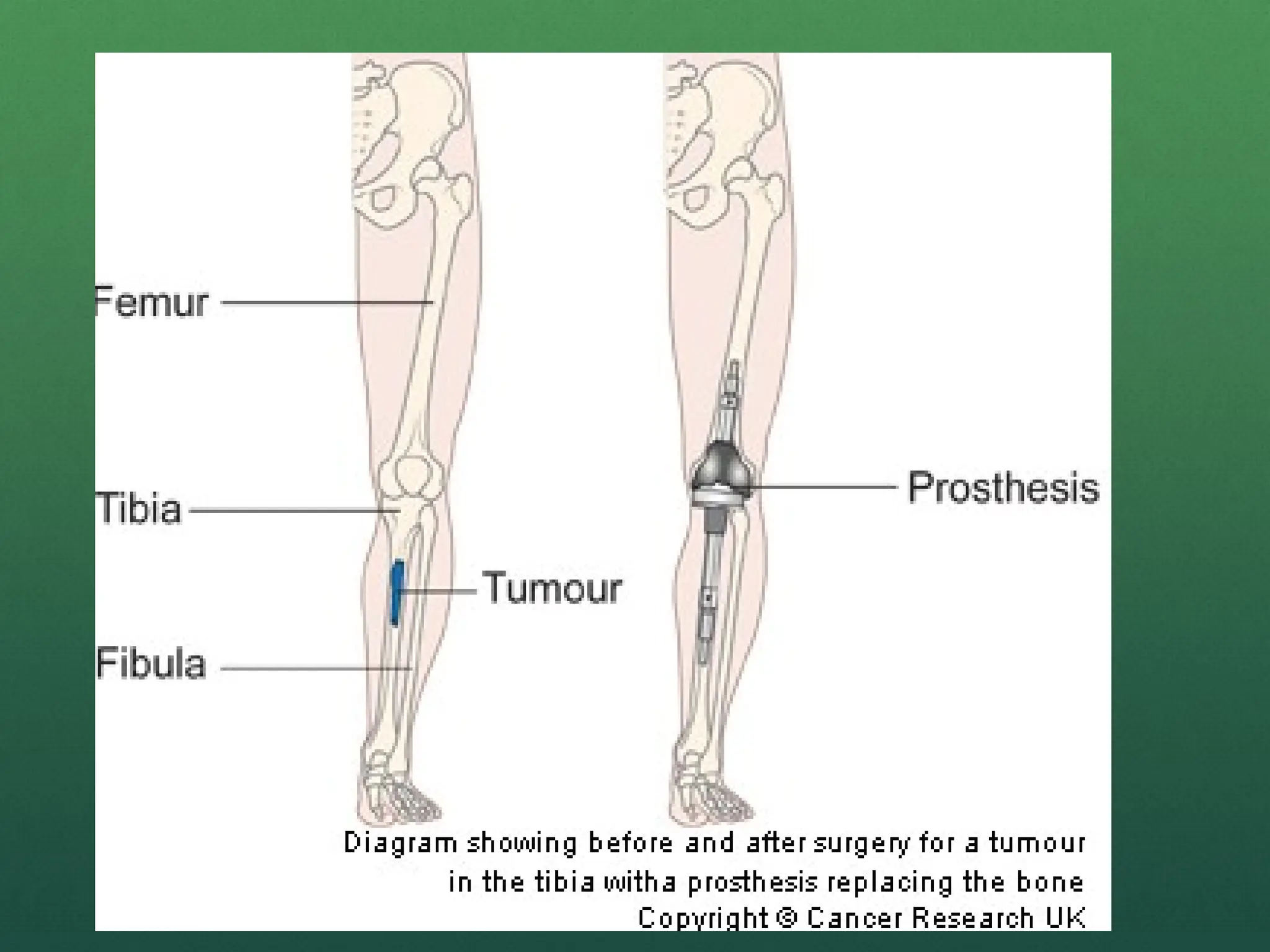
Resection with reconstruction/endoprosthesis
Limb salvage surgery
Resection of metastatic lesion (lobectomy in lung)
19.
Limb salvage surgery
Principle is to eradicate the bone tumor, retain integrity of
skeletal system and preserve the limb with useful function.
After resection, skeletal reconstruction done by bone
grafting(auto or allograft) or by endoprosthesis (modular or
custom made).
Prosthetic reconstruction is more effective
As compared to the radical amputation and external
prosthetic fitting or limb sparing surgery with bone grafting
this treatment is more effective in early mobilization.
22.
Chondrosarcoma
Definition:
Malignanttumor of chondroblasts cells
Second most common malignant matrix producing tumor
Etiology:
The tumor may arise de novo (primary) or secondary to
preexisting enchondroma, exostosis (osteochondromas) or
Paget’s disease
Primary chodrosarcoma is very uncommon, arises centrally in
the bone and found in children
23.
Chondrosarcoma subclassified according to –
Site – Central (intramedullary)
Peripheral (juxtacortical and surface)
Histologically - Conventional (hyaline/myxoid)
Clear cell
Dedifferentiated
Mesenchymal
Conventional central tumors constitute about 90% of
chondrosarcoma
24.
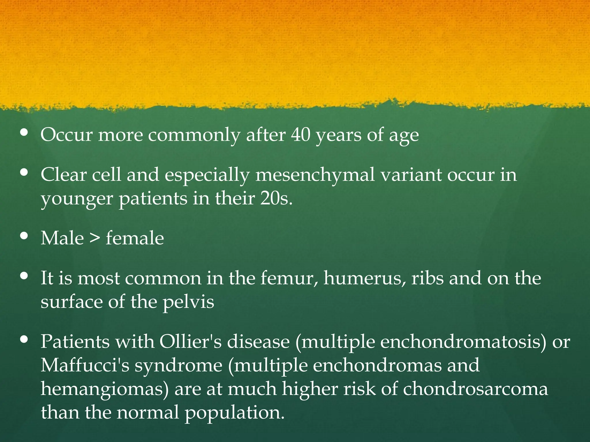
Occur morecommonly after 40 years of age
Clear cell and especially mesenchymal variant occur in
younger patients in their 20s.
Male > female
It is most common in the femur, humerus, ribs and on the
surface of the pelvis
Patients with Ollier's disease (multiple enchondromatosis) or
Maffucci's syndrome (multiple enchondromas and
hemangiomas) are at much higher risk of chondrosarcoma
than the normal population.
25.
Clinical features
Presentationof chondrosarcoma depends on the grade of the
tumor.
A high-grade, fast growing tumor can present with excruciating
pain.
A low grade, more indolent tumor is more likely to present as an
older patient complaining of hip pain and swelling.
Pelvic tumors present with urinary frequency or obstruction or
may masquerade as "groin muscle pulls".
26.
Investigations
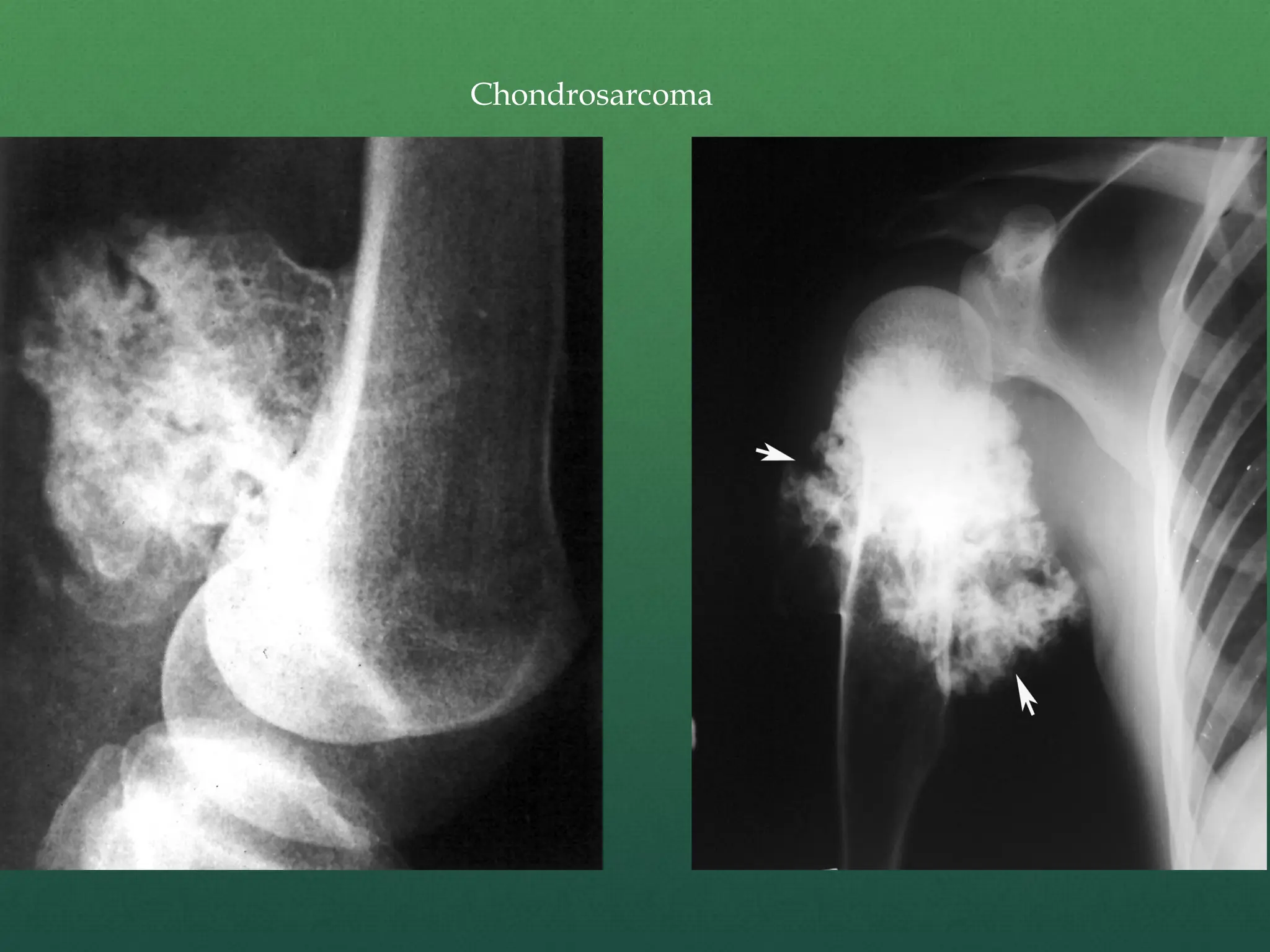
X –ray –
Chondrosarcoma is a fusiform, lucent defect with
scalloping of the inner cortex and periosteal reaction.
Extension into the soft tissue may be present as well as
punctate or stippled calcification of the cartilage
matrix.
C.T. scan–
Helpful in defining the integrity of the cortex and distribution of
calcification.
MRI –
Surgical planning as it demonstrates the intraosseus and soft
tissue involvement of the tumor.
MRI is also helpful in evaluating possible malignant degeneration
of osteochondromas by allowing accurate measurements of the
cartilage cap
29.
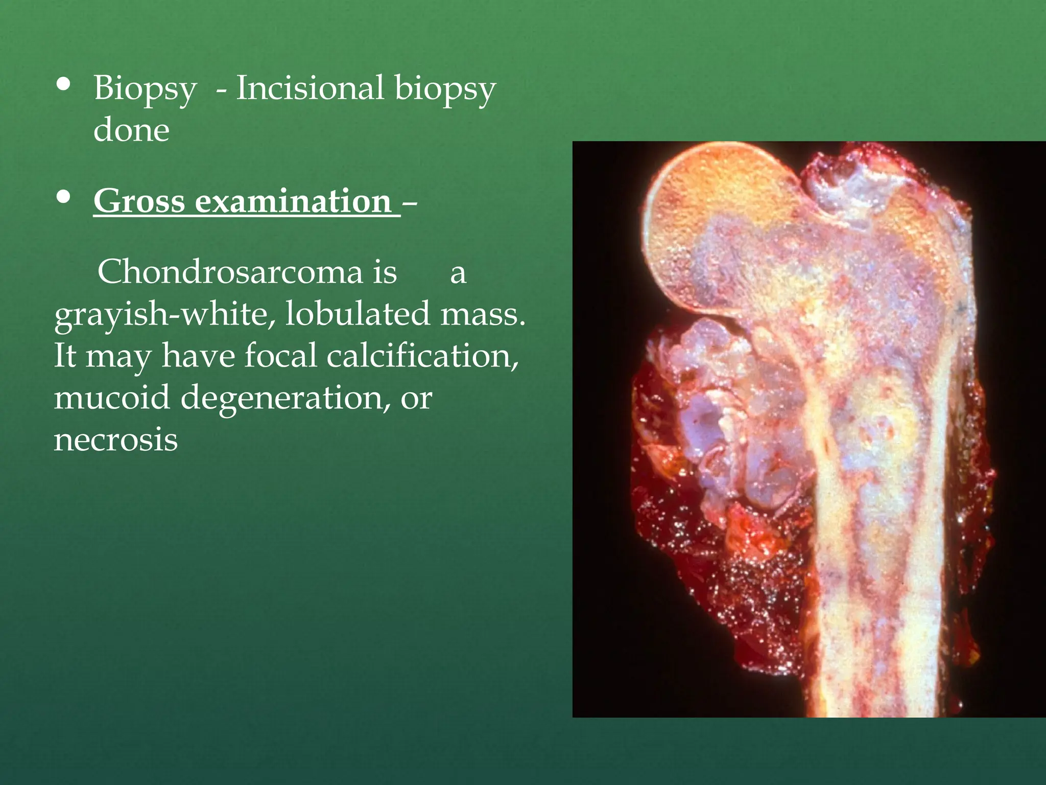
Biopsy -Incisional biopsy
done
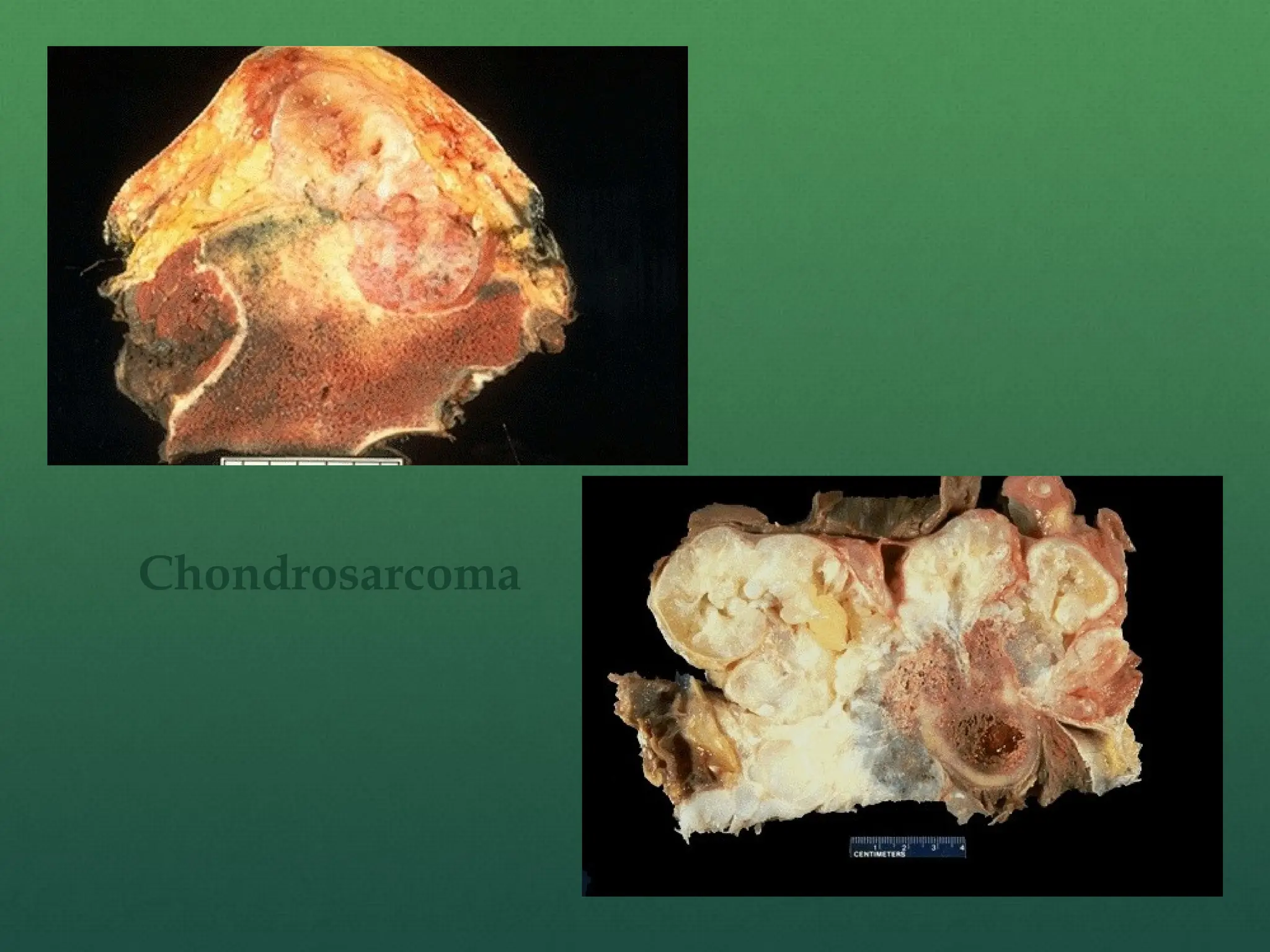
Gross examination –
Chondrosarcoma is a
grayish-white, lobulated mass.
It may have focal calcification,
mucoid degeneration, or
necrosis
Treatment
Treatment ofchondrosarcoma is wide surgical
excision. There is a very limited role for
chemotherapy or radiation.
Low grade tm. – Limb salvage surgery(WLE)
High grade tm. - Amputation
32.
Ewing’s sarcoma
Malignantneoplasm of undifferentiated cells arising within
the bone marrow cavity
Ewing's sarcoma is a highly malignant tumor that is a type
of peripheral primitive neuroectodermal tumor
Found in the lower extremity more than the upper
extremity, but any long tubular bone may be affected.
Most common sites are the diaphysis and metaphysis of the
femur followed by the tibia and humerus.
Most common in the first and second decade
Ratio of male to female is 3:2.
33.
Clinical features
Presentedwith pain, swelling and tenderness
Erythema and warmth of the local area are sometimes seen
Osteomyelitis is often the initial diagnosis based on
intermittent fevers, leukocytosis, anemia and an increased
ESR.
Contain glycogen granule so can cause hyperglycemia
34.
Investigation
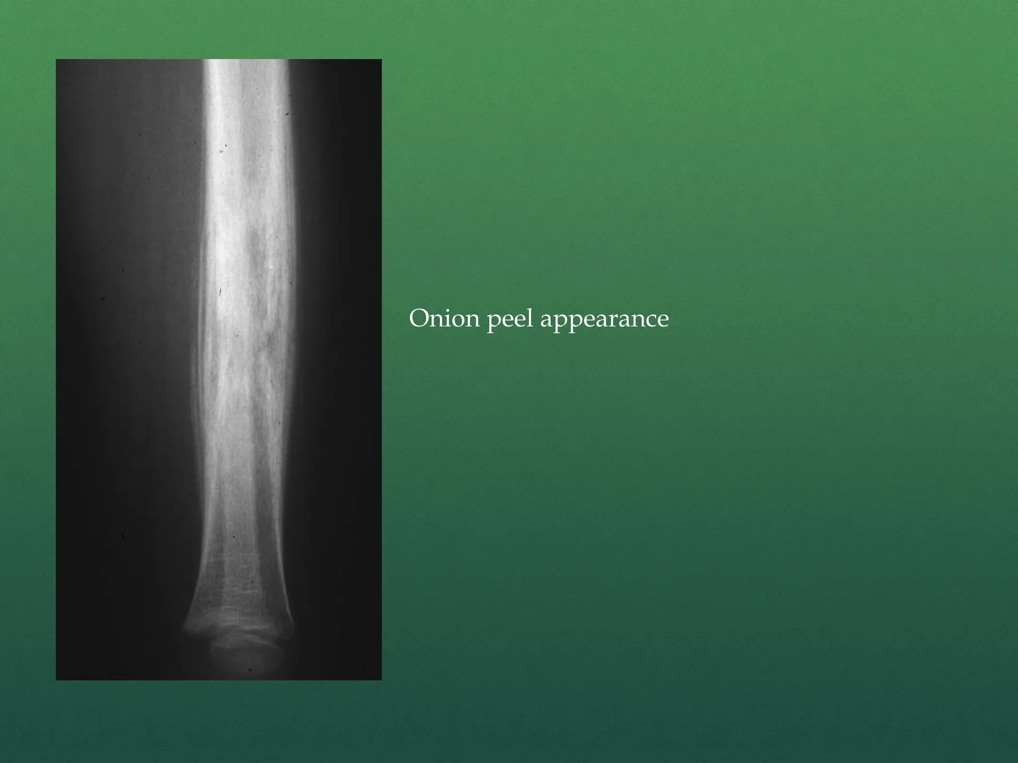
X-ray:
Concentric,onion-skin layering of new periosteal bone
This appearance is caused by and splitting and
thickening of the cortex by tumor cells.
The lesion is usually lytic and central.
CT is helpful in defining bone destruction.
MRI is essential to elucidate the soft tissue involvement
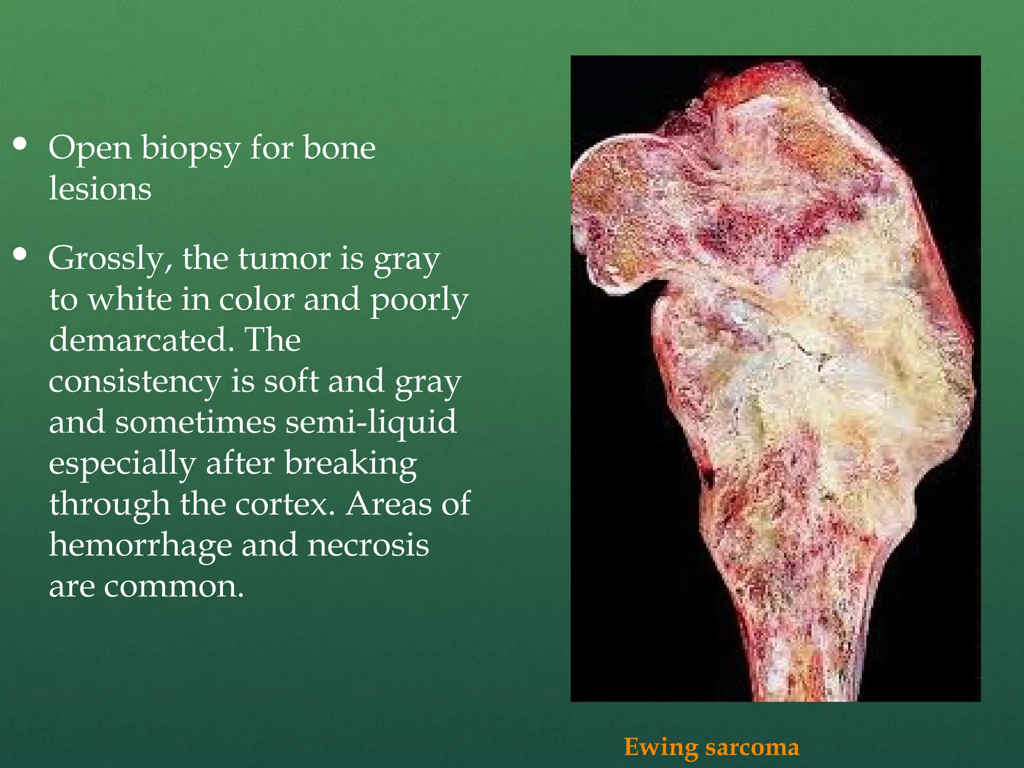
Open biopsyfor bone
lesions
Grossly, the tumor is gray
to white in color and poorly
demarcated. The
consistency is soft and gray
and sometimes semi-liquid
especially after breaking
through the cortex. Areas of
hemorrhage and necrosis
are common.
Ewing sarcoma
38.
Treatment
Chemotherapy –A – Actinomycin D
B – Bleomycin
C – Cylophosphamide
D – Doxirubicin
Surgery followed by adjuvant chemotherapy.
Radiotherapy
39.
Poor Prognostic Factor
High grade tumour
Age > 12 yrs
Male
H/O fever and increase TLC
Proximal lesion
Larger lesion
Metastasis
Chemoresistant
40.
MULTIPLE MYELOMA/
PLASMACYTOMA
Multiplemyeloma is a malignant tumor of plasma cells that
causes widespread osteolytic bone damage.
Multiple myeloma is the most common primary tumor of bone.
Found in the spine, skull, ribs, sternum and pelvis but may
affect any bone with hematopoietic red marrow.
There are chromosomal abnormalities that are associated with
MM, such as 14q32 and deletion of chromosome 13, and these
findings are more likely to be found in cases with poor
outcome.
Other diseases, such as solitary plasmacytoma and monoclonal
gammopathy are associate with MM.
41.
Average ageof the patients at diagnosis is 65 years.
Men are slightly more likely to get multiple
myeloma than women.
42.
Causes
Multiple myelomamay occur spontaneously
On exposure to ionizing radiation and the pesticide
dioxin
Infection with some viruses (HIV and human
herpes 8) has also been associated with multiple
myeloma.
No known risk factors are inherited.
43.
SYMPTOMS
Usually bonepain is main complain. Other symptoms include:
Fatigue
Feeling ill
Fever
Night sweats
Weight loss is not common in the early stages.
Patients are pale with diffuse bone tenderness, especially
around the sternum (breastbone) and pelvis (hips).
44.
Spine isthe most common location for a pathological
fracture. It can also happen in the ribs and pelvis.
Compression of the spinal cord in 10%-15%This causes pain
in the back and legs and numbness and weakness in the legs.
Patients who have high levels of calcium in the blood may
experience nausea, fatigue, confusion, constipation, and
frequent urination.
Patients with anemia may experience fatigue, weakness, and
shortness of breath with exercise.
In advanced cases, patients typically have recurrent
infections and can have kidney failure.
45.
Investigations
Blood andurine tests –
Monoclonal immunoglobulin (Ig G)is found on serum
electrophoresis and on urinalysis. Immunoglobulin (Ig) is the
protein that is produced by the tumor cells. Light chain
subunits of immunoglobulin are called Bence Jones proteins
and are present in urine.
Bone marrow aspiration and/or biopsy –
A procedure that involves taking a small amount of bone
marrow fluid (aspiration) and/or solid bone marrow tissue
(called a core biopsy), usually from the hip bones, to be
examined for the number, size, and maturity of blood cells
and/or abnormal cells.
46.
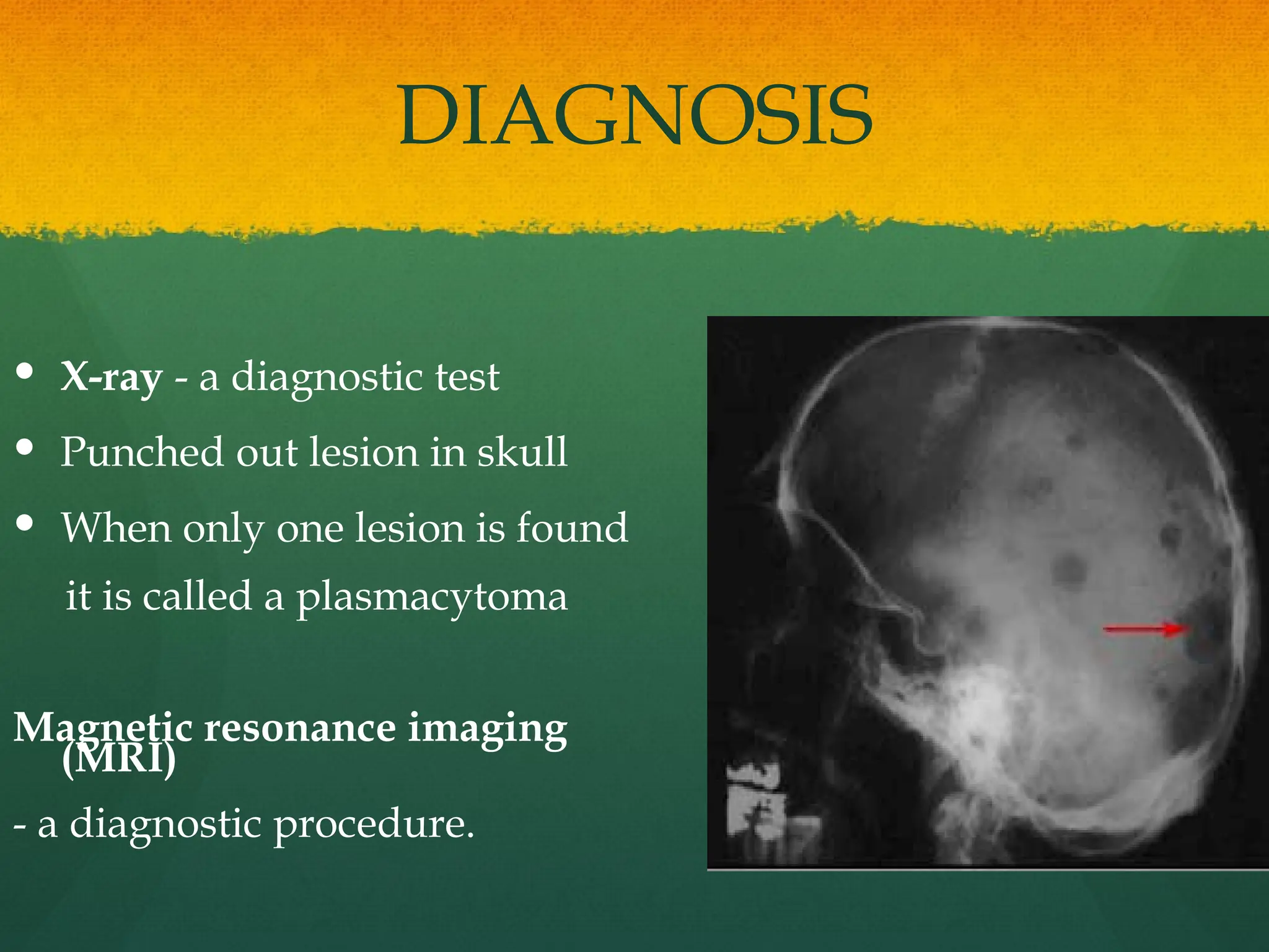
DIAGNOSIS
X-ray -a diagnostic test
Punched out lesion in skull
When only one lesion is found
it is called a plasmacytoma
Magnetic resonance imaging
(MRI)
- a diagnostic procedure.
CHEMOTHERAPY
The standardtreatment medications are melphalan and
prednisone.
The median survival rate is three years with this treatment
alone.
For patients in whom this therapy is ineffective, alternatives
include:
VBMCP (vincristine, carmustine, melphalan,
cyclophosphamide and prednisone)
VAD (vincristine, adriamycin and dexamethasone)
49.
A recentadvancement in the treatment of multiple
myeloma has increased, response rates and survival.
This treatment consists of high-dose chemotherapy,
followed by autologous stem cell transplantation.
With this treatment, patients have a 20 percent chance
of living longer than 10 years.
This stem cell transplantation involves:
1. Harvesting a patient's own blood cells
2. Conditioning them with very high doses of
melphalan
3. Re-infusing the blood cells back into the patient
SUPPORTIVE CARE
Supportivecare is critical. Supportive care includes managing the bone
disease, anemia, infections, kidney failure, and pain associated with
multiple myeloma.
Bisphosphonates (medication) can prevent destructive bone lesions and
spine fractures.
Erythropoetin or occasional blood transfusions can manage anemia.
Antibody infusions and vaccinations can help patients with recurrent
infections.
Corticosteroids and hydration can be used to treat high blood calcium
concentrations (from bone loss) and dehydration.
Narcotics can decrease the pain associated with bone lesions.
Operative intervention may be required to stabilize and control the pain
associated with bone fractures.
52.
SURGICAL
TREATMENT
Surgery willnot cure multiple myeloma. Surgery is
used to treat fractures and impending fractures in
the spine, pelvis, hip, and shoulder. The goal of
these surgeries is to decrease pain and maintain
function.
Internal fixation augmented with cement is
frequently recommended, as are joint replacements
and vertebroplasties (for spine fractures).
Operative intervention does not alter the survival
rate, but it does increase the quality of life.
53.
Bone Metastasis
Itis catastrophic complication for most patient of
cancer.
Usually occur in elderly age group
Clinical features –
Pain
Metastatic destruction of bone reduces load bearing
capacity.
Usually manifests as complication like pathological
fracture, paraplegia (medullary compression), pressure
symptoms.
54.
M/C cause– Lung Carcinoma
Male – Prostate Carcinoma
Female – Breast Carcinoma
M/C Area – D-L Spine
In prostate carcinoma –
Pelvis > Lumber spine > Dorsal spine
55.
M/C lesion– Lytic lesion
Purely osteoblastic sec. – Prostate
- Carcinoid
Metastasis do not cross elbow and knee
56.
Bone tm.Which have bony metastasis –
1. Osteosarcoma
2. Neuroblastoma
3. Ewing sarcoma