SlideShare a Scribd company logo
1 of 1
Download to read offline
Enhancing Adipose-derived Stem Cell-based Cartilage Regeneration using Macroporous Microribbon Scaffolds
Heather Rogan1
, MS; Krista Chew1
; Fan Yang1, 2
, PhD
1
Dept. Bioengineering, 2
Dept. Orthopaedic Surgery, Stanford University
DISCLOSURES: The authors declare no competing interests.
INTRODUCTION: Articular cartilage has a limited capacity to repair itself. Cartilage repair by stem cells has been widely explored in order to treat focal
cartilage defects and prevent progression to osteoarthritis. Adipose-derived stem cells (ADSCs) represent a promising autologous cell source for cartilage
repair givens its abundance, ease of isolation, and chondrogenic potential. However, delivery of stem cells alone often leads to fibrocartilage formation with
inferior mechanical properties. Hydrogels are attractive matrices for cell delivery given their injectability and tunable biochemical and mechanical
properties. However, conventional hydrogels generally lack the flexibility when subject to mechanical load, and lack the macroporosity desirable for
efficient cell proliferation, migration and matrix deposition. To overcome these limitations, we have recently reported development of microribbon (μRB)-
based hydrogels with cartilage-mimicking shock-absorbing capacity upon cyclic mechanical compression. Furthermore, how μRB scaffolds influence
chondrogenesis of ADSCs in 3D remains unknown. The goal of this study is to investigate the potential of gelatin-based μRB hydrogels as 3D matrices for
supporting chondrogenesis of ADSCs in vitro, and compare the efficacy to conventional hydrogels for inducing chondrogenesis.
METHODS: Human ADSCs were isolated from liposuction aspirates with informed consent for use in medical research, as otherwise discarded tissues
under an IRB approved protocol by Stanford University. hADSCs were encapsulated in μRB or conventional hydrogel (HG) scaffolds and cultured for 21
days in chondrogenic medium containing 10 ng/ml TGF-β3. μRB scaffolds were synthesized from gelatin; briefly, gelatin was dissolved in dimethyl
sulfoxide, wet spun into ethanol, flattened with acetone, chopped, methacrylated, fixed, washed, and lyophilized prior to use at 7.5% (w/w). HG scaffolds
contained 5% polyethylene (glycol) diacrylate (PEGDA) and 3% methacrylated chondroitin sulfate (CS-MA). Outcomes were analyzed using biochemical
assays (DNA and sGAG), histology (Safranin O), and immunostaining (type II and X collagen). An unpaired t test was used for statistical analysis to
compare μRB and HG scaffolds (n = 3).
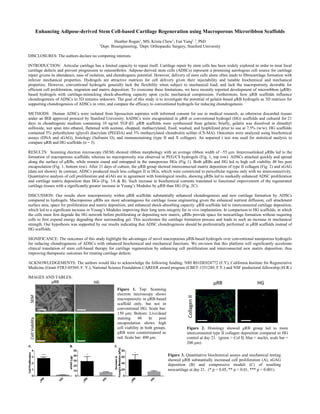
RESULTS: Scanning electron microscopy (SEM) showed ribbon morphology with an average ribbon width of ~55 µm. Intercrosslinked μRBs led to the
formation of macroporous scaffolds, whereas no macroporosity was observed in PEG/CS hydrogels (Fig. 1, top row). ADSCs attached quickly and spread
along the surface of μRBs, while remain round and entrapped in the nanoporous HGs (Fig 1). Both μRBs and HG led to high cell viability 48 hrs post
encapsulation (Fig. 1, bottom row). After 21 days of culture, the µRB group showed interconnected matrix deposition of type II collagen (Fig. 2) and sGAG
(data not shown). In contrast, ADSCs produced much less collagen II in HGs, which were constricted to pericellular regions only with no interconnectivity.
Quantitative analysis of cell proliferation and sGAG are in agreement with histological results, showing μRBs led to markedly enhanced ADSC proliferation
and cartilage matrix deposition than HGs (Fig. 3A & B). Such increase in biochemical contents translated to functional improvement of the regenerated
cartilage tissues with a significantly greater increase in Young’s Modulus by µRB than HG (Fig. 2C).
DISCUSSION: Our results show macroporosity within μRB scaffolds substantially enhanced chondrogenesis and new cartilage formation by ADSCs
compared to hydrogels. Macroporous μRBs are more advantageous for cartilage tissue engineering given the enhanced nutrient diffusion, cell attachment
surface area, space for proliferation and matrix deposition, and enhanced shock-absorbing capacity. μRB scaffolds led to interconnected cartilage deposition,
which led to a significant increase in Young’s Modulus improving their long term integrity for in vivo implantation. In comparison to HG scaffolds, in which
the cells must first degrade the HG network before proliferating or depositing new matrix, μRBs provide space for neocartilage formation without requiring
cells to first expend energy degrading their surrounding gel. This accelerates the cartilage formation process and leads to such an increase in mechanical
strength. Our hypothesis was supported by our results indicating that ADSC chondrogenesis should be preferentially performed in μRB scaffolds instead of
HG scaffolds.
SIGNIFICANCE: The outcomes of this study highlight the advantages of novel macroporous μRB-based hydrogels over conventional nanoporous hydrogels
for inducing chondrogenesis of ADSCs with enhanced biochemical and mechanical functions. We envision that this platform will significantly accelerate
clinical translation of stem cell-based therapy for cartilage regeneration by enhancing cell proliferation and interconnected new matrix deposition, thus
improving therapeutic outcomes for treating cartilage defects.
ACKNOWLEDGEMENTS: The authors would like to acknowledge the following funding: NIH R01DE024772 (F.Y), California Institute for Regenerative
Medicine (Grant #TR3-05569, F. Y.), National Science Foundation CAREER award program (CBET-1351289, F.Y.) and NSF predoctoral fellowship (H.R.)
IMAGES AND TABLES:
Figure 3. Quantitative biochemical assays and mechanical testing
showed μRB substantially increased cell proliferation (A), sGAG
deposition (B) and compressive moduli (C) of resulting
neocartilage at day 21. (* p < 0.05, ** p < 0.01, *** p < 0.001).
Figure 2. Histology showed μRB group led to more
interconnected type II collagen deposition compared to HG
control at day 21. (green = Col II, blue = nuclei, scale bar =
200 μm)
Figure 1. Top: Scanning
electron microscopy shows
macroporosity in μRB-based
scaffold only, but not in
conventional HG. Scale bar:
150 μm; Bottom: Live/dead
staining 48 hr post
encapsulation shows high
cell viability in both groups.
μRB were counterstained as
red. Scale bar: 400 μm.

More Related Content

What's hot

ten.tea.2008.0014
ten.tea.2008.0014ten.tea.2008.0014
ten.tea.2008.0014Anne Kwei
 
Genes and Tissue Culture Technology - Presentation
Genes and Tissue Culture Technology - PresentationGenes and Tissue Culture Technology - Presentation
Genes and Tissue Culture Technology - PresentationBenjamin Lee
 
CELL-DERIVED EXTRACELLUALR MATRIX (ECM) IN TISSUE ENGINEERING [SCT60103 GENE ...
CELL-DERIVED EXTRACELLUALR MATRIX (ECM) IN TISSUE ENGINEERING [SCT60103 GENE ...CELL-DERIVED EXTRACELLUALR MATRIX (ECM) IN TISSUE ENGINEERING [SCT60103 GENE ...
CELL-DERIVED EXTRACELLUALR MATRIX (ECM) IN TISSUE ENGINEERING [SCT60103 GENE ...Preveena Ravi
 
Periodontal regeneration
Periodontal regenerationPeriodontal regeneration
Periodontal regenerationR Viswa Chandra
 
Orthopaedic tissue engineering
Orthopaedic tissue engineeringOrthopaedic tissue engineering
Orthopaedic tissue engineeringrajusvmc
 
1 s2.0-s1742706114003110-main(4)
1 s2.0-s1742706114003110-main(4)1 s2.0-s1742706114003110-main(4)
1 s2.0-s1742706114003110-main(4)ayuprasiska
 
Tissue engineering in orthopaedics
Tissue engineering in orthopaedicsTissue engineering in orthopaedics
Tissue engineering in orthopaedicsLibin Thomas
 
Articular Cartilage Repair
Articular Cartilage RepairArticular Cartilage Repair
Articular Cartilage RepairAaron Sparshott
 
Bone Presentation Final5 5 2008
Bone Presentation Final5 5 2008Bone Presentation Final5 5 2008
Bone Presentation Final5 5 2008niyati_patel18
 
A new polymer-free drug-eluting stent with nanocarriers eluting sirolimus fro...
A new polymer-free drug-eluting stent with nanocarriers eluting sirolimus fro...A new polymer-free drug-eluting stent with nanocarriers eluting sirolimus fro...
A new polymer-free drug-eluting stent with nanocarriers eluting sirolimus fro...Paul Schoenhagen
 
Effect of Seeding Density of Mesenchymal Stem Cells on Cell Proliferation and...
Effect of Seeding Density of Mesenchymal Stem Cells on Cell Proliferation and...Effect of Seeding Density of Mesenchymal Stem Cells on Cell Proliferation and...
Effect of Seeding Density of Mesenchymal Stem Cells on Cell Proliferation and...Jaideep Sahni
 
Aligned nanofibrillar collagen scaffolds – Guiding lymphangiogenesis for trea...
Aligned nanofibrillar collagen scaffolds – Guiding lymphangiogenesis for trea...Aligned nanofibrillar collagen scaffolds – Guiding lymphangiogenesis for trea...
Aligned nanofibrillar collagen scaffolds – Guiding lymphangiogenesis for trea...Mary Fickling
 
Engineering bone tissue using human Embryonic Stem Cells
Engineering bone tissue using human Embryonic Stem CellsEngineering bone tissue using human Embryonic Stem Cells
Engineering bone tissue using human Embryonic Stem CellsBalaganesh Kuruba
 
Biomaterials for tissue engineering slideshare
Biomaterials for tissue engineering slideshareBiomaterials for tissue engineering slideshare
Biomaterials for tissue engineering slideshareBukar Abdullahi
 

What's hot (20)

ten.tea.2008.0014
ten.tea.2008.0014ten.tea.2008.0014
ten.tea.2008.0014
 
Tissue assembly in microgravity
Tissue assembly in microgravityTissue assembly in microgravity
Tissue assembly in microgravity
 
Tissue engineering
Tissue engineeringTissue engineering
Tissue engineering
 
Journal club
Journal clubJournal club
Journal club
 
Genes and Tissue Culture Technology - Presentation
Genes and Tissue Culture Technology - PresentationGenes and Tissue Culture Technology - Presentation
Genes and Tissue Culture Technology - Presentation
 
CELL-DERIVED EXTRACELLUALR MATRIX (ECM) IN TISSUE ENGINEERING [SCT60103 GENE ...
CELL-DERIVED EXTRACELLUALR MATRIX (ECM) IN TISSUE ENGINEERING [SCT60103 GENE ...CELL-DERIVED EXTRACELLUALR MATRIX (ECM) IN TISSUE ENGINEERING [SCT60103 GENE ...
CELL-DERIVED EXTRACELLUALR MATRIX (ECM) IN TISSUE ENGINEERING [SCT60103 GENE ...
 
Periodontal regeneration
Periodontal regenerationPeriodontal regeneration
Periodontal regeneration
 
Orthopaedic tissue engineering
Orthopaedic tissue engineeringOrthopaedic tissue engineering
Orthopaedic tissue engineering
 
1 s2.0-s1742706114003110-main(4)
1 s2.0-s1742706114003110-main(4)1 s2.0-s1742706114003110-main(4)
1 s2.0-s1742706114003110-main(4)
 
Tissue engineering in orthopaedics
Tissue engineering in orthopaedicsTissue engineering in orthopaedics
Tissue engineering in orthopaedics
 
paper 2
paper 2paper 2
paper 2
 
Platelet concentrates
Platelet concentratesPlatelet concentrates
Platelet concentrates
 
Articular Cartilage Repair
Articular Cartilage RepairArticular Cartilage Repair
Articular Cartilage Repair
 
Bone Presentation Final5 5 2008
Bone Presentation Final5 5 2008Bone Presentation Final5 5 2008
Bone Presentation Final5 5 2008
 
PSN poster
PSN posterPSN poster
PSN poster
 
A new polymer-free drug-eluting stent with nanocarriers eluting sirolimus fro...
A new polymer-free drug-eluting stent with nanocarriers eluting sirolimus fro...A new polymer-free drug-eluting stent with nanocarriers eluting sirolimus fro...
A new polymer-free drug-eluting stent with nanocarriers eluting sirolimus fro...
 
Effect of Seeding Density of Mesenchymal Stem Cells on Cell Proliferation and...
Effect of Seeding Density of Mesenchymal Stem Cells on Cell Proliferation and...Effect of Seeding Density of Mesenchymal Stem Cells on Cell Proliferation and...
Effect of Seeding Density of Mesenchymal Stem Cells on Cell Proliferation and...
 
Aligned nanofibrillar collagen scaffolds – Guiding lymphangiogenesis for trea...
Aligned nanofibrillar collagen scaffolds – Guiding lymphangiogenesis for trea...Aligned nanofibrillar collagen scaffolds – Guiding lymphangiogenesis for trea...
Aligned nanofibrillar collagen scaffolds – Guiding lymphangiogenesis for trea...
 
Engineering bone tissue using human Embryonic Stem Cells
Engineering bone tissue using human Embryonic Stem CellsEngineering bone tissue using human Embryonic Stem Cells
Engineering bone tissue using human Embryonic Stem Cells
 
Biomaterials for tissue engineering slideshare
Biomaterials for tissue engineering slideshareBiomaterials for tissue engineering slideshare
Biomaterials for tissue engineering slideshare
 

Similar to ORS Abstract

Collagen II Microbeads Final
Collagen II Microbeads FinalCollagen II Microbeads Final
Collagen II Microbeads FinalDavid R. Mertz
 
2007 Bailey Tissue Engineering
2007 Bailey Tissue Engineering2007 Bailey Tissue Engineering
2007 Bailey Tissue EngineeringMark Bailey
 
Layer by-layer decorated herbal cell compatible scaffolds for bone tissue eng...
Layer by-layer decorated herbal cell compatible scaffolds for bone tissue eng...Layer by-layer decorated herbal cell compatible scaffolds for bone tissue eng...
Layer by-layer decorated herbal cell compatible scaffolds for bone tissue eng...Advancells Stem Cell Therapies
 
Tissue Engineering Poster
Tissue Engineering PosterTissue Engineering Poster
Tissue Engineering PosterShasta Rizzi
 
2014 Acta Biomaterialia
2014 Acta Biomaterialia2014 Acta Biomaterialia
2014 Acta BiomaterialiaHelen Cox
 
Intra_articular injection of synovial mesenchymal stem cells improves cartila...
Intra_articular injection of synovial mesenchymal stem cells improves cartila...Intra_articular injection of synovial mesenchymal stem cells improves cartila...
Intra_articular injection of synovial mesenchymal stem cells improves cartila...Christina Jablonski
 
Biotechnology and gene expression profiling for mechanistic understanding of ...
Biotechnology and gene expression profiling for mechanistic understanding of ...Biotechnology and gene expression profiling for mechanistic understanding of ...
Biotechnology and gene expression profiling for mechanistic understanding of ...Hana Fayed
 
White paper publications in 3 dimensional tissue engineering
White paper publications in 3 dimensional tissue engineeringWhite paper publications in 3 dimensional tissue engineering
White paper publications in 3 dimensional tissue engineeringAdvancells Stem Cell Therapies
 
UROPProposalA_Marks
UROPProposalA_MarksUROPProposalA_Marks
UROPProposalA_MarksAndrea Marks
 
Layer by layer deposited osteoinductive scaffolds for bone tissue engineering
Layer by layer deposited osteoinductive scaffolds for bone tissue engineeringLayer by layer deposited osteoinductive scaffolds for bone tissue engineering
Layer by layer deposited osteoinductive scaffolds for bone tissue engineeringAdvancells Stem Cell Therapies
 
The Effect of Intra-articular Injection of Autologous Microfragmented Fat Tis...
The Effect of Intra-articular Injection of Autologous Microfragmented Fat Tis...The Effect of Intra-articular Injection of Autologous Microfragmented Fat Tis...
The Effect of Intra-articular Injection of Autologous Microfragmented Fat Tis...Lipogems Equine & Lipogems Canine
 
Bioprinting of osteocandral tissue
Bioprinting of osteocandral tissueBioprinting of osteocandral tissue
Bioprinting of osteocandral tissuepradeepyadav380
 
Genus BMAC Abstract
Genus BMAC AbstractGenus BMAC Abstract
Genus BMAC AbstractJoe Moore
 
Effect of stemregenin1 and sb431542 small molecules on ex vivo expansion of u...
Effect of stemregenin1 and sb431542 small molecules on ex vivo expansion of u...Effect of stemregenin1 and sb431542 small molecules on ex vivo expansion of u...
Effect of stemregenin1 and sb431542 small molecules on ex vivo expansion of u...Liberty University (LU)
 
Effect of Surface Engineering on Stem Cells.pdf
Effect of Surface Engineering on Stem Cells.pdfEffect of Surface Engineering on Stem Cells.pdf
Effect of Surface Engineering on Stem Cells.pdfaman15nanavaty
 
Bone Marrow Stromal Stem cells (bMSCs) therapy for musculoskeletal problems ...
Bone Marrow Stromal Stem cells (bMSCs) therapy for musculoskeletal problems ...Bone Marrow Stromal Stem cells (bMSCs) therapy for musculoskeletal problems ...
Bone Marrow Stromal Stem cells (bMSCs) therapy for musculoskeletal problems ...Asosiasi Sel Punca Indonesia
 
A Novel Alternative To Cryopreservation For The Short-Term Storage Of Stem Ce...
A Novel Alternative To Cryopreservation For The Short-Term Storage Of Stem Ce...A Novel Alternative To Cryopreservation For The Short-Term Storage Of Stem Ce...
A Novel Alternative To Cryopreservation For The Short-Term Storage Of Stem Ce...Cynthia King
 
3D cell culture engineering
3D cell culture engineering3D cell culture engineering
3D cell culture engineeringN Poorin
 

Similar to ORS Abstract (20)

Collagen II Microbeads Final
Collagen II Microbeads FinalCollagen II Microbeads Final
Collagen II Microbeads Final
 
Introductiondata
IntroductiondataIntroductiondata
Introductiondata
 
Collagen/HA
Collagen/HACollagen/HA
Collagen/HA
 
2007 Bailey Tissue Engineering
2007 Bailey Tissue Engineering2007 Bailey Tissue Engineering
2007 Bailey Tissue Engineering
 
Layer by-layer decorated herbal cell compatible scaffolds for bone tissue eng...
Layer by-layer decorated herbal cell compatible scaffolds for bone tissue eng...Layer by-layer decorated herbal cell compatible scaffolds for bone tissue eng...
Layer by-layer decorated herbal cell compatible scaffolds for bone tissue eng...
 
Tissue Engineering Poster
Tissue Engineering PosterTissue Engineering Poster
Tissue Engineering Poster
 
2014 Acta Biomaterialia
2014 Acta Biomaterialia2014 Acta Biomaterialia
2014 Acta Biomaterialia
 
Intra_articular injection of synovial mesenchymal stem cells improves cartila...
Intra_articular injection of synovial mesenchymal stem cells improves cartila...Intra_articular injection of synovial mesenchymal stem cells improves cartila...
Intra_articular injection of synovial mesenchymal stem cells improves cartila...
 
Biotechnology and gene expression profiling for mechanistic understanding of ...
Biotechnology and gene expression profiling for mechanistic understanding of ...Biotechnology and gene expression profiling for mechanistic understanding of ...
Biotechnology and gene expression profiling for mechanistic understanding of ...
 
White paper publications in 3 dimensional tissue engineering
White paper publications in 3 dimensional tissue engineeringWhite paper publications in 3 dimensional tissue engineering
White paper publications in 3 dimensional tissue engineering
 
UROPProposalA_Marks
UROPProposalA_MarksUROPProposalA_Marks
UROPProposalA_Marks
 
Layer by layer deposited osteoinductive scaffolds for bone tissue engineering
Layer by layer deposited osteoinductive scaffolds for bone tissue engineeringLayer by layer deposited osteoinductive scaffolds for bone tissue engineering
Layer by layer deposited osteoinductive scaffolds for bone tissue engineering
 
The Effect of Intra-articular Injection of Autologous Microfragmented Fat Tis...
The Effect of Intra-articular Injection of Autologous Microfragmented Fat Tis...The Effect of Intra-articular Injection of Autologous Microfragmented Fat Tis...
The Effect of Intra-articular Injection of Autologous Microfragmented Fat Tis...
 
Bioprinting of osteocandral tissue
Bioprinting of osteocandral tissueBioprinting of osteocandral tissue
Bioprinting of osteocandral tissue
 
Genus BMAC Abstract
Genus BMAC AbstractGenus BMAC Abstract
Genus BMAC Abstract
 
Effect of stemregenin1 and sb431542 small molecules on ex vivo expansion of u...
Effect of stemregenin1 and sb431542 small molecules on ex vivo expansion of u...Effect of stemregenin1 and sb431542 small molecules on ex vivo expansion of u...
Effect of stemregenin1 and sb431542 small molecules on ex vivo expansion of u...
 
Effect of Surface Engineering on Stem Cells.pdf
Effect of Surface Engineering on Stem Cells.pdfEffect of Surface Engineering on Stem Cells.pdf
Effect of Surface Engineering on Stem Cells.pdf
 
Bone Marrow Stromal Stem cells (bMSCs) therapy for musculoskeletal problems ...
Bone Marrow Stromal Stem cells (bMSCs) therapy for musculoskeletal problems ...Bone Marrow Stromal Stem cells (bMSCs) therapy for musculoskeletal problems ...
Bone Marrow Stromal Stem cells (bMSCs) therapy for musculoskeletal problems ...
 
A Novel Alternative To Cryopreservation For The Short-Term Storage Of Stem Ce...
A Novel Alternative To Cryopreservation For The Short-Term Storage Of Stem Ce...A Novel Alternative To Cryopreservation For The Short-Term Storage Of Stem Ce...
A Novel Alternative To Cryopreservation For The Short-Term Storage Of Stem Ce...
 
3D cell culture engineering
3D cell culture engineering3D cell culture engineering
3D cell culture engineering
 

ORS Abstract

  • 1. Enhancing Adipose-derived Stem Cell-based Cartilage Regeneration using Macroporous Microribbon Scaffolds Heather Rogan1 , MS; Krista Chew1 ; Fan Yang1, 2 , PhD 1 Dept. Bioengineering, 2 Dept. Orthopaedic Surgery, Stanford University DISCLOSURES: The authors declare no competing interests. INTRODUCTION: Articular cartilage has a limited capacity to repair itself. Cartilage repair by stem cells has been widely explored in order to treat focal cartilage defects and prevent progression to osteoarthritis. Adipose-derived stem cells (ADSCs) represent a promising autologous cell source for cartilage repair givens its abundance, ease of isolation, and chondrogenic potential. However, delivery of stem cells alone often leads to fibrocartilage formation with inferior mechanical properties. Hydrogels are attractive matrices for cell delivery given their injectability and tunable biochemical and mechanical properties. However, conventional hydrogels generally lack the flexibility when subject to mechanical load, and lack the macroporosity desirable for efficient cell proliferation, migration and matrix deposition. To overcome these limitations, we have recently reported development of microribbon (μRB)- based hydrogels with cartilage-mimicking shock-absorbing capacity upon cyclic mechanical compression. Furthermore, how μRB scaffolds influence chondrogenesis of ADSCs in 3D remains unknown. The goal of this study is to investigate the potential of gelatin-based μRB hydrogels as 3D matrices for supporting chondrogenesis of ADSCs in vitro, and compare the efficacy to conventional hydrogels for inducing chondrogenesis. METHODS: Human ADSCs were isolated from liposuction aspirates with informed consent for use in medical research, as otherwise discarded tissues under an IRB approved protocol by Stanford University. hADSCs were encapsulated in μRB or conventional hydrogel (HG) scaffolds and cultured for 21 days in chondrogenic medium containing 10 ng/ml TGF-β3. μRB scaffolds were synthesized from gelatin; briefly, gelatin was dissolved in dimethyl sulfoxide, wet spun into ethanol, flattened with acetone, chopped, methacrylated, fixed, washed, and lyophilized prior to use at 7.5% (w/w). HG scaffolds contained 5% polyethylene (glycol) diacrylate (PEGDA) and 3% methacrylated chondroitin sulfate (CS-MA). Outcomes were analyzed using biochemical assays (DNA and sGAG), histology (Safranin O), and immunostaining (type II and X collagen). An unpaired t test was used for statistical analysis to compare μRB and HG scaffolds (n = 3). RESULTS: Scanning electron microscopy (SEM) showed ribbon morphology with an average ribbon width of ~55 µm. Intercrosslinked μRBs led to the formation of macroporous scaffolds, whereas no macroporosity was observed in PEG/CS hydrogels (Fig. 1, top row). ADSCs attached quickly and spread along the surface of μRBs, while remain round and entrapped in the nanoporous HGs (Fig 1). Both μRBs and HG led to high cell viability 48 hrs post encapsulation (Fig. 1, bottom row). After 21 days of culture, the µRB group showed interconnected matrix deposition of type II collagen (Fig. 2) and sGAG (data not shown). In contrast, ADSCs produced much less collagen II in HGs, which were constricted to pericellular regions only with no interconnectivity. Quantitative analysis of cell proliferation and sGAG are in agreement with histological results, showing μRBs led to markedly enhanced ADSC proliferation and cartilage matrix deposition than HGs (Fig. 3A & B). Such increase in biochemical contents translated to functional improvement of the regenerated cartilage tissues with a significantly greater increase in Young’s Modulus by µRB than HG (Fig. 2C). DISCUSSION: Our results show macroporosity within μRB scaffolds substantially enhanced chondrogenesis and new cartilage formation by ADSCs compared to hydrogels. Macroporous μRBs are more advantageous for cartilage tissue engineering given the enhanced nutrient diffusion, cell attachment surface area, space for proliferation and matrix deposition, and enhanced shock-absorbing capacity. μRB scaffolds led to interconnected cartilage deposition, which led to a significant increase in Young’s Modulus improving their long term integrity for in vivo implantation. In comparison to HG scaffolds, in which the cells must first degrade the HG network before proliferating or depositing new matrix, μRBs provide space for neocartilage formation without requiring cells to first expend energy degrading their surrounding gel. This accelerates the cartilage formation process and leads to such an increase in mechanical strength. Our hypothesis was supported by our results indicating that ADSC chondrogenesis should be preferentially performed in μRB scaffolds instead of HG scaffolds. SIGNIFICANCE: The outcomes of this study highlight the advantages of novel macroporous μRB-based hydrogels over conventional nanoporous hydrogels for inducing chondrogenesis of ADSCs with enhanced biochemical and mechanical functions. We envision that this platform will significantly accelerate clinical translation of stem cell-based therapy for cartilage regeneration by enhancing cell proliferation and interconnected new matrix deposition, thus improving therapeutic outcomes for treating cartilage defects. ACKNOWLEDGEMENTS: The authors would like to acknowledge the following funding: NIH R01DE024772 (F.Y), California Institute for Regenerative Medicine (Grant #TR3-05569, F. Y.), National Science Foundation CAREER award program (CBET-1351289, F.Y.) and NSF predoctoral fellowship (H.R.) IMAGES AND TABLES: Figure 3. Quantitative biochemical assays and mechanical testing showed μRB substantially increased cell proliferation (A), sGAG deposition (B) and compressive moduli (C) of resulting neocartilage at day 21. (* p < 0.05, ** p < 0.01, *** p < 0.001). Figure 2. Histology showed μRB group led to more interconnected type II collagen deposition compared to HG control at day 21. (green = Col II, blue = nuclei, scale bar = 200 μm) Figure 1. Top: Scanning electron microscopy shows macroporosity in μRB-based scaffold only, but not in conventional HG. Scale bar: 150 μm; Bottom: Live/dead staining 48 hr post encapsulation shows high cell viability in both groups. μRB were counterstained as red. Scale bar: 400 μm.