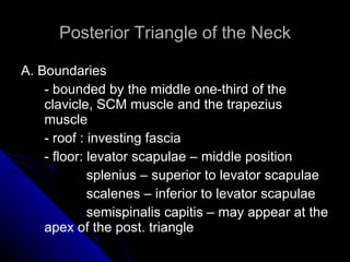
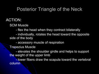
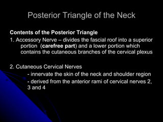
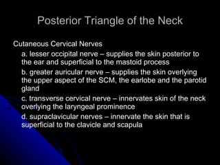
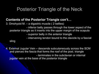
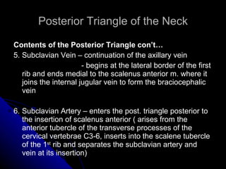
Posterior Triangle of the Neck Maribel G. Castro-Enano, M.D. CPU College of Medicine
Surface Anatomy of the Neck Spinous process of the  axis  – first bony prominence that can be felt in the median plane Spinous process of the  vertebra prominens  (C7) – easily palpable when the neck is flexed Thyroid cartilage (Adam’s apple) – at the level of C4; contains 2 quadrilateral plates called  laminae   Hyoid bone – u-shaped bone at the level of the body of C3; lie superior to the thyroid cartilage Transverse process of the  atlas  – felt by deep palpation between the angle of the mandible and a point about 1cm anteroinferior to the tip of the mastoid process
Surface Anatomy of the Neck Cricoid cartilage – inferior  to the thyroid cartilage; lies at the level of C6 Tracheal rings – palpable in the inferior part of the neck; moves superiorly during swallowing Jugular notch – depression in the manubrium of the sternum; easily palpable between the medial ends of the clavicle Sternocleidomastoid muscle – has  sternal and clavicular heads; divides lateral side of the neck into anterior and posterior triangles Supraclavicular fossa – triangular depression between the sternal and clavicular heads of the SCM; contains the pressure point for the subclavian artery
Surface Anatomy of the Neck Medial ends of the  clavicle  – clearly visible at the root of the neck Inferior margin of the  mandible , the  mastoid process  of the temporal bone, and the  external occipital protuberance  – bony landmarks of the superior limit of the neck
Superficial Structures Superficial Fascia – contains the  platysma Platysma  – wide, thin subcutaneous sheet of striated muscle that ascends to the face from the anterior part of the neck   - covers the superior part of the anterior cervical triangle and the anteroinferior part of the posterior cervical triangle       Inferior Attachment – fascia and skin over the pectoralis major (second rib) and deltoid muscles     Superior Attachment – inferior border of mandible and skin of the lower face   Innervation – cervical branch of CN VII   Action – muscle of facial exrpression; tense the subcutaneous tissue of the neck and assist in depressing the lower lip   - produces skin ridges in the neck, releasing pressure of the skin on the the underlying veins
Superficial Structures 1. Superficial or Enveloping Layer - surrounds the whole neck - starts off at the back where it is attached to the  ligamentum nuchae   -  encloses the trapezius and SCM muscles   - contains the accessory nerve as it passes posteriorly and inferiorly from the SCM to the trapezius
Superficial Structures 2. Prevertebral Fascia    - part of the fascia that envelopes the vertebral column   - envelopes the prevertebral and postvertebral muscles of the neck   - covers the subclavian vessels  and the roots of the brachial plexus    - drawn into the axilla on the brachial plexus and subclavian artery as the  axillary sheath * provides an anatomical course for infections within the posterior compartment of the neck to travel into the axilla
Superficial Structures 3. Carotid Sheath   - anterior to the prevertebral fascia   - invests the common and internal carotid arteries, the internal jugular vein and the vagus nerve
Superficial Structures 5. Infrahyoid Fascia   - encloses the infrahyoid muscles ( sternothyroid, sternohyoid, thyrohoid and cricohyoid m.)   -  attaches above either into the hyoid or thyroid muscles and below into the posterior aspect of the manubrium
Fascial Layers
Superficial Structures 4. Pretracheal Fascia   - has 2 layers, the anterior and posterior layers a. anterior layer – superiorly attached to the hyoid bone up to the border of the mandible   - inferiorly found anterior to the larynx, trachea and thyroid gland and end in front of the great vessels b. posterior layer – posterior to the pharynx and esophagus   - covers the posterior aspect of the esophagus   - the anterior and posterior layers are both attached the the skull
Posterior Triangle of the Neck A. Boundaries - bounded by the middle one-third of the clavicle, SCM muscle and the trapezius muscle - roof : investing fascia - floor: levator scapulae – middle position   splenius – superior to levator scapulae   scalenes – inferior to levator scapulae   semispinalis capitis – may appear at the apex of the post. triangle
 
Posterior Triangle of the Neck ACTION: SCM Muscle - flex the head when they contract bilaterally - individually, rotates the head toward the opposite side of the body - accessory muscle of respiration  Trapezius Muscle - elevates the shoulder girdle and helps to support the weight of the upper limb - lower fibers draw the scapula toward the vertebral column
Posterior Triangle of the Neck A. Boundaries con’t… - the apex of the triangle projects superiorly behind the ear to the level of the  superior nuchal line  of the occipital bone, where the SCM and trapezius muscles meet
Posterior Triangle of the Neck SCM and Trapezius Muscles - have a continuous superior attachment from the  mastoid process  to the  inion   along the superior nuchal line - inferior attachments SCM – medial third of clavicle and sternum trapezius – lateral third of clavicle, acromion and spine of the scapula - innervated by CN XI
Posterior Triangle of the Neck Contents of the Posterior Triangle 1. Accessory Nerve – divides the fascial roof into a superior portion  ( carefree part ) and a lower portion which contains the cutaneous branches of the cervical plexus 2. Cutaneous Cervical Nerves - innervate the skin of the neck and shoulder region - derived from the anterior rami of cervical nerves 2, 3 and 4
Posterior Triangle of the Neck Cutaneous Cervical Nerves a. lesser occipital nerve – supplies the skin posterior to the ear and superficial to the mastoid process b. greater auricular nerve – supplies the skin overlying the upper aspect of the SCM, the earlobe and the parotid gland c. transverse cervical nerve – innervates skin of the neck overlying the laryngeal prominence d. supraclavicular nerves – innervate the skin that is superficial to the clavicle and scapula
Posterior Triangle of the Neck Contents of the Posterior Triangle con’t… 3. Omohyoid M. – a digastric muscle ( 2 bellies)   - inferior belly passes through the lower aspect of the posterior triangle as it inserts into the upper margin of the scapula   - superior belly in the anterior triangle   - intervening tendon bound to the clavicle by a fascial sling 4. External Jugular Vein – descends subcutaneously across the SCM and pierces the fascia that forms the roof of the post. triangle   - terminates in the subclavian or internal jugular vein at the base of the posterior triangle
Posterior Triangle of the Neck Contents of the Posterior Triangle con’t… 5. Subclavian Vein – continuation of the axillary vein - begins at the lateral border of the first rib and ends medial to the scalenus anterior m. where it joins the internal jugular vein to form the braciocephalic vein 6. Subclavian Artery – enters the post. triangle posterior to the insertion of scalenus anterior ( arises from the anterior tubercle of the transverse processes of the cervical vertebrae C3-6, inserts into the scalene tubercle of the 1 st  rib and separates the subclavian artery and vein at its insertion)
Posterior Triangle of the Neck - subclavian artery in the post.  triangle may give off a  dorsal scapular artery  which traverses through the trunks of the brachial plexus to supply the medial border of the scapula and the rhomboids * Important Relationships Around the Subclavian A. 1. Inferiorly – 1st rib and pleura of apex of lung 2. Posteriorly – scalenus medius and lower trunk of brachial plexus 3. Anteriorly – scalenus anterior
Posterior Triangle of the Neck 7. Suprascapular Artery – arise from the first part of the subclavian a.   - supply the supraspinatus and infraspinatus fossae structures of the scapula 8. Transverse Cervical Artery – arise from the first part of the subclavian a.  - innervate the posterior surface of the trapezius through a superficial branch; deep branches supply the rhomboids and medial border of the scapula
Posterior Triangle of the Neck 9. Lateral group of the inferior deep cervical lymph nodes - on the posterior margin of the SCM at the level of CN XI - drain the back of the scalp and neck and drain into the jugular lymphatic trunks 10. Brachial Plexus – formed by the ventral rami of C5,6,7,8 and T1 (C4 and T2)
 
Posterior Triangle of the Neck THE BRACHIAL PLEXUS 1. upper trunk – C5 and C6  anterior and  2. middle trunk – C7  posterior divisions 3. lower trunk – C8 and T1 * posterior cord – 3 posterior divisions * lateral cord – anterior divisions of the upper and middle trunks * medial cord – anterior division of the lower trunk
The Brachial Plexus
Posterior Triangle of the Neck THE BRACHIAL PLEXUS cont…… * suprascapular nerve –  major nerve that arises from the upper trunk of the brachial plexus (C5,6); passes through the suprascapular notch to innervate and  supraspinatus  and  infraspinatus m. *Branches from the  roots  of the brachial plexus innervate the following: Rhomboids – dorsal scapular nerve, C5 Serratus anterior – long thoracic n., C5,6,7 Diaphragm -  phrenic n., C3,4,5 Adjacent prevertebral muscles * subclavius muscle – branch from the upper trunk of the brachial plexus in the posterior triangle
Good Afternoon!

Posterior triangle

  • 1.
    Posterior Triangle ofthe Neck Maribel G. Castro-Enano, M.D. CPU College of Medicine
  • 2.
    Surface Anatomy ofthe Neck Spinous process of the axis – first bony prominence that can be felt in the median plane Spinous process of the vertebra prominens (C7) – easily palpable when the neck is flexed Thyroid cartilage (Adam’s apple) – at the level of C4; contains 2 quadrilateral plates called laminae Hyoid bone – u-shaped bone at the level of the body of C3; lie superior to the thyroid cartilage Transverse process of the atlas – felt by deep palpation between the angle of the mandible and a point about 1cm anteroinferior to the tip of the mastoid process
  • 3.
    Surface Anatomy ofthe Neck Cricoid cartilage – inferior to the thyroid cartilage; lies at the level of C6 Tracheal rings – palpable in the inferior part of the neck; moves superiorly during swallowing Jugular notch – depression in the manubrium of the sternum; easily palpable between the medial ends of the clavicle Sternocleidomastoid muscle – has sternal and clavicular heads; divides lateral side of the neck into anterior and posterior triangles Supraclavicular fossa – triangular depression between the sternal and clavicular heads of the SCM; contains the pressure point for the subclavian artery
  • 4.
    Surface Anatomy ofthe Neck Medial ends of the clavicle – clearly visible at the root of the neck Inferior margin of the mandible , the mastoid process of the temporal bone, and the external occipital protuberance – bony landmarks of the superior limit of the neck
  • 5.
    Superficial Structures SuperficialFascia – contains the platysma Platysma – wide, thin subcutaneous sheet of striated muscle that ascends to the face from the anterior part of the neck - covers the superior part of the anterior cervical triangle and the anteroinferior part of the posterior cervical triangle Inferior Attachment – fascia and skin over the pectoralis major (second rib) and deltoid muscles Superior Attachment – inferior border of mandible and skin of the lower face Innervation – cervical branch of CN VII Action – muscle of facial exrpression; tense the subcutaneous tissue of the neck and assist in depressing the lower lip - produces skin ridges in the neck, releasing pressure of the skin on the the underlying veins
  • 6.
    Superficial Structures 1.Superficial or Enveloping Layer - surrounds the whole neck - starts off at the back where it is attached to the ligamentum nuchae - encloses the trapezius and SCM muscles - contains the accessory nerve as it passes posteriorly and inferiorly from the SCM to the trapezius
  • 7.
    Superficial Structures 2.Prevertebral Fascia - part of the fascia that envelopes the vertebral column - envelopes the prevertebral and postvertebral muscles of the neck - covers the subclavian vessels and the roots of the brachial plexus - drawn into the axilla on the brachial plexus and subclavian artery as the axillary sheath * provides an anatomical course for infections within the posterior compartment of the neck to travel into the axilla
  • 8.
    Superficial Structures 3.Carotid Sheath - anterior to the prevertebral fascia - invests the common and internal carotid arteries, the internal jugular vein and the vagus nerve
  • 9.
    Superficial Structures 5.Infrahyoid Fascia - encloses the infrahyoid muscles ( sternothyroid, sternohyoid, thyrohoid and cricohyoid m.) - attaches above either into the hyoid or thyroid muscles and below into the posterior aspect of the manubrium
  • 10.
  • 11.
    Superficial Structures 4.Pretracheal Fascia - has 2 layers, the anterior and posterior layers a. anterior layer – superiorly attached to the hyoid bone up to the border of the mandible - inferiorly found anterior to the larynx, trachea and thyroid gland and end in front of the great vessels b. posterior layer – posterior to the pharynx and esophagus - covers the posterior aspect of the esophagus - the anterior and posterior layers are both attached the the skull
  • 12.
    Posterior Triangle ofthe Neck A. Boundaries - bounded by the middle one-third of the clavicle, SCM muscle and the trapezius muscle - roof : investing fascia - floor: levator scapulae – middle position splenius – superior to levator scapulae scalenes – inferior to levator scapulae semispinalis capitis – may appear at the apex of the post. triangle
  • 13.
  • 14.
    Posterior Triangle ofthe Neck ACTION: SCM Muscle - flex the head when they contract bilaterally - individually, rotates the head toward the opposite side of the body - accessory muscle of respiration Trapezius Muscle - elevates the shoulder girdle and helps to support the weight of the upper limb - lower fibers draw the scapula toward the vertebral column
  • 15.
    Posterior Triangle ofthe Neck A. Boundaries con’t… - the apex of the triangle projects superiorly behind the ear to the level of the superior nuchal line of the occipital bone, where the SCM and trapezius muscles meet
  • 16.
    Posterior Triangle ofthe Neck SCM and Trapezius Muscles - have a continuous superior attachment from the mastoid process to the inion along the superior nuchal line - inferior attachments SCM – medial third of clavicle and sternum trapezius – lateral third of clavicle, acromion and spine of the scapula - innervated by CN XI
  • 17.
    Posterior Triangle ofthe Neck Contents of the Posterior Triangle 1. Accessory Nerve – divides the fascial roof into a superior portion ( carefree part ) and a lower portion which contains the cutaneous branches of the cervical plexus 2. Cutaneous Cervical Nerves - innervate the skin of the neck and shoulder region - derived from the anterior rami of cervical nerves 2, 3 and 4
  • 18.
    Posterior Triangle ofthe Neck Cutaneous Cervical Nerves a. lesser occipital nerve – supplies the skin posterior to the ear and superficial to the mastoid process b. greater auricular nerve – supplies the skin overlying the upper aspect of the SCM, the earlobe and the parotid gland c. transverse cervical nerve – innervates skin of the neck overlying the laryngeal prominence d. supraclavicular nerves – innervate the skin that is superficial to the clavicle and scapula
  • 19.
    Posterior Triangle ofthe Neck Contents of the Posterior Triangle con’t… 3. Omohyoid M. – a digastric muscle ( 2 bellies) - inferior belly passes through the lower aspect of the posterior triangle as it inserts into the upper margin of the scapula - superior belly in the anterior triangle - intervening tendon bound to the clavicle by a fascial sling 4. External Jugular Vein – descends subcutaneously across the SCM and pierces the fascia that forms the roof of the post. triangle - terminates in the subclavian or internal jugular vein at the base of the posterior triangle
  • 20.
    Posterior Triangle ofthe Neck Contents of the Posterior Triangle con’t… 5. Subclavian Vein – continuation of the axillary vein - begins at the lateral border of the first rib and ends medial to the scalenus anterior m. where it joins the internal jugular vein to form the braciocephalic vein 6. Subclavian Artery – enters the post. triangle posterior to the insertion of scalenus anterior ( arises from the anterior tubercle of the transverse processes of the cervical vertebrae C3-6, inserts into the scalene tubercle of the 1 st rib and separates the subclavian artery and vein at its insertion)
  • 21.
    Posterior Triangle ofthe Neck - subclavian artery in the post. triangle may give off a dorsal scapular artery which traverses through the trunks of the brachial plexus to supply the medial border of the scapula and the rhomboids * Important Relationships Around the Subclavian A. 1. Inferiorly – 1st rib and pleura of apex of lung 2. Posteriorly – scalenus medius and lower trunk of brachial plexus 3. Anteriorly – scalenus anterior
  • 22.
    Posterior Triangle ofthe Neck 7. Suprascapular Artery – arise from the first part of the subclavian a. - supply the supraspinatus and infraspinatus fossae structures of the scapula 8. Transverse Cervical Artery – arise from the first part of the subclavian a. - innervate the posterior surface of the trapezius through a superficial branch; deep branches supply the rhomboids and medial border of the scapula
  • 23.
    Posterior Triangle ofthe Neck 9. Lateral group of the inferior deep cervical lymph nodes - on the posterior margin of the SCM at the level of CN XI - drain the back of the scalp and neck and drain into the jugular lymphatic trunks 10. Brachial Plexus – formed by the ventral rami of C5,6,7,8 and T1 (C4 and T2)
  • 24.
  • 25.
    Posterior Triangle ofthe Neck THE BRACHIAL PLEXUS 1. upper trunk – C5 and C6 anterior and 2. middle trunk – C7 posterior divisions 3. lower trunk – C8 and T1 * posterior cord – 3 posterior divisions * lateral cord – anterior divisions of the upper and middle trunks * medial cord – anterior division of the lower trunk
  • 26.
  • 27.
    Posterior Triangle ofthe Neck THE BRACHIAL PLEXUS cont…… * suprascapular nerve – major nerve that arises from the upper trunk of the brachial plexus (C5,6); passes through the suprascapular notch to innervate and supraspinatus and infraspinatus m. *Branches from the roots of the brachial plexus innervate the following: Rhomboids – dorsal scapular nerve, C5 Serratus anterior – long thoracic n., C5,6,7 Diaphragm - phrenic n., C3,4,5 Adjacent prevertebral muscles * subclavius muscle – branch from the upper trunk of the brachial plexus in the posterior triangle
  • 28.