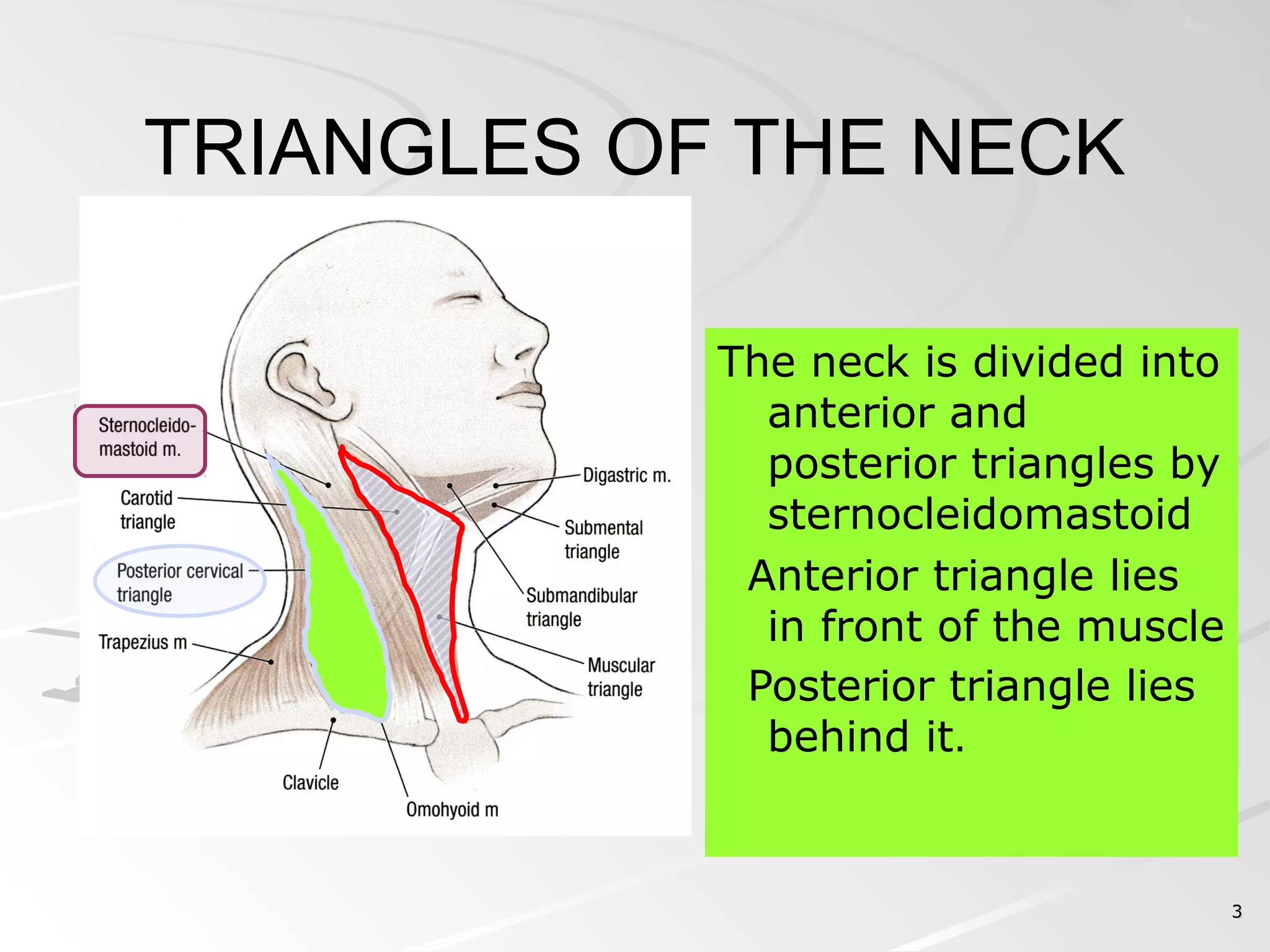
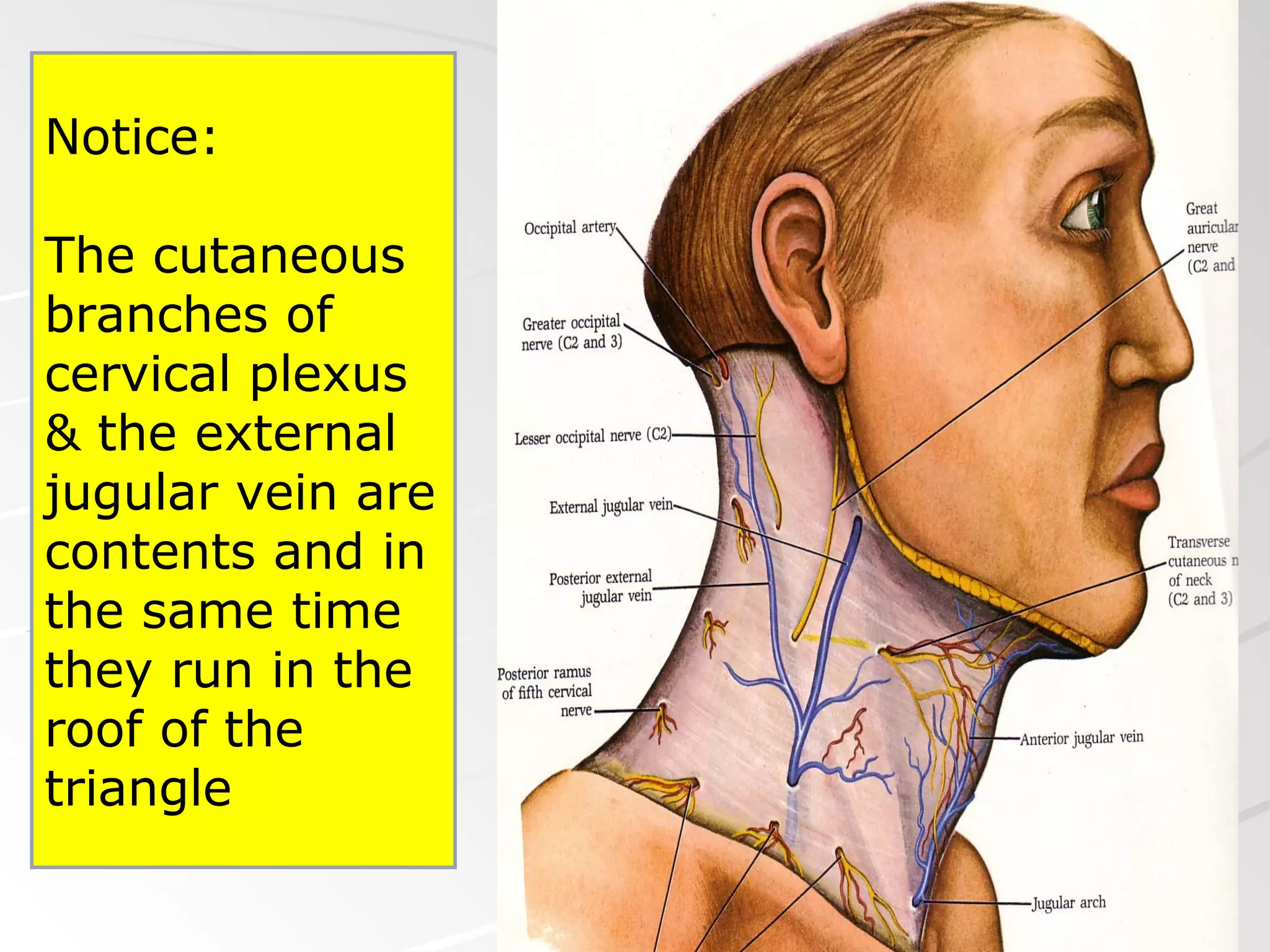
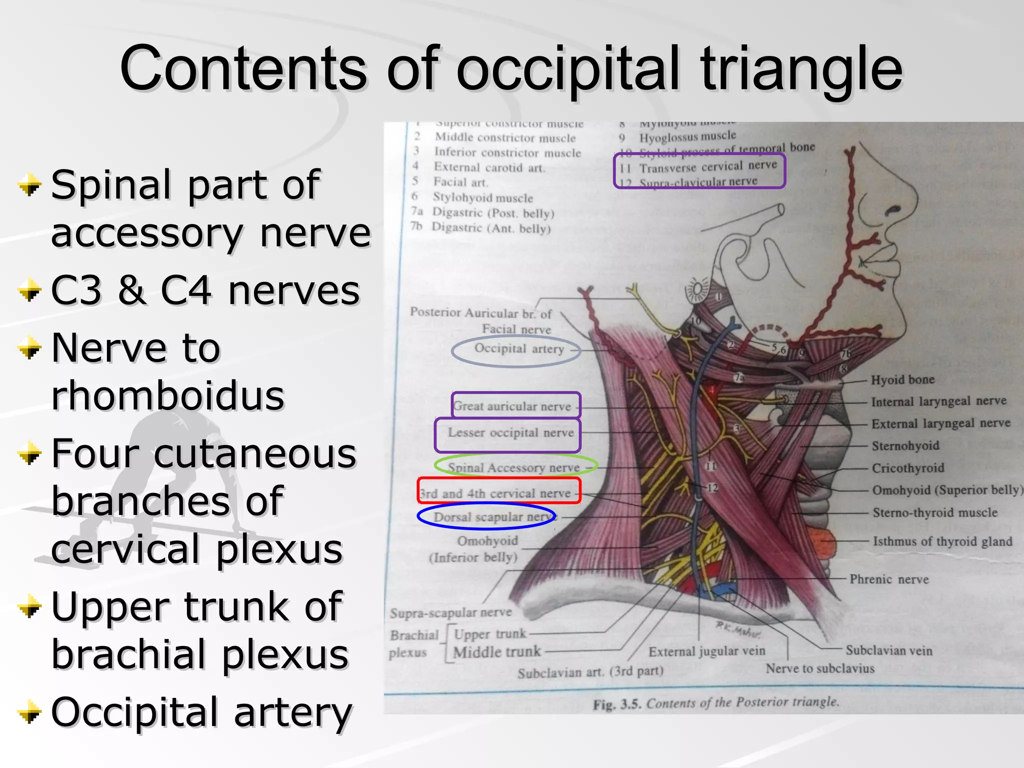
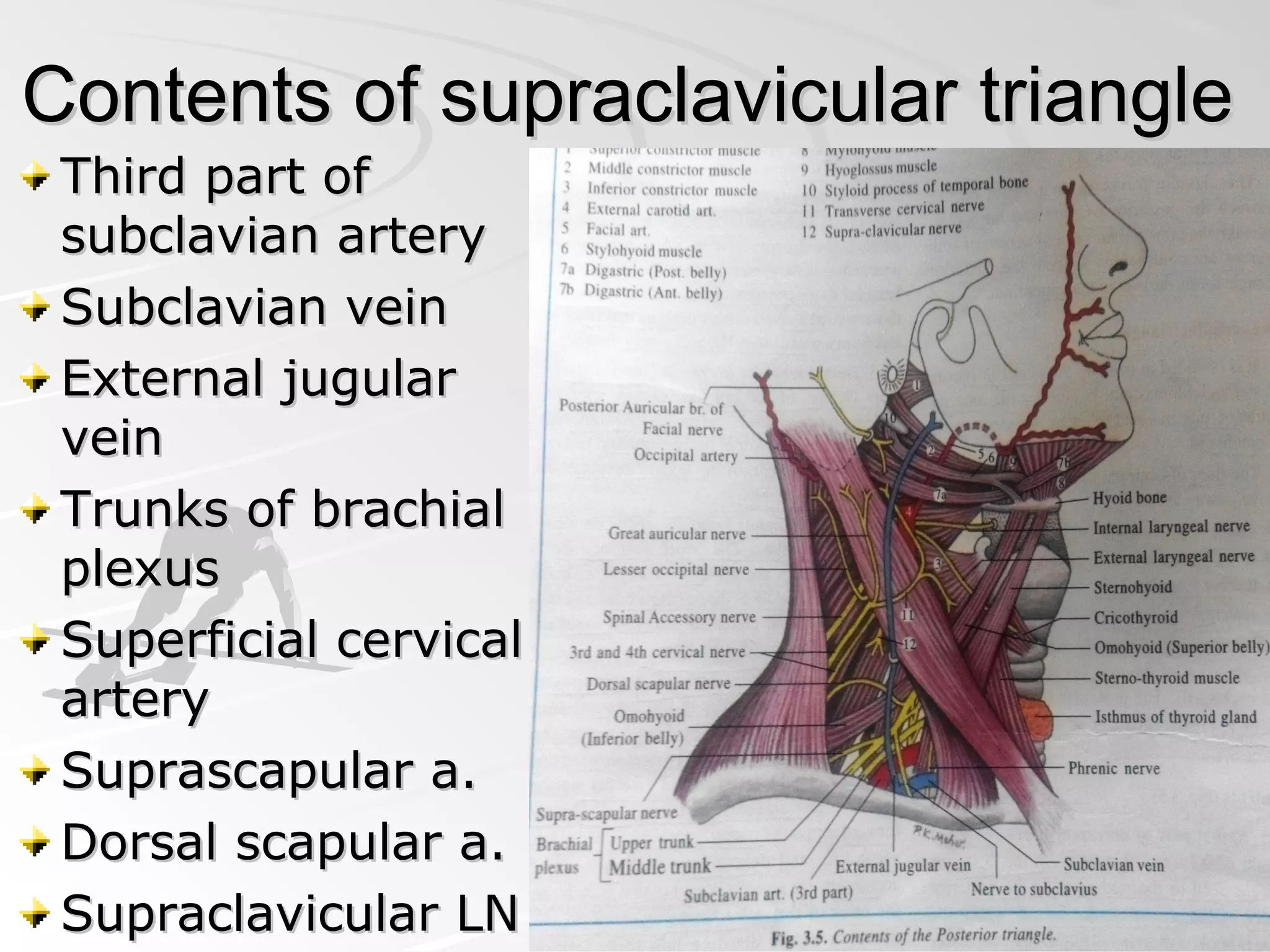
Posterior triangle ofPosterior triangle of
neckneck
Dr Pratik Mistry
22
STERNOCLEIDOMASTOID
Sternocleidomastoid
is a strap muscle.
It extends obliquely
across the side of
the neck.
It forms a prominent
surface landmark.
It divides the side of
the neck into
anterior & posterior
triangles
33
The neck is divided into
anterior and
posterior triangles by
sternocleidomastoid
Anterior triangle lies
in front of the muscle
Posterior triangle lies
behind it.
TRIANGLES OF THE NECK
Torticollis (wry neck)Torticollis (wry neck)
55
Boundaries of the
Posterior triangle:
Anteriorly: posterior
border of sternomastoid
Posteriorly: anterior
border of trapezius
Inferiorly: middle third
of the clavicle.
66
OMOHYOID MUSCLE
The omohyoid
muscle has:
 Inferior belly
 Intermediate
tendon
 Superior
belly.
77
The inferior belly
of omohyoid
subdivides the
posterior triangle
into:
 a large occipital
triangle above
a small
supraclavicular
triangle below.
88
The roof of the triangle is
covered by:
 Skin.
 superficial fascia, which
contains:
 platysma,
 cutaneous branches of
cervical plexus
 External jugular vein
 Investing layer of deep
cervical fascia.
1010
PLATYSMA MUSCLE
The platysma can
be seen as a sheet
of muscle by
asking the patient
to clench the jaws
firmly.
It extends from
the body of the
mandible
downward over
the clavicle onto
the anterior
thoracic wall.
1111
Running across
the triangle in its
covering:
the cutaneous
branches of
cervical plexus
1313
BRANCHES OF THE CERVICAL PLEXUS
The cervical plexus is
formed by the anterior
rami of C1,C2,C3 &
C4.
Several cutaneous
nerves emerge from
under the middle of
the posterior border
of the sternomastoid
muscle.
They innervate parts
of the skin in the head
and neck region.
1414
LESSER OCCIPITAL NERVE
The lesser occipital
nerve is a branch of
C2.
It hooks around the
accessory nerve and
ascends along the
posterior border of the
sternomastoid muscle
It supply the skin over
the lateral part of the
occipital region and
the medial surface of
the auricle.
1515
GREAT AURICULAR NERVE
The great auricular
nerve is a branch of
C2 & C3.
It ascends across
the sternomastoid
where it divides into
branches that
supply the skin over
the angle of the
mandible, the
parotid gland, and
the auricle.
1616
TRANSVERSE CERVICAL NERVE
Transverse cervical nerve
of the neck is a branch of
C2 and C3.
It emerges from behind
the middle of the
posterior border of the
sternomastoid.
It passes forward across
that muscle and divides
into branches that supply
the skin on the anterior
and lateral surfaces of the
neck, from the body of
the mandible to the
sternum.
1717
SUPRACLAVICULAR NERVES
The supraclavicular
nerves are branches
of C3 and C4.
They emerge from
beneath the posterior
border of the
sternocleidomastoid
muscle and descend
across the side of the
neck.
They pass onto the
chest wall and
shoulder region, down
to the level of the
second rib.
1818
EXTERNAL JUGULAR VEIN
The External
jugular vein
begins just
behind the angle
of the mandible
by the union of
the posterior
auricular vein
with the
posterior division
of the
retromandibular
vein.
2020
TRIBUTARIES Posterior auricular
vein.
Posterior division of
the retromandibular
vein.
Transverse cervical
vein.
Suprascapular vein.
Anterior jugular vein.
Posterior external
jugular vein, a small
vein that drains the
posterior part of the
scalp and neck and
joins the external
jugular vein about
halfway along its
course.
Notice:
The cutaneous
branches of
cervical plexus
& the external
jugular vein are
contents and in
the same time
they run in the
roof of the
triangle
Parts of Deep
cervical
fascia:
Investing
layer.
Prevertebral
layer.
Pretracheal
layer.
Carotid
sheath.
2323
The FLOOR of the
triangle is covered by the
prevertebral layer of
deep cervical fascia.
It is formed from below
upward by the:
– Scalenus medius.
– Scalenus posterior
– Levator scapulae
– Splenius capitis, &
Semispinalis capitis,
A small part of the
scalenus anterior may
be present, but it is
usually overlapped and
hidden by the
sternocleidomastoid.
Contents of occipital triangleContents of occipital triangle
Spinal part ofSpinal part of
accessory nerveaccessory nerve
C3 & C4 nervesC3 & C4 nerves
Nerve toNerve to
rhomboidusrhomboidus
Four cutaneousFour cutaneous
branches ofbranches of
cervical plexuscervical plexus
Upper trunk ofUpper trunk of
brachial plexusbrachial plexus
Occipital arteryOccipital artery
2525
ACCESSORY NERVE
(SPINAL PART)
The spinal part of
the accessory nerve
enters the posterior
triangle by emerging
from beneath the
middle of the
posterior border of
sternomastoid.
It runs downward
and laterally across
the posterior triangle
on the levator
scapulae muscle
2626
OCCIPITAL ARTERY It is a branch of the
external carotid
artery.
It enters the posterior
triangle at its superior
angle, appearing
between the
sternomastoid &
trapezius muscles.
Then, it ascends in a
tortuous course over
the back of the scalp,
accompanied by the
greater occipital
nerve.
Contents of supraclavicular triangleContents of supraclavicular triangle
Third part ofThird part of
subclavian arterysubclavian artery
Subclavian veinSubclavian vein
External jugularExternal jugular
veinvein
Trunks of brachialTrunks of brachial
plexusplexus
Superficial cervicalSuperficial cervical
arteryartery
Suprascapular a.Suprascapular a.
Dorsal scapular a.Dorsal scapular a.
Supraclavicular LNSupraclavicular LN
2828
THIRD PART OF THE SUBCLAVIAN ARTERY
The subclavian artery
is divided into three
parts by the scalenus
anterior muscle, which
crosses in front of the
artery.
 First part medial to
scalenus anterior.
 Second part lies
behind the muscle.
 Third part extends
from the lateral border
of the muscle to the
outer border of the first
rib; where, it is
continuous as the
axillary artery.
2929
The third part of the subclavian artery enters the
anteroinferior angle of the posterior triangle and
disappears behind the middle of the clavicle.
3030
BRACHIAL PLEXUS
The brachial
plexus is formed
from the
anterior rami of
the C 5th
, 6th
, 7th
,&
8th
&T 1st
.
It lies in the
subclavian
triangle of the
posterior
triangle.
3131
The trunks of the
brachial plexus
enter the posterior
triangle of the
neck through the
interval between
the scalenus
anterior and the
scalenus medius
muscles.
Branches of supraclavicular part ofBranches of supraclavicular part of
brachial plexus in the trianglebrachial plexus in the triangle
Nerve toNerve to
rhomboidsrhomboids
Nerve to serratusNerve to serratus
anterioranterior
Nerve to subclaviusNerve to subclavius
SuprascapularSuprascapular
nervenerve
3333
SUPERFICIAL CERVICAL ARTERY
It is a branch of
the thyrocervical
trunk, of the first
part of the
subclavian artery.
It runs across the
lower part of the
posterior triangle
and disappears
deep to the
trapezius muscle.
3434
SUPASCAPULAR ARTERY
The suprascapular
artery is also a
branch of the
thyrocervical trunk.
It runs across the
lower part of the
posterior triangle.
It follows the
suprascapular nerve
into the supraspinous
fossa and takes part
in the anastomosis
around the scapula.
Supraclavicular lymph nodesSupraclavicular lymph nodes
In relation withIn relation with
inferior belly ofinferior belly of
omohyoidomohyoid
Drain lymph fromDrain lymph from
back of scalp, neckback of scalp, neck
and from upperand from upper
deep cervical,deep cervical,
axillary nodesaxillary nodes
Empty into theEmpty into the
jugular lymphjugular lymph
trunktrunk
Clinical anatomyClinical anatomy
Subclavian steal syndromeSubclavian steal syndrome
Cervical ribCervical rib
Swellings in the posteriorSwellings in the posterior
trianglestriangles
Tuberculosis of cervical vertebraTuberculosis of cervical vertebra
Enlarged supraclavicular lymphEnlarged supraclavicular lymph
nodes (Virchow's nodes) as seen innodes (Virchow's nodes) as seen in
many infections like TB, Lymphoma,many infections like TB, Lymphoma,
malignancies of breast, stomach,malignancies of breast, stomach,
testis and other abdominal organstestis and other abdominal organs
External jugular veinExternal jugular vein
Injury to EJV in theInjury to EJV in the
supraclavicularsupraclavicular
space cause airspace cause air
embolism andembolism and
sudden death.sudden death.
JVPJVP
Raised in rightRaised in right
sided heart failuresided heart failure
THANK YOUTHANK YOU

posterior triangle of neck

  • 1.
    Posterior triangle ofPosteriortriangle of neckneck Dr Pratik Mistry
  • 2.
    22 STERNOCLEIDOMASTOID Sternocleidomastoid is a strapmuscle. It extends obliquely across the side of the neck. It forms a prominent surface landmark. It divides the side of the neck into anterior & posterior triangles
  • 3.
    33 The neck isdivided into anterior and posterior triangles by sternocleidomastoid Anterior triangle lies in front of the muscle Posterior triangle lies behind it. TRIANGLES OF THE NECK
  • 4.
  • 5.
    55 Boundaries of the Posteriortriangle: Anteriorly: posterior border of sternomastoid Posteriorly: anterior border of trapezius Inferiorly: middle third of the clavicle.
  • 6.
    66 OMOHYOID MUSCLE The omohyoid musclehas:  Inferior belly  Intermediate tendon  Superior belly.
  • 7.
    77 The inferior belly ofomohyoid subdivides the posterior triangle into:  a large occipital triangle above a small supraclavicular triangle below.
  • 8.
    88 The roof ofthe triangle is covered by:  Skin.  superficial fascia, which contains:  platysma,  cutaneous branches of cervical plexus  External jugular vein  Investing layer of deep cervical fascia.
  • 10.
    1010 PLATYSMA MUSCLE The platysmacan be seen as a sheet of muscle by asking the patient to clench the jaws firmly. It extends from the body of the mandible downward over the clavicle onto the anterior thoracic wall.
  • 11.
    1111 Running across the trianglein its covering: the cutaneous branches of cervical plexus
  • 13.
    1313 BRANCHES OF THECERVICAL PLEXUS The cervical plexus is formed by the anterior rami of C1,C2,C3 & C4. Several cutaneous nerves emerge from under the middle of the posterior border of the sternomastoid muscle. They innervate parts of the skin in the head and neck region.
  • 14.
    1414 LESSER OCCIPITAL NERVE Thelesser occipital nerve is a branch of C2. It hooks around the accessory nerve and ascends along the posterior border of the sternomastoid muscle It supply the skin over the lateral part of the occipital region and the medial surface of the auricle.
  • 15.
    1515 GREAT AURICULAR NERVE Thegreat auricular nerve is a branch of C2 & C3. It ascends across the sternomastoid where it divides into branches that supply the skin over the angle of the mandible, the parotid gland, and the auricle.
  • 16.
    1616 TRANSVERSE CERVICAL NERVE Transversecervical nerve of the neck is a branch of C2 and C3. It emerges from behind the middle of the posterior border of the sternomastoid. It passes forward across that muscle and divides into branches that supply the skin on the anterior and lateral surfaces of the neck, from the body of the mandible to the sternum.
  • 17.
    1717 SUPRACLAVICULAR NERVES The supraclavicular nervesare branches of C3 and C4. They emerge from beneath the posterior border of the sternocleidomastoid muscle and descend across the side of the neck. They pass onto the chest wall and shoulder region, down to the level of the second rib.
  • 18.
    1818 EXTERNAL JUGULAR VEIN TheExternal jugular vein begins just behind the angle of the mandible by the union of the posterior auricular vein with the posterior division of the retromandibular vein.
  • 20.
    2020 TRIBUTARIES Posterior auricular vein. Posteriordivision of the retromandibular vein. Transverse cervical vein. Suprascapular vein. Anterior jugular vein. Posterior external jugular vein, a small vein that drains the posterior part of the scalp and neck and joins the external jugular vein about halfway along its course.
  • 21.
    Notice: The cutaneous branches of cervicalplexus & the external jugular vein are contents and in the same time they run in the roof of the triangle
  • 22.
  • 23.
    2323 The FLOOR ofthe triangle is covered by the prevertebral layer of deep cervical fascia. It is formed from below upward by the: – Scalenus medius. – Scalenus posterior – Levator scapulae – Splenius capitis, & Semispinalis capitis, A small part of the scalenus anterior may be present, but it is usually overlapped and hidden by the sternocleidomastoid.
  • 24.
    Contents of occipitaltriangleContents of occipital triangle Spinal part ofSpinal part of accessory nerveaccessory nerve C3 & C4 nervesC3 & C4 nerves Nerve toNerve to rhomboidusrhomboidus Four cutaneousFour cutaneous branches ofbranches of cervical plexuscervical plexus Upper trunk ofUpper trunk of brachial plexusbrachial plexus Occipital arteryOccipital artery
  • 25.
    2525 ACCESSORY NERVE (SPINAL PART) Thespinal part of the accessory nerve enters the posterior triangle by emerging from beneath the middle of the posterior border of sternomastoid. It runs downward and laterally across the posterior triangle on the levator scapulae muscle
  • 26.
    2626 OCCIPITAL ARTERY Itis a branch of the external carotid artery. It enters the posterior triangle at its superior angle, appearing between the sternomastoid & trapezius muscles. Then, it ascends in a tortuous course over the back of the scalp, accompanied by the greater occipital nerve.
  • 27.
    Contents of supraclaviculartriangleContents of supraclavicular triangle Third part ofThird part of subclavian arterysubclavian artery Subclavian veinSubclavian vein External jugularExternal jugular veinvein Trunks of brachialTrunks of brachial plexusplexus Superficial cervicalSuperficial cervical arteryartery Suprascapular a.Suprascapular a. Dorsal scapular a.Dorsal scapular a. Supraclavicular LNSupraclavicular LN
  • 28.
    2828 THIRD PART OFTHE SUBCLAVIAN ARTERY The subclavian artery is divided into three parts by the scalenus anterior muscle, which crosses in front of the artery.  First part medial to scalenus anterior.  Second part lies behind the muscle.  Third part extends from the lateral border of the muscle to the outer border of the first rib; where, it is continuous as the axillary artery.
  • 29.
    2929 The third partof the subclavian artery enters the anteroinferior angle of the posterior triangle and disappears behind the middle of the clavicle.
  • 30.
    3030 BRACHIAL PLEXUS The brachial plexusis formed from the anterior rami of the C 5th , 6th , 7th ,& 8th &T 1st . It lies in the subclavian triangle of the posterior triangle.
  • 31.
    3131 The trunks ofthe brachial plexus enter the posterior triangle of the neck through the interval between the scalenus anterior and the scalenus medius muscles.
  • 32.
    Branches of supraclavicularpart ofBranches of supraclavicular part of brachial plexus in the trianglebrachial plexus in the triangle Nerve toNerve to rhomboidsrhomboids Nerve to serratusNerve to serratus anterioranterior Nerve to subclaviusNerve to subclavius SuprascapularSuprascapular nervenerve
  • 33.
    3333 SUPERFICIAL CERVICAL ARTERY Itis a branch of the thyrocervical trunk, of the first part of the subclavian artery. It runs across the lower part of the posterior triangle and disappears deep to the trapezius muscle.
  • 34.
    3434 SUPASCAPULAR ARTERY The suprascapular arteryis also a branch of the thyrocervical trunk. It runs across the lower part of the posterior triangle. It follows the suprascapular nerve into the supraspinous fossa and takes part in the anastomosis around the scapula.
  • 35.
    Supraclavicular lymph nodesSupraclavicularlymph nodes In relation withIn relation with inferior belly ofinferior belly of omohyoidomohyoid Drain lymph fromDrain lymph from back of scalp, neckback of scalp, neck and from upperand from upper deep cervical,deep cervical, axillary nodesaxillary nodes Empty into theEmpty into the jugular lymphjugular lymph trunktrunk
  • 36.
    Clinical anatomyClinical anatomy Subclaviansteal syndromeSubclavian steal syndrome
  • 37.
  • 38.
    Swellings in theposteriorSwellings in the posterior trianglestriangles Tuberculosis of cervical vertebraTuberculosis of cervical vertebra Enlarged supraclavicular lymphEnlarged supraclavicular lymph nodes (Virchow's nodes) as seen innodes (Virchow's nodes) as seen in many infections like TB, Lymphoma,many infections like TB, Lymphoma, malignancies of breast, stomach,malignancies of breast, stomach, testis and other abdominal organstestis and other abdominal organs
  • 39.
    External jugular veinExternaljugular vein Injury to EJV in theInjury to EJV in the supraclavicularsupraclavicular space cause airspace cause air embolism andembolism and sudden death.sudden death. JVPJVP Raised in rightRaised in right sided heart failuresided heart failure
  • 41.