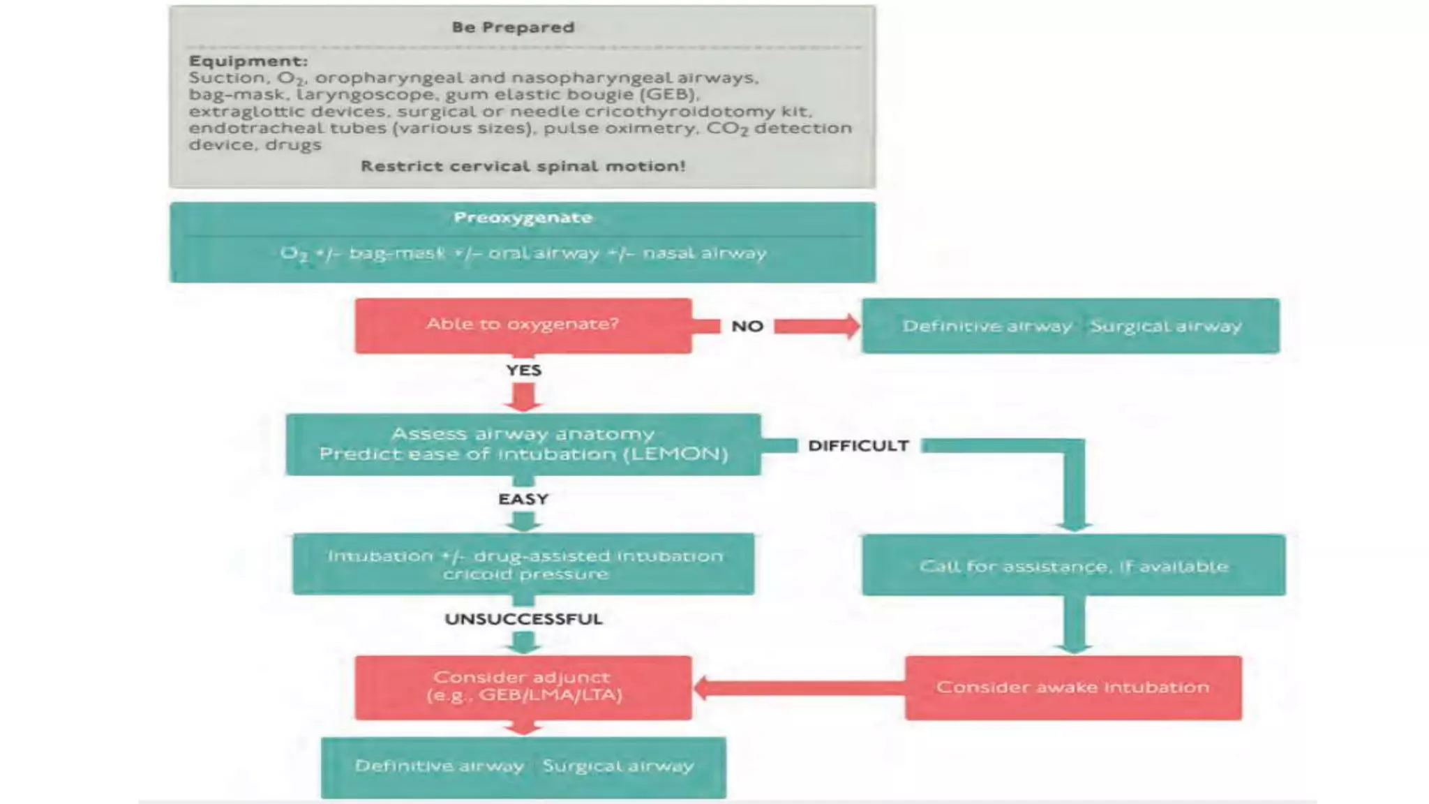
SlideShare a Scribd company logo
1 of 58
Polytrauma- Assessment and management
till discharge
(ATLS Recent guideline)
Presenter: Dr. Bokinala Naveen
Moderator: Dr Saurabh Taneja
• Preparation
• Triage
• Primary survey
• Need for patient transfer
• Secondary survey
• Adjuncts
• Continued post resuscitation monitoring and evaluation
• Definitive care
Definition
• The term ‘polytrauma’ was first used by Tscherne et al., in 1966 for
patients that demonstrated a combination of at least 2 ‘severe injuries
of the head, chest or abdomen’, or ‘one of them in association with an
extremity injury’.
• Trentz in 1999 defined Polytrauma is a syndrome of multiple injuries
exceeding a defined severity (Injury Severity Score [ISS] >17) with
sequential systemic reactions that can lead to dysfunction or failure of
remote organs and vital systems, which have not themselves been
directly injured.
• Oestern, Hans-Jörg; Trentz, Otmar; Uranues, Selman (2014). General Trauma Care and Related Aspects || Polytrauma: Pathophysiology, Priorities, and
Management. , 10.1007/978-3-540-88124-7(Chapter 5), 69–76. doi:10.1007/978-3-540-88124-7_5
Injury severity score
Baker SP, O'Neill B, Haddon W Jr, Long WB. The injury severity score: a method for describing patients with multiple injuries and evaluating emergency care.
The Journal of Trauma. 1974 Mar;14(3):187-196. PMID: 4814394.
Trimodal distribution of trauma deaths (ATLS 10th ed)
First Peak
• Within minutes
• Apnea due to TBI or
high spinal cord injury
or rupture of the
heart, aorta or other
large blood vessels.
• Very few of these
patients survive
Second Peak
• Minutes to hours
• Injuries like SDH/EDH,
hemopneumothorax,
ruptured spleen,
lacerations of the
liver, pelvic fractures,
and/or injuries
associated with
significant blood loss.
Third peak
• Days to weeks
• Sepsis and Multiple
organ system
dysfunctions.
Gunst M, Ghaemmaghami V, Gruszecki A, et al. Changing epidemiology of trauma deaths leads to a bimodal
distribution. Proc (Baylor Univ Med Cent), 2010;23(4):349–354
Pre Hospital set up and levels of Trauma centres
• The prehospital set up is to
notify the receiving hospital
before personnel transport
the patient from the scene.
• This allows for mobilization
of the hospital’s trauma team
members so that all
necessary personnel and
resources are present in the
emergency department (ED)
at the time of the patient’s
arrival.
• Scoop and run
• Multiple vs mass casualty
Pre Hospital phase – admit to Level 1 trauma set up
1. GCS <13
2. Systolic BP <90
3. RR <10 or >29 / needs ventilator support
Anatomical injury
1. Penetrating trauma : head, neck, torso and extremities proximal to elbow/knee
2. Flial chest, pelvic fractures
3. 2 or more proximal long bone fractures
4. Proximal amputation (Wrist/Ankle)
5. Crushed, degloved, mangled, pulseless extremity
6. Depressed skull fractures
7. Paralysis
Based on the mechanism of injury & high-energy impact –
Transfer to Level 2 trauma setup
Falls
• Adults > 20 feet or 6 meters (2 stories)
• Children >10 feet or 3 meters (2-3 times the height of child)
High risk motor vehicle crash
• Intrusion, including roof >12 inches from occupant side
• Ejection (partial or complete from the vehicle)
• Death in same passenger compartment
• Vehicle telemetry data consistent with injury
Auto vs pedestrian/bicyclist thrown, run over with significant impact (>32kmph)
Motor cycle crash >32kmph
Special patient or system considerations
Older adults
• Risk of injury/death more in age >55 years
• Systolic BP <110mmHg in age >55 years
• Low impact mechanism (ground-level fall)
Patients on anticoagulation are at high risk for bleed after injury
Children preferably to a pediatric capable trauma center
Burns with trauma to a trauma center or else burns facility
Pregnant >20 weeks
Primary Survey
Rapid examination to identify and treat life threatening conditions
• A – Airway maintenance with C-spine protection
• B – Breathing and ventilation
• C – Circulation with hemorrhage control
• D – Disability and neurological status
• E – Exposure/ Environmental control
10 second assessment : Ask the patient to identify himself, name and
what happened
A-Airway
• Check for airway patency and suction the accumulated secretions/ blood
• Inspect for foreign bodies; identify facial, mandibular, and/or tracheal/laryngeal
fractures causing airway obstruction.
• Hoarseness, subcutaneous emphysema, palpable fracture suggests laryngeal
trauma
• Patients with facial burns and those with potential inhalation injury are at risk for
insidious respiratory compromise.
• For this reason, consider preemptive intubation
in burn patients.
• Patients with severe head injuries who have an altered level of
consciousness or GCS score of 8 or lower usually require the placement
of a definitive cuffed airway
• Initially, the jaw-thrust or chin-lift maneuver often suffices as an initial
intervention.
• If the patient is unconscious and has no gag reflex, the placement of an
oropharyngeal airway can be helpful temporarily.
• Establish a patent airway while restricting cervical spine motion.
• Use Cervical collar when necessary
Canadian C-spine rule
To be more selective in use of radiography in alert and stable
trauma patients.
Design: Prospective cohort study conducted from October 1996 to April
1999, sample of 8924 adults (mean age, 37 years) with blunt trauma to the
head/neck, stable vital signs, and a GCS of 15.
151 patients had (1.7%) had C-spine injury.
The resultant model and final Canadian C-Spine Rule comprises 3 main
questions:
(1) is there any high-risk factor present that mandates radiography
(2) is there any low-risk factor present that allows safe assessment of range
of motion
(3) is the patient able to actively rotate neck 45 degrees to the left and
right?
By cross-validation, this rule had 100% sensitivity (95% confidence interval
[CI], 98%-100%) and 42.5% specificity (95% CI, 40%-44%) for identifying
151 clinically important C-spine injuries. The potential radiography ordering
rate would be 58.2%.
The Canadian C-Spine rule of radiography in alert and stable trauma patients. JAMA
2001;286:1841–1848.
NEXUS STUDY
Cervical-spine radiography does not need to be performed on
selected blunt trauma patients who are awake, alert, non
intoxicated, do not complain of midline neck pain, and have no
tenderness over the bony cervical spine.
• Study population:1000 patients in the UCLA Emergency Medicine
Center with a chief complaint of blunt trauma, for whom cervical-
spine films were ordered and for whom prospective data
questionnaires were completed.
• Results:
• 27 patients with cervical-spine fracture were among the 974
patients. All 27 patients with fracture had at least one of the
following four characteristics: midline neck tenderness, evidence of
intoxication, altered level of alertness, or a severely painful injury
elsewhere. Three hundred fifty-three of 947 (37.3%) patients without
cervical-spine fracture had none of these findings.
• Conclusion: Cervical-spine radiology may not be necessary in
patients without spinous tenderness in the neck, intoxication,
altered level of alertness, or other severely painful injury.
Hoffman JR, Schriger DL, Mower W, Luo JS, Zucker M. Low-risk criteria for cervical-spine eadiography in blunt trauma: a prospective study. Ann Emerg Med. 1992
Dec;21(12):1454- 60
Burns
• Risk for upper airway obstruction increases with burn size and depth, burns to the head and
face, inhalation injury, associated trauma, and burns inside the mouth causing edema and pose
a greater risk for airway compromise
• Pharyngeal thermal injuries can produce marked upper airway edema, and early protection of
the airway is critical.
• The clinical manifestations of inhalation injury may be subtle and frequently do not appear in
the first 24 hours.
• If the provider waits for x-ray evidence of pulmonary injury or changes in blood gas
determinations, airway edema can preclude intubation, and a surgical airway may be required.
• Examine the patient’s oropharynx for signs of inflammation, mucosal injury, soot in
the pharynx, and edema, taking care not to injure the area further.
• A carboxyhemoglobin level greater than 10% in a patient who was involved in a fire
also suggests inhalation injury.
• Transfer to a burn center is indicated for patients suspected of experiencing
inhalation injury; however, if the transport time is prolonged, intubate the patient
before transport.
• Circumferential burns of the neck can lead to swelling of the tissues around the
airway; therefore, early intubation is also indicated for full thickness circumferential
neck burns
American Burn Life Support (ABLS) indications for early intubation include:
• Signs of airway obstruction (hoarseness, stridor, accessory respiratory muscle use,
sternal retraction)
• Extent of the burn (total body surface area burn > 40%–50%)
• Extensive and deep facial burns
• Burns inside the mouth
• Significant edema or risk for edema
• Difficulty swallowing
• Signs of respiratory compromise: inability to clear secretions, respiratory fatigue, poor
oxygenation or ventilation
• Decreased level of consciousness where airway protective reflexes are impaired
• Anticipated patient transfer of large burn with airway issue without qualified personnel to
intubate en route
Breathing
Look
• Rate and depth of
respiration
• See for engorged neck
veins
• Tracheal deviation
• U/L and B/L chest
movements
• Use of accessory muscles
Listen
• Chest bilaterally
• Hoarseness
• Dysphonia
Feel
• Chest for dullness
(hemothorax) or hyper-
resonance(pneumothorax)
• Crepitus(Emphysema)
• Deformity
Tension pneumothorax
• Tension pneumothorax develops when a “one-way valve” air leak
occurs from the lung or through the chest Wall.
• Air is forced into the pleural space with no means of escape,
eventually collapsing the affected lung.
• Chest pain, Air hunger, Tachypnea, Respiratory distress,
Tachycardia, Hypotension, Tracheal deviation away from the side
of the injury, Unilateral absence of breath sounds, Elevated
hemithorax without respiratory movement, Neck vein distention
Cyanosis (late manifestation)
• The mediastinum is displaced to the opposite side,
decreasing venous return and compressing the
opposite lung.
• Shock results from marked decrease in venous return,
causing a reduction in cardiac output
• A simple pneumothorax can be converted to a tension
pneumothorax when a patient is intubated and
positive pressure ventilation is provided before
decompressing the pneumothorax with a chest tube.
• Needle decompression (5th ICS anterior to MAL)
converts tension pneumothorax to a simple
pneumothorax and ICD to be placed as early as
possible
• Tension pneumothorax can be managed initially
by rapidly applying the finger decompression
technique.
Flail chest
• When a segment of the chest wall does not have bony continuity with the rest of the thoracic cage.
• This condition usually results from trauma associated with multiple rib fractures (i.e., 2 or more
adjacent ribs fractured in 2 or more places), although it can also occur when there is a costochondral
separation of a single rib from the thorax
• Decreased respiratory effort, combined with contusion and atelectasis, may limit movement of the
chest wall
• Definitive treatment is oxygen therapy, adequate ventilation, and cautious fluid resuscitation.
• Analgesia with IV narcotics or local anesthetic administration
• Options for administering local anesthetics include intermittent intercostal nerve block(s) and
transcutaneous intrapleural, extrapleural, or epidural anesthesia.
Open Pneumothorax
• Large injuries to the chest wall that remain open results in an open
pneumothorax(sucking chest wound).
• Equilibration between intrathoracic pressure and atmospheric
pressure is immediate.
• Air tends to follow the path of least resistance, when the opening in
the chest wall is approximately two-thirds the diameter of the
trachea or greater, air passes preferentially through the chest wall
defect with each inspiration.
• Effective ventilation is impaired, leading to hypoxia and hypercarbia.
• The clinical signs and symptoms are pain, difficulty breathing,
tachypnea, decreased breath sounds on the affected side, and noisy
movement of air through the chest wall injury.
• Close the defect with a sterile dressing large enough to overlap the
wound’s edges.
• Any occlusive dressing (e.g. plastic wrap or petrolatum gauze) may
be used as temporary measure to enable rapid assessment to
continue.
• Tape it securely on only three sides to provide a flutter-valve effect
• As the patient breathes in, the dressing occludes the wound,
preventing air from entering. During exhalation, the open end of
the dressing allows air to escape from the pleural space.
• Taping all four edges of the dressing can cause air to accumulate in
the thoracic cavity, resulting in a tension pneumothorax unless a
chest tube is in place.
• Place a chest tube remote from the wound as soon as possible.
Subsequent definitive surgical closure of the wound is frequently
required.
Circulation- Shock
• Any injured patient who is cool to the touch and is tachycardic should be
considered to be in shock until proven otherwise
• Identify the probable cause of shock and adjust treatment accordingly
• Check peripheral pulses, heart rate, BP, pulse pressure, capillary refill, cyanosis
• Most injured patients have hypovolemic shock due to blood loss
• Shock may be either hemorrhagic or non hemorrhagic
• 2 large bore peripheral IV’s (at least 18 gauge) and control bleeding
Focused Assessment with Sonography for Trauma
(FAST & eFAST)
Look for blood : 4 places other than floor
A. The chest
B. The abdomen
C. The pelvis and retroperitoneum
D. Major long bones and soft tissues.
• C
• B
• D`2
Hemorrhagic shock
• Hemorrhage is the most common cause of shock in trauma patients.
• The trauma patient’s response to blood loss is made more complex by fluid shifts among
the fluid compartments in the body, particularly in the extracellular fluid compartment.
• Soft tissue injury, even without severe hemorrhage, can result in shifts of fluid to the
extracellular compartment.
• The response to blood loss must be considered in the context of these fluid shifts. Also
consider the changes associated with severe, prolonged shock and the pathophysiologic
results of resuscitation and reperfusion.
Hemorr hagic Shock
Initial Resuscitation
Cardiogenic Shock
• Cardiac injury, cardiac tamponade, an air embolus, or, rarely, myocardial infarction may cause
• Suspect a blunt cardiac injury when the mechanism of injury to the thorax involves rapid
deceleration.
• All patients with blunt thoracic trauma need continuous electrocardiographic (ECG)
monitoring to detect injury patterns and dysrhythmias.
• The shock state may be secondary to myocardial infarction in the elderly and other high-risk
patients, such as those with cocaine intoxication.
• Cardiac enzyme levels may assist in diagnosing and treating injured patients in the
emergency department (ED), as acute myocardial ischemia may be the precipitating event.
Cardiac Tamponade
• Cardiac tamponade is most commonly encountered in penetrating thoracic trauma, it can also result
from blunt injury to the thorax.
• Tachycardia, muffled heart sounds, and dilated, engorged neck veins with hypotension and insufficient
response to fluid therapy suggest cardiac tamponade –becks traid (Dilated RA, RV)
• Absence of these classic findings does not exclude.
• Tension pneumothorax can mimic cardiac tamponade, with findings of distended neck veins and
hypotension in both. However, absent breath sounds and hyper-resonant percussion are not present
with tamponade.
• Echocardiography can be used , FAST to identify pericardial fluid,
• Cardiac tamponade is best managed by formal
operative intervention, as pericardiocentesis is
at best only a temporizing maneuver
• Tension pneumothorax
Neurogenic Shock
• Isolated intracranial injuries do not cause shock unless the brainstem is injured.
• Cervical and upper thoracic spinal cord injuries can produce hypotension due to loss of
sympathetic tone
• The classic presentation of neurogenic shock is hypotension without tachycardia or
cutaneous vasoconstriction.
• A narrowed pulse pressure is not seen in neurogenic shock.
• The failure of fluid resuscitation to restore organ perfusion and tissue oxygenation
suggests either continuing hemorrhage or neurogenic shock.
• Advanced techniques for monitoring intravascular volume status and cardiac output may
be helpful in managing this complex problem
Massive transfusion
1. Replacement of one entire blood volume within 24 h
2. Transfusion of >10 units of packed red blood cells (PRBCs) in 24 h
3. Transfusion of >20 units of PRBCs in 24 h
4. Transfusion of >4 units of PRBCs in 1 h when on-going need is foreseeable
5. Replacement of 50% of total blood volume (TBV) within 3 h.
Definitions of MBT suggested for use in children are
• Transfusion of >50% TBV in 3 h,
• Transfusion >100% TBV in 24 h or transfusion support to replace on-going blood
loss of >10% TBV/min
Patil V, Shetmahajan M. Massive transfusion and massive transfusion protocol. Indian journal of anaesthesia. 2014 Sep;58(5):590.
Criteria to trigger the activation of an MTP should include one or more of the following:
 Persistent hemodynamic instability
 Active bleeding requiring operation or angioembolization
 Blood transfusion in the trauma bay
If MTP triggers are met:
• Begin universal blood product infusion rather than crystalloid orcolloid solutions.
• Transfuse universal RBC and plasma in a ratio between 1:1 and 1:2 (plasmato RBC).
• Transfuse one single donor apheresisor random donor platelet pool for each six units of RBC.
• Blood products should be automatically sent by the transfusionservice in established ratios.
• Subsequent coolers should be delivered at 15-minute intervals until the MTP has been terminated.
• The goal is to keep at least one MTPcooler ahead for the duration of the MTP activation.
ACS TQIP MASSIVE TRANSFUSIONIN TRAUMA GUIDELINES
Endpoints of Transfusion
 The ratio-driven massive transfusion may be discontinued or downgraded to goal-directed transfusion
based on the laboratory findings if surgical bleeding has been controlled by the surgeon in the
operating room OR there is radiographic and physiologicevidence of bleeding control after
angioembolization.
 The MTP should be discontinued when there is recognition that further resuscitation is futile.
 The following should be used as guides to cease therapy with blood and blood components in a
patientwho is not actively bleeding and still in the acute resuscitation phase
 RBC transfusions for hemoglobin ≥10 g/dL
 Plasma transfusion for prothrombin time (PT) <18 seconds
 Plasma transfusion for activated partial thromboplastin time (aPTT) <35 seconds
 Platelet transfusions for platelet count >1.5 lac/ml
 Cryoprecipitate or fibrinogen concentrate for fibrinogen level >180 mg/dL
ACS TQIP MASSIVE TRANSFUSIONIN TRAUMA GUIDELINES
• If standard thrombelastography (TEG®) is available, the following cut-
points for transfusion triggers may also be used:
• Plasma for r-value >9 minutes
• Plasma and/or cryoprecipitate (fibrinogen concentrate) for k-time >4
minutes
• Cryoprecipitate (or fibrinogen concentrate) and/or plasm for α-angle
<60 degrees
• Platelets for mA <55 mm
• Anti-fibrinolytics for LY30 >7.5 %
ACS TQIP MASSIVE TRANSFUSIONIN TRAUMA GUIDELINES
If rapid TEG is available, the following cut-points for
transfusion triggers may also be used:
• Plasma for ACT >128 seconds
• Plasma and/or cryoprecipitate (fibrinogen concentrate) for k-
time >2.5 minutes
• Cryoprecipitate (fibrinogen concentrate) and/or plasma for α-
angle <60 degrees
• Platelets for mA <55 mm
• Antifibrinolytics for LY30 >3 %
Disability
• Level of consciousness, pupillary size and
reaction, lateralizing signs and spinal cord
injury level ( ability to move all extremities)
• Glasco Coma Scale
• Careful in head injury patients
Exposure
• Completely undress the patient and inspect the
entire patient from head to toe both front and
back
• Maintain spinal precautions during logrolling
• Inspect both axilla and perineum
• Warm blankets.
Secondary survey
• The secondary survey does not begin until the primary survey (ABCDE) is completed,
resuscitative efforts are underway, and improvement of the patient’s vital functions has
been demonstrated
• Complete history and physical examination
• Reassessment of all vital signs
AMPLE History
Allergies
Medications
Past medical history (LMP, transfusions)
Last meal
Events leading up to trauma
Physical examination
• Stepwise, organized
• Every patient, the same way, every time
• Superior to inferior; proximal to distal
• Look--Listen—Feel
 Neurologic
 Head
 Maxillofacial
 Cervical spine and neck
 Chest
 Abdomen
 Perineum/rectum/vagina
 Musculoskeletal
ADJUNCTS TO SECONDARY SURVEY
X ray examination
• Haemothorax or simple pneumothorax.
• Spine fractures
• Rib, Sternum, and Scapular Fractures
• Limb fractures
• Suspected of having smoke inhalation injury
• Appearance of an elevated right diaphragm on
a chest x-ray may be the only finding of a right-
sided injury.
CT Brain
• To rule out head injury
CT scans of head, chest, abdomen and spine
Contrast angiography
• Contrast-enhanced CT that demonstrates ruptured gastrointestinal tract, intraperitoneal
bladder injury, renal pedicle injury, or severe visceral parenchymal injury after blunt or
penetrating trauma
• Abdominal CT scan with IV contrast can document the presence and extent of a blunt
renal injury
• Suspected urinary system injuries are best evaluated by CECT. Intravenous pyelogram
(IVP) provides an alternative
• Some fractures traverse the carotid canals and can damage the carotid arteries
(dissection, pseudoaneurysm, or thrombosis.
• For detecting fractures of the posterior elements (pedicles, lamina, and spinous
processes) and determining the degree of canal compromise caused by burst fractures.
• CT angiography may be used to evaluate extremity vascular injuries
• Do not perform CT scanning if it delays the transfer of a patient to a higher level of care.
Bronchoscopy
• Tracheobronchial injury is a fatal condition
• The majority of tracheobronchial tree injuries occur within 1 inch (2.54 cm)
of the carina.
• High mortality rate from associated injuries, inadequate airway, or
development of a tension pneumothorax or tension pneumopericardium
• Patients typically present with hemoptysis, cervical subcutaneous
emphysema, tension pneumothorax, and/or cyanosis
• Smoke inhalation is a potential cause of airway obstruction from
particulate and chemical injury.
Esophagoscopy for suspected Oesophageal tear/ injury
Course of stay
Evaluate and manage
• DVT
• Infections
• Surgical management
• Nutritional management
• Pain relief
• Long term rehabilitation
New recommendations
Initial assessment Restriction to only 1 L of crystalloid fluid during initial assessment.
Airway & ventilation Drug-assisted intubation has now replaced rapid sequence intubation (RSI).
Video laryngoscopy highlighted as useful.
Shock Early use of blood products advocated.
Tranexamic acid is now recommended within 3 hours.
Thoracic trauma Flail chest replaced by tracheobronchial tree injury as a life-threatening injury.
5
th
ICS for needle thoracocentesis in adults
Modified FAST recommended for identification of pneumothorax.
Traumatic circulatory arrest algorithm (thoracotomy/pericardiotomy if no ROSC)
Chest size tube smaller 28-32F (previously 36-40F)
Abdominal & pelvic trauma Prostate examination no longer recommended as part of the evaluation.
Preperitoneal pelvic packing included in hemorrhage protocol.
Head trauma Anticoagulation reversal table is now included in the guidance.
Revised version of the GCS introduced.
Spine & spinal cord trauma CCR and NEXUS guidelines are now recommended.
“Spinal immobilisation” has been replaced with “spinal motion restriction.”
Prolonged backboard usage (>2 hours) should be avoided.
Musculoskeletal trauma The use of a tourniquet to control severe extremity bleeding is now recommended.
Antibiotics dosing regimens for open fractures introduced. Use weight based regimen
Thermal injuries New fluid resuscitation formula (2 ml/kg/%TBSA)
Paediatric trauma The PECARN traumatic brain injury algorithm now recommended.
(CT head- LOC >5sec, abnormal behavior, scalp hematoma, vomiting, headache)
Geriatric trauma Lower threshold for imaging in the elderly population is now recommended.
High-risk pre-existing conditions highlighted.(Cirrhosis, Coagulopathy, COPD, IHD, DM)
Trauma in pregnancy Vaginal fluid pH greater than 4.5 is an indicator of amniotic fluid leakage.
Transfer to definitive care CT scans should now be avoided in the primary hospital.
SBAR communication tool now recommended.
THANK YOU
Polytrauma- Assessment and management till discharge.pptx

More Related Content

What's hot

Crush injury-and-crush-syndrome
Crush injury-and-crush-syndromeCrush injury-and-crush-syndrome
Crush injury-and-crush-syndromedrmahbub88
 
Advanced trauma and life support (atls)
Advanced trauma and life support (atls)Advanced trauma and life support (atls)
Advanced trauma and life support (atls)anu_sandhya
 
Golden hour -Introduction & Literature Review
Golden hour -Introduction & Literature ReviewGolden hour -Introduction & Literature Review
Golden hour -Introduction & Literature ReviewDr.Sharad H. Gajuryal
 
advanced trauma life support
advanced trauma life supportadvanced trauma life support
advanced trauma life supportDr. SHEETAL KAPSE
 
Polytrauma part 7 (Management)
Polytrauma part 7 (Management)Polytrauma part 7 (Management)
Polytrauma part 7 (Management)fathi neana
 
Management polytrauma
Management polytraumaManagement polytrauma
Management polytraumaorthoprince
 
ATLS (Advanced Trauma Life Support)
ATLS (Advanced Trauma Life Support)ATLS (Advanced Trauma Life Support)
ATLS (Advanced Trauma Life Support)Srijan Roychowdhury
 
Polytrauma in orthopaedics
Polytrauma in orthopaedicsPolytrauma in orthopaedics
Polytrauma in orthopaedicsNamithRangaswamy
 
Advanced trauma life support
Advanced trauma life supportAdvanced trauma life support
Advanced trauma life supportyakubuahmed1
 
12 trauma – initial assessement and management
12 trauma – initial assessement and management12 trauma – initial assessement and management
12 trauma – initial assessement and managementDang Thanh Tuan
 
Acetabulum fractures
Acetabulum fracturesAcetabulum fractures
Acetabulum fracturesmithilesh216
 

What's hot (20)

Crush injury-and-crush-syndrome
Crush injury-and-crush-syndromeCrush injury-and-crush-syndrome
Crush injury-and-crush-syndrome
 
Advanced trauma and life support (atls)
Advanced trauma and life support (atls)Advanced trauma and life support (atls)
Advanced trauma and life support (atls)
 
POLYTRAUMA
POLYTRAUMAPOLYTRAUMA
POLYTRAUMA
 
Polytrauma
PolytraumaPolytrauma
Polytrauma
 
Golden hour -Introduction & Literature Review
Golden hour -Introduction & Literature ReviewGolden hour -Introduction & Literature Review
Golden hour -Introduction & Literature Review
 
Prehospital care 'n' trauma life support
Prehospital care 'n' trauma life support Prehospital care 'n' trauma life support
Prehospital care 'n' trauma life support
 
advanced trauma life support
advanced trauma life supportadvanced trauma life support
advanced trauma life support
 
Polytrauma part 7 (Management)
Polytrauma part 7 (Management)Polytrauma part 7 (Management)
Polytrauma part 7 (Management)
 
Management of Trauma
Management of TraumaManagement of Trauma
Management of Trauma
 
Sacral fractures
Sacral fractures Sacral fractures
Sacral fractures
 
Management polytrauma
Management polytraumaManagement polytrauma
Management polytrauma
 
Atls presentation
Atls presentationAtls presentation
Atls presentation
 
ATLS (Advanced Trauma Life Support)
ATLS (Advanced Trauma Life Support)ATLS (Advanced Trauma Life Support)
ATLS (Advanced Trauma Life Support)
 
Spine trauma basics
Spine trauma basicsSpine trauma basics
Spine trauma basics
 
Polytrauma in orthopaedics
Polytrauma in orthopaedicsPolytrauma in orthopaedics
Polytrauma in orthopaedics
 
Geriatric trauma
Geriatric traumaGeriatric trauma
Geriatric trauma
 
Advanced trauma life support
Advanced trauma life supportAdvanced trauma life support
Advanced trauma life support
 
POLYTRAUMAfinal.pptx
POLYTRAUMAfinal.pptxPOLYTRAUMAfinal.pptx
POLYTRAUMAfinal.pptx
 
12 trauma – initial assessement and management
12 trauma – initial assessement and management12 trauma – initial assessement and management
12 trauma – initial assessement and management
 
Acetabulum fractures
Acetabulum fracturesAcetabulum fractures
Acetabulum fractures
 

Similar to Polytrauma- Assessment and management till discharge.pptx

Initial Management of the Trauma Patient.pptx
Initial Management of the Trauma Patient.pptxInitial Management of the Trauma Patient.pptx
Initial Management of the Trauma Patient.pptxHadi Munib
 
Dr radhey shyam(polytrauma management)
Dr radhey shyam(polytrauma management)Dr radhey shyam(polytrauma management)
Dr radhey shyam(polytrauma management)rsd8106
 
Cervical spine and airway in trauma
Cervical spine and airway in traumaCervical spine and airway in trauma
Cervical spine and airway in traumashivani gaba
 
Anesthesia for Trauma
Anesthesia for Trauma Anesthesia for Trauma
Anesthesia for Trauma Saeid Safari
 
Primary assessment in maxillofacial trauma.pptx
Primary assessment in maxillofacial trauma.pptxPrimary assessment in maxillofacial trauma.pptx
Primary assessment in maxillofacial trauma.pptxKathirvelGopalakrish
 
ATLS , polytrauma and Triage.pptx
ATLS , polytrauma and Triage.pptxATLS , polytrauma and Triage.pptx
ATLS , polytrauma and Triage.pptxAngelLucas14
 
PRIMARY MANAGEMENT OF TRAUMA.pptx
PRIMARY MANAGEMENT OF TRAUMA.pptxPRIMARY MANAGEMENT OF TRAUMA.pptx
PRIMARY MANAGEMENT OF TRAUMA.pptxRAGHUNATHKARMAKER1
 
Trauma goldlin-160901182439 (1) (1)
Trauma goldlin-160901182439 (1) (1)Trauma goldlin-160901182439 (1) (1)
Trauma goldlin-160901182439 (1) (1)Rizwan Rajput
 
Trauma Nursing
Trauma NursingTrauma Nursing
Trauma NursingA Y
 
Evaluation of Trauma - AMR.pptx
Evaluation of Trauma - AMR.pptxEvaluation of Trauma - AMR.pptx
Evaluation of Trauma - AMR.pptxssuser0c1992
 
Retrosternal SZISACON anaesthesia periop
Retrosternal SZISACON anaesthesia periopRetrosternal SZISACON anaesthesia periop
Retrosternal SZISACON anaesthesia periopUmaKumar14
 
Preliminary care in maxillofacial injuries
Preliminary care in maxillofacial injuriesPreliminary care in maxillofacial injuries
Preliminary care in maxillofacial injuriesDr. SHEETAL KAPSE
 
Decision making in Polytrauma.pptx
Decision making in Polytrauma.pptxDecision making in Polytrauma.pptx
Decision making in Polytrauma.pptxCHANDRAKANT SABALE
 
Trauma management in_primary_care_setting
Trauma management in_primary_care_settingTrauma management in_primary_care_setting
Trauma management in_primary_care_settingbausher willayat
 

Similar to Polytrauma- Assessment and management till discharge.pptx (20)

Initial Management of the Trauma Patient.pptx
Initial Management of the Trauma Patient.pptxInitial Management of the Trauma Patient.pptx
Initial Management of the Trauma Patient.pptx
 
Dr radhey shyam(polytrauma management)
Dr radhey shyam(polytrauma management)Dr radhey shyam(polytrauma management)
Dr radhey shyam(polytrauma management)
 
Thoracic trauma
Thoracic traumaThoracic trauma
Thoracic trauma
 
Cervical spine and airway in trauma
Cervical spine and airway in traumaCervical spine and airway in trauma
Cervical spine and airway in trauma
 
Anesthesia for Trauma
Anesthesia for Trauma Anesthesia for Trauma
Anesthesia for Trauma
 
Primary assessment in maxillofacial trauma.pptx
Primary assessment in maxillofacial trauma.pptxPrimary assessment in maxillofacial trauma.pptx
Primary assessment in maxillofacial trauma.pptx
 
ATLS , polytrauma and Triage.pptx
ATLS , polytrauma and Triage.pptxATLS , polytrauma and Triage.pptx
ATLS , polytrauma and Triage.pptx
 
PRIMARY MANAGEMENT OF TRAUMA.pptx
PRIMARY MANAGEMENT OF TRAUMA.pptxPRIMARY MANAGEMENT OF TRAUMA.pptx
PRIMARY MANAGEMENT OF TRAUMA.pptx
 
Acute trauma management
Acute trauma managementAcute trauma management
Acute trauma management
 
Jurnal.pptx
Jurnal.pptxJurnal.pptx
Jurnal.pptx
 
Trauma goldlin-160901182439 (1) (1)
Trauma goldlin-160901182439 (1) (1)Trauma goldlin-160901182439 (1) (1)
Trauma goldlin-160901182439 (1) (1)
 
Trauma Nursing
Trauma NursingTrauma Nursing
Trauma Nursing
 
Evaluation of Trauma - AMR.pptx
Evaluation of Trauma - AMR.pptxEvaluation of Trauma - AMR.pptx
Evaluation of Trauma - AMR.pptx
 
Polytrauma.pptx
Polytrauma.pptxPolytrauma.pptx
Polytrauma.pptx
 
Retrosternal SZISACON anaesthesia periop
Retrosternal SZISACON anaesthesia periopRetrosternal SZISACON anaesthesia periop
Retrosternal SZISACON anaesthesia periop
 
Preliminary care in maxillofacial injuries
Preliminary care in maxillofacial injuriesPreliminary care in maxillofacial injuries
Preliminary care in maxillofacial injuries
 
Decision making in Polytrauma.pptx
Decision making in Polytrauma.pptxDecision making in Polytrauma.pptx
Decision making in Polytrauma.pptx
 
Approach to the emergency patient
Approach to the emergency patientApproach to the emergency patient
Approach to the emergency patient
 
Basic concepts of resuscitation in trauma patients
Basic concepts of resuscitation in trauma patientsBasic concepts of resuscitation in trauma patients
Basic concepts of resuscitation in trauma patients
 
Trauma management in_primary_care_setting
Trauma management in_primary_care_settingTrauma management in_primary_care_setting
Trauma management in_primary_care_setting
 

Recently uploaded

Call Girl Number in Panvel Mumbai📲 9833363713 💞 Full Night Enjoy
Call Girl Number in Panvel Mumbai📲 9833363713 💞 Full Night EnjoyCall Girl Number in Panvel Mumbai📲 9833363713 💞 Full Night Enjoy
Call Girl Number in Panvel Mumbai📲 9833363713 💞 Full Night Enjoybabeytanya
 
VIP Call Girls Indore Kirti 💚😋 9256729539 🚀 Indore Escorts
VIP Call Girls Indore Kirti 💚😋  9256729539 🚀 Indore EscortsVIP Call Girls Indore Kirti 💚😋  9256729539 🚀 Indore Escorts
VIP Call Girls Indore Kirti 💚😋 9256729539 🚀 Indore Escortsaditipandeya
 
Vip Call Girls Anna Salai Chennai 👉 8250192130 ❣️💯 Top Class Girls Available
Vip Call Girls Anna Salai Chennai 👉 8250192130 ❣️💯 Top Class Girls AvailableVip Call Girls Anna Salai Chennai 👉 8250192130 ❣️💯 Top Class Girls Available
Vip Call Girls Anna Salai Chennai 👉 8250192130 ❣️💯 Top Class Girls AvailableNehru place Escorts
 
Best Rate (Hyderabad) Call Girls Jahanuma ⟟ 8250192130 ⟟ High Class Call Girl...
Best Rate (Hyderabad) Call Girls Jahanuma ⟟ 8250192130 ⟟ High Class Call Girl...Best Rate (Hyderabad) Call Girls Jahanuma ⟟ 8250192130 ⟟ High Class Call Girl...
Best Rate (Hyderabad) Call Girls Jahanuma ⟟ 8250192130 ⟟ High Class Call Girl...astropune
 
High Profile Call Girls Coimbatore Saanvi☎️ 8250192130 Independent Escort Se...
High Profile Call Girls Coimbatore Saanvi☎️  8250192130 Independent Escort Se...High Profile Call Girls Coimbatore Saanvi☎️  8250192130 Independent Escort Se...
High Profile Call Girls Coimbatore Saanvi☎️ 8250192130 Independent Escort Se...narwatsonia7
 
(👑VVIP ISHAAN ) Russian Call Girls Service Navi Mumbai🖕9920874524🖕Independent...
(👑VVIP ISHAAN ) Russian Call Girls Service Navi Mumbai🖕9920874524🖕Independent...(👑VVIP ISHAAN ) Russian Call Girls Service Navi Mumbai🖕9920874524🖕Independent...
(👑VVIP ISHAAN ) Russian Call Girls Service Navi Mumbai🖕9920874524🖕Independent...Taniya Sharma
 
Russian Call Girls in Pune Riya 9907093804 Short 1500 Night 6000 Best call gi...
Russian Call Girls in Pune Riya 9907093804 Short 1500 Night 6000 Best call gi...Russian Call Girls in Pune Riya 9907093804 Short 1500 Night 6000 Best call gi...
Russian Call Girls in Pune Riya 9907093804 Short 1500 Night 6000 Best call gi...Miss joya
 
💎VVIP Kolkata Call Girls Parganas🩱7001035870🩱Independent Girl ( Ac Rooms Avai...
💎VVIP Kolkata Call Girls Parganas🩱7001035870🩱Independent Girl ( Ac Rooms Avai...💎VVIP Kolkata Call Girls Parganas🩱7001035870🩱Independent Girl ( Ac Rooms Avai...
💎VVIP Kolkata Call Girls Parganas🩱7001035870🩱Independent Girl ( Ac Rooms Avai...Taniya Sharma
 
Low Rate Call Girls Pune Esha 9907093804 Short 1500 Night 6000 Best call girl...
Low Rate Call Girls Pune Esha 9907093804 Short 1500 Night 6000 Best call girl...Low Rate Call Girls Pune Esha 9907093804 Short 1500 Night 6000 Best call girl...
Low Rate Call Girls Pune Esha 9907093804 Short 1500 Night 6000 Best call girl...Miss joya
 
Call Girls Service Bellary Road Just Call 7001305949 Enjoy College Girls Service
Call Girls Service Bellary Road Just Call 7001305949 Enjoy College Girls ServiceCall Girls Service Bellary Road Just Call 7001305949 Enjoy College Girls Service
Call Girls Service Bellary Road Just Call 7001305949 Enjoy College Girls Servicenarwatsonia7
 
Call Girls Service In Shyam Nagar Whatsapp 8445551418 Independent Escort Service
Call Girls Service In Shyam Nagar Whatsapp 8445551418 Independent Escort ServiceCall Girls Service In Shyam Nagar Whatsapp 8445551418 Independent Escort Service
Call Girls Service In Shyam Nagar Whatsapp 8445551418 Independent Escort Serviceparulsinha
 
Artifacts in Nuclear Medicine with Identifying and resolving artifacts.
Artifacts in Nuclear Medicine with Identifying and resolving artifacts.Artifacts in Nuclear Medicine with Identifying and resolving artifacts.
Artifacts in Nuclear Medicine with Identifying and resolving artifacts.MiadAlsulami
 
Bangalore Call Girls Majestic 📞 9907093804 High Profile Service 100% Safe
Bangalore Call Girls Majestic 📞 9907093804 High Profile Service 100% SafeBangalore Call Girls Majestic 📞 9907093804 High Profile Service 100% Safe
Bangalore Call Girls Majestic 📞 9907093804 High Profile Service 100% Safenarwatsonia7
 
Call Girls In Andheri East Call 9920874524 Book Hot And Sexy Girls
Call Girls In Andheri East Call 9920874524 Book Hot And Sexy GirlsCall Girls In Andheri East Call 9920874524 Book Hot And Sexy Girls
Call Girls In Andheri East Call 9920874524 Book Hot And Sexy Girlsnehamumbai
 
College Call Girls Pune Mira 9907093804 Short 1500 Night 6000 Best call girls...
College Call Girls Pune Mira 9907093804 Short 1500 Night 6000 Best call girls...College Call Girls Pune Mira 9907093804 Short 1500 Night 6000 Best call girls...
College Call Girls Pune Mira 9907093804 Short 1500 Night 6000 Best call girls...Miss joya
 
VIP Mumbai Call Girls Hiranandani Gardens Just Call 9920874524 with A/C Room ...
VIP Mumbai Call Girls Hiranandani Gardens Just Call 9920874524 with A/C Room ...VIP Mumbai Call Girls Hiranandani Gardens Just Call 9920874524 with A/C Room ...
VIP Mumbai Call Girls Hiranandani Gardens Just Call 9920874524 with A/C Room ...Garima Khatri
 
Call Girls Colaba Mumbai ❤️ 9920874524 👈 Cash on Delivery
Call Girls Colaba Mumbai ❤️ 9920874524 👈 Cash on DeliveryCall Girls Colaba Mumbai ❤️ 9920874524 👈 Cash on Delivery
Call Girls Colaba Mumbai ❤️ 9920874524 👈 Cash on Deliverynehamumbai
 
Call Girl Bangalore Nandini 7001305949 Independent Escort Service Bangalore
Call Girl Bangalore Nandini 7001305949 Independent Escort Service BangaloreCall Girl Bangalore Nandini 7001305949 Independent Escort Service Bangalore
Call Girl Bangalore Nandini 7001305949 Independent Escort Service Bangalorenarwatsonia7
 

Recently uploaded (20)

Call Girl Number in Panvel Mumbai📲 9833363713 💞 Full Night Enjoy
Call Girl Number in Panvel Mumbai📲 9833363713 💞 Full Night EnjoyCall Girl Number in Panvel Mumbai📲 9833363713 💞 Full Night Enjoy
Call Girl Number in Panvel Mumbai📲 9833363713 💞 Full Night Enjoy
 
VIP Call Girls Indore Kirti 💚😋 9256729539 🚀 Indore Escorts
VIP Call Girls Indore Kirti 💚😋  9256729539 🚀 Indore EscortsVIP Call Girls Indore Kirti 💚😋  9256729539 🚀 Indore Escorts
VIP Call Girls Indore Kirti 💚😋 9256729539 🚀 Indore Escorts
 
Vip Call Girls Anna Salai Chennai 👉 8250192130 ❣️💯 Top Class Girls Available
Vip Call Girls Anna Salai Chennai 👉 8250192130 ❣️💯 Top Class Girls AvailableVip Call Girls Anna Salai Chennai 👉 8250192130 ❣️💯 Top Class Girls Available
Vip Call Girls Anna Salai Chennai 👉 8250192130 ❣️💯 Top Class Girls Available
 
Best Rate (Hyderabad) Call Girls Jahanuma ⟟ 8250192130 ⟟ High Class Call Girl...
Best Rate (Hyderabad) Call Girls Jahanuma ⟟ 8250192130 ⟟ High Class Call Girl...Best Rate (Hyderabad) Call Girls Jahanuma ⟟ 8250192130 ⟟ High Class Call Girl...
Best Rate (Hyderabad) Call Girls Jahanuma ⟟ 8250192130 ⟟ High Class Call Girl...
 
High Profile Call Girls Coimbatore Saanvi☎️ 8250192130 Independent Escort Se...
High Profile Call Girls Coimbatore Saanvi☎️  8250192130 Independent Escort Se...High Profile Call Girls Coimbatore Saanvi☎️  8250192130 Independent Escort Se...
High Profile Call Girls Coimbatore Saanvi☎️ 8250192130 Independent Escort Se...
 
(👑VVIP ISHAAN ) Russian Call Girls Service Navi Mumbai🖕9920874524🖕Independent...
(👑VVIP ISHAAN ) Russian Call Girls Service Navi Mumbai🖕9920874524🖕Independent...(👑VVIP ISHAAN ) Russian Call Girls Service Navi Mumbai🖕9920874524🖕Independent...
(👑VVIP ISHAAN ) Russian Call Girls Service Navi Mumbai🖕9920874524🖕Independent...
 
Russian Call Girls in Pune Riya 9907093804 Short 1500 Night 6000 Best call gi...
Russian Call Girls in Pune Riya 9907093804 Short 1500 Night 6000 Best call gi...Russian Call Girls in Pune Riya 9907093804 Short 1500 Night 6000 Best call gi...
Russian Call Girls in Pune Riya 9907093804 Short 1500 Night 6000 Best call gi...
 
sauth delhi call girls in Bhajanpura 🔝 9953056974 🔝 escort Service
sauth delhi call girls in Bhajanpura 🔝 9953056974 🔝 escort Servicesauth delhi call girls in Bhajanpura 🔝 9953056974 🔝 escort Service
sauth delhi call girls in Bhajanpura 🔝 9953056974 🔝 escort Service
 
💎VVIP Kolkata Call Girls Parganas🩱7001035870🩱Independent Girl ( Ac Rooms Avai...
💎VVIP Kolkata Call Girls Parganas🩱7001035870🩱Independent Girl ( Ac Rooms Avai...💎VVIP Kolkata Call Girls Parganas🩱7001035870🩱Independent Girl ( Ac Rooms Avai...
💎VVIP Kolkata Call Girls Parganas🩱7001035870🩱Independent Girl ( Ac Rooms Avai...
 
Low Rate Call Girls Pune Esha 9907093804 Short 1500 Night 6000 Best call girl...
Low Rate Call Girls Pune Esha 9907093804 Short 1500 Night 6000 Best call girl...Low Rate Call Girls Pune Esha 9907093804 Short 1500 Night 6000 Best call girl...
Low Rate Call Girls Pune Esha 9907093804 Short 1500 Night 6000 Best call girl...
 
Call Girls Service Bellary Road Just Call 7001305949 Enjoy College Girls Service
Call Girls Service Bellary Road Just Call 7001305949 Enjoy College Girls ServiceCall Girls Service Bellary Road Just Call 7001305949 Enjoy College Girls Service
Call Girls Service Bellary Road Just Call 7001305949 Enjoy College Girls Service
 
Escort Service Call Girls In Sarita Vihar,, 99530°56974 Delhi NCR
Escort Service Call Girls In Sarita Vihar,, 99530°56974 Delhi NCREscort Service Call Girls In Sarita Vihar,, 99530°56974 Delhi NCR
Escort Service Call Girls In Sarita Vihar,, 99530°56974 Delhi NCR
 
Call Girls Service In Shyam Nagar Whatsapp 8445551418 Independent Escort Service
Call Girls Service In Shyam Nagar Whatsapp 8445551418 Independent Escort ServiceCall Girls Service In Shyam Nagar Whatsapp 8445551418 Independent Escort Service
Call Girls Service In Shyam Nagar Whatsapp 8445551418 Independent Escort Service
 
Artifacts in Nuclear Medicine with Identifying and resolving artifacts.
Artifacts in Nuclear Medicine with Identifying and resolving artifacts.Artifacts in Nuclear Medicine with Identifying and resolving artifacts.
Artifacts in Nuclear Medicine with Identifying and resolving artifacts.
 
Bangalore Call Girls Majestic 📞 9907093804 High Profile Service 100% Safe
Bangalore Call Girls Majestic 📞 9907093804 High Profile Service 100% SafeBangalore Call Girls Majestic 📞 9907093804 High Profile Service 100% Safe
Bangalore Call Girls Majestic 📞 9907093804 High Profile Service 100% Safe
 
Call Girls In Andheri East Call 9920874524 Book Hot And Sexy Girls
Call Girls In Andheri East Call 9920874524 Book Hot And Sexy GirlsCall Girls In Andheri East Call 9920874524 Book Hot And Sexy Girls
Call Girls In Andheri East Call 9920874524 Book Hot And Sexy Girls
 
College Call Girls Pune Mira 9907093804 Short 1500 Night 6000 Best call girls...
College Call Girls Pune Mira 9907093804 Short 1500 Night 6000 Best call girls...College Call Girls Pune Mira 9907093804 Short 1500 Night 6000 Best call girls...
College Call Girls Pune Mira 9907093804 Short 1500 Night 6000 Best call girls...
 
VIP Mumbai Call Girls Hiranandani Gardens Just Call 9920874524 with A/C Room ...
VIP Mumbai Call Girls Hiranandani Gardens Just Call 9920874524 with A/C Room ...VIP Mumbai Call Girls Hiranandani Gardens Just Call 9920874524 with A/C Room ...
VIP Mumbai Call Girls Hiranandani Gardens Just Call 9920874524 with A/C Room ...
 
Call Girls Colaba Mumbai ❤️ 9920874524 👈 Cash on Delivery
Call Girls Colaba Mumbai ❤️ 9920874524 👈 Cash on DeliveryCall Girls Colaba Mumbai ❤️ 9920874524 👈 Cash on Delivery
Call Girls Colaba Mumbai ❤️ 9920874524 👈 Cash on Delivery
 
Call Girl Bangalore Nandini 7001305949 Independent Escort Service Bangalore
Call Girl Bangalore Nandini 7001305949 Independent Escort Service BangaloreCall Girl Bangalore Nandini 7001305949 Independent Escort Service Bangalore
Call Girl Bangalore Nandini 7001305949 Independent Escort Service Bangalore
 

Polytrauma- Assessment and management till discharge.pptx

  • 1. Polytrauma- Assessment and management till discharge (ATLS Recent guideline) Presenter: Dr. Bokinala Naveen Moderator: Dr Saurabh Taneja
  • 2. • Preparation • Triage • Primary survey • Need for patient transfer • Secondary survey • Adjuncts • Continued post resuscitation monitoring and evaluation • Definitive care
  • 3. Definition • The term ‘polytrauma’ was first used by Tscherne et al., in 1966 for patients that demonstrated a combination of at least 2 ‘severe injuries of the head, chest or abdomen’, or ‘one of them in association with an extremity injury’. • Trentz in 1999 defined Polytrauma is a syndrome of multiple injuries exceeding a defined severity (Injury Severity Score [ISS] >17) with sequential systemic reactions that can lead to dysfunction or failure of remote organs and vital systems, which have not themselves been directly injured. • Oestern, Hans-Jörg; Trentz, Otmar; Uranues, Selman (2014). General Trauma Care and Related Aspects || Polytrauma: Pathophysiology, Priorities, and Management. , 10.1007/978-3-540-88124-7(Chapter 5), 69–76. doi:10.1007/978-3-540-88124-7_5
  • 4. Injury severity score Baker SP, O'Neill B, Haddon W Jr, Long WB. The injury severity score: a method for describing patients with multiple injuries and evaluating emergency care. The Journal of Trauma. 1974 Mar;14(3):187-196. PMID: 4814394.
  • 5. Trimodal distribution of trauma deaths (ATLS 10th ed) First Peak • Within minutes • Apnea due to TBI or high spinal cord injury or rupture of the heart, aorta or other large blood vessels. • Very few of these patients survive Second Peak • Minutes to hours • Injuries like SDH/EDH, hemopneumothorax, ruptured spleen, lacerations of the liver, pelvic fractures, and/or injuries associated with significant blood loss. Third peak • Days to weeks • Sepsis and Multiple organ system dysfunctions.
  • 6. Gunst M, Ghaemmaghami V, Gruszecki A, et al. Changing epidemiology of trauma deaths leads to a bimodal distribution. Proc (Baylor Univ Med Cent), 2010;23(4):349–354
  • 7. Pre Hospital set up and levels of Trauma centres • The prehospital set up is to notify the receiving hospital before personnel transport the patient from the scene. • This allows for mobilization of the hospital’s trauma team members so that all necessary personnel and resources are present in the emergency department (ED) at the time of the patient’s arrival. • Scoop and run • Multiple vs mass casualty
  • 8. Pre Hospital phase – admit to Level 1 trauma set up 1. GCS <13 2. Systolic BP <90 3. RR <10 or >29 / needs ventilator support Anatomical injury 1. Penetrating trauma : head, neck, torso and extremities proximal to elbow/knee 2. Flial chest, pelvic fractures 3. 2 or more proximal long bone fractures 4. Proximal amputation (Wrist/Ankle) 5. Crushed, degloved, mangled, pulseless extremity 6. Depressed skull fractures 7. Paralysis
  • 9. Based on the mechanism of injury & high-energy impact – Transfer to Level 2 trauma setup Falls • Adults > 20 feet or 6 meters (2 stories) • Children >10 feet or 3 meters (2-3 times the height of child) High risk motor vehicle crash • Intrusion, including roof >12 inches from occupant side • Ejection (partial or complete from the vehicle) • Death in same passenger compartment • Vehicle telemetry data consistent with injury Auto vs pedestrian/bicyclist thrown, run over with significant impact (>32kmph) Motor cycle crash >32kmph
  • 10. Special patient or system considerations Older adults • Risk of injury/death more in age >55 years • Systolic BP <110mmHg in age >55 years • Low impact mechanism (ground-level fall) Patients on anticoagulation are at high risk for bleed after injury Children preferably to a pediatric capable trauma center Burns with trauma to a trauma center or else burns facility Pregnant >20 weeks
  • 11. Primary Survey Rapid examination to identify and treat life threatening conditions • A – Airway maintenance with C-spine protection • B – Breathing and ventilation • C – Circulation with hemorrhage control • D – Disability and neurological status • E – Exposure/ Environmental control 10 second assessment : Ask the patient to identify himself, name and what happened
  • 12. A-Airway • Check for airway patency and suction the accumulated secretions/ blood • Inspect for foreign bodies; identify facial, mandibular, and/or tracheal/laryngeal fractures causing airway obstruction. • Hoarseness, subcutaneous emphysema, palpable fracture suggests laryngeal trauma • Patients with facial burns and those with potential inhalation injury are at risk for insidious respiratory compromise. • For this reason, consider preemptive intubation in burn patients.
  • 13. • Patients with severe head injuries who have an altered level of consciousness or GCS score of 8 or lower usually require the placement of a definitive cuffed airway • Initially, the jaw-thrust or chin-lift maneuver often suffices as an initial intervention. • If the patient is unconscious and has no gag reflex, the placement of an oropharyngeal airway can be helpful temporarily. • Establish a patent airway while restricting cervical spine motion. • Use Cervical collar when necessary
  • 14. Canadian C-spine rule To be more selective in use of radiography in alert and stable trauma patients. Design: Prospective cohort study conducted from October 1996 to April 1999, sample of 8924 adults (mean age, 37 years) with blunt trauma to the head/neck, stable vital signs, and a GCS of 15. 151 patients had (1.7%) had C-spine injury. The resultant model and final Canadian C-Spine Rule comprises 3 main questions: (1) is there any high-risk factor present that mandates radiography (2) is there any low-risk factor present that allows safe assessment of range of motion (3) is the patient able to actively rotate neck 45 degrees to the left and right? By cross-validation, this rule had 100% sensitivity (95% confidence interval [CI], 98%-100%) and 42.5% specificity (95% CI, 40%-44%) for identifying 151 clinically important C-spine injuries. The potential radiography ordering rate would be 58.2%. The Canadian C-Spine rule of radiography in alert and stable trauma patients. JAMA 2001;286:1841–1848.
  • 15. NEXUS STUDY Cervical-spine radiography does not need to be performed on selected blunt trauma patients who are awake, alert, non intoxicated, do not complain of midline neck pain, and have no tenderness over the bony cervical spine. • Study population:1000 patients in the UCLA Emergency Medicine Center with a chief complaint of blunt trauma, for whom cervical- spine films were ordered and for whom prospective data questionnaires were completed. • Results: • 27 patients with cervical-spine fracture were among the 974 patients. All 27 patients with fracture had at least one of the following four characteristics: midline neck tenderness, evidence of intoxication, altered level of alertness, or a severely painful injury elsewhere. Three hundred fifty-three of 947 (37.3%) patients without cervical-spine fracture had none of these findings. • Conclusion: Cervical-spine radiology may not be necessary in patients without spinous tenderness in the neck, intoxication, altered level of alertness, or other severely painful injury. Hoffman JR, Schriger DL, Mower W, Luo JS, Zucker M. Low-risk criteria for cervical-spine eadiography in blunt trauma: a prospective study. Ann Emerg Med. 1992 Dec;21(12):1454- 60
  • 16. Burns • Risk for upper airway obstruction increases with burn size and depth, burns to the head and face, inhalation injury, associated trauma, and burns inside the mouth causing edema and pose a greater risk for airway compromise • Pharyngeal thermal injuries can produce marked upper airway edema, and early protection of the airway is critical. • The clinical manifestations of inhalation injury may be subtle and frequently do not appear in the first 24 hours. • If the provider waits for x-ray evidence of pulmonary injury or changes in blood gas determinations, airway edema can preclude intubation, and a surgical airway may be required.
  • 17. • Examine the patient’s oropharynx for signs of inflammation, mucosal injury, soot in the pharynx, and edema, taking care not to injure the area further. • A carboxyhemoglobin level greater than 10% in a patient who was involved in a fire also suggests inhalation injury. • Transfer to a burn center is indicated for patients suspected of experiencing inhalation injury; however, if the transport time is prolonged, intubate the patient before transport. • Circumferential burns of the neck can lead to swelling of the tissues around the airway; therefore, early intubation is also indicated for full thickness circumferential neck burns
  • 18. American Burn Life Support (ABLS) indications for early intubation include: • Signs of airway obstruction (hoarseness, stridor, accessory respiratory muscle use, sternal retraction) • Extent of the burn (total body surface area burn > 40%–50%) • Extensive and deep facial burns • Burns inside the mouth • Significant edema or risk for edema • Difficulty swallowing • Signs of respiratory compromise: inability to clear secretions, respiratory fatigue, poor oxygenation or ventilation • Decreased level of consciousness where airway protective reflexes are impaired • Anticipated patient transfer of large burn with airway issue without qualified personnel to intubate en route
  • 19.
  • 20. Breathing Look • Rate and depth of respiration • See for engorged neck veins • Tracheal deviation • U/L and B/L chest movements • Use of accessory muscles Listen • Chest bilaterally • Hoarseness • Dysphonia Feel • Chest for dullness (hemothorax) or hyper- resonance(pneumothorax) • Crepitus(Emphysema) • Deformity
  • 21. Tension pneumothorax • Tension pneumothorax develops when a “one-way valve” air leak occurs from the lung or through the chest Wall. • Air is forced into the pleural space with no means of escape, eventually collapsing the affected lung. • Chest pain, Air hunger, Tachypnea, Respiratory distress, Tachycardia, Hypotension, Tracheal deviation away from the side of the injury, Unilateral absence of breath sounds, Elevated hemithorax without respiratory movement, Neck vein distention Cyanosis (late manifestation)
  • 22. • The mediastinum is displaced to the opposite side, decreasing venous return and compressing the opposite lung. • Shock results from marked decrease in venous return, causing a reduction in cardiac output • A simple pneumothorax can be converted to a tension pneumothorax when a patient is intubated and positive pressure ventilation is provided before decompressing the pneumothorax with a chest tube. • Needle decompression (5th ICS anterior to MAL) converts tension pneumothorax to a simple pneumothorax and ICD to be placed as early as possible • Tension pneumothorax can be managed initially by rapidly applying the finger decompression technique.
  • 23. Flail chest • When a segment of the chest wall does not have bony continuity with the rest of the thoracic cage. • This condition usually results from trauma associated with multiple rib fractures (i.e., 2 or more adjacent ribs fractured in 2 or more places), although it can also occur when there is a costochondral separation of a single rib from the thorax • Decreased respiratory effort, combined with contusion and atelectasis, may limit movement of the chest wall • Definitive treatment is oxygen therapy, adequate ventilation, and cautious fluid resuscitation. • Analgesia with IV narcotics or local anesthetic administration • Options for administering local anesthetics include intermittent intercostal nerve block(s) and transcutaneous intrapleural, extrapleural, or epidural anesthesia.
  • 24. Open Pneumothorax • Large injuries to the chest wall that remain open results in an open pneumothorax(sucking chest wound). • Equilibration between intrathoracic pressure and atmospheric pressure is immediate. • Air tends to follow the path of least resistance, when the opening in the chest wall is approximately two-thirds the diameter of the trachea or greater, air passes preferentially through the chest wall defect with each inspiration. • Effective ventilation is impaired, leading to hypoxia and hypercarbia. • The clinical signs and symptoms are pain, difficulty breathing, tachypnea, decreased breath sounds on the affected side, and noisy movement of air through the chest wall injury.
  • 25. • Close the defect with a sterile dressing large enough to overlap the wound’s edges. • Any occlusive dressing (e.g. plastic wrap or petrolatum gauze) may be used as temporary measure to enable rapid assessment to continue. • Tape it securely on only three sides to provide a flutter-valve effect • As the patient breathes in, the dressing occludes the wound, preventing air from entering. During exhalation, the open end of the dressing allows air to escape from the pleural space. • Taping all four edges of the dressing can cause air to accumulate in the thoracic cavity, resulting in a tension pneumothorax unless a chest tube is in place. • Place a chest tube remote from the wound as soon as possible. Subsequent definitive surgical closure of the wound is frequently required.
  • 26. Circulation- Shock • Any injured patient who is cool to the touch and is tachycardic should be considered to be in shock until proven otherwise • Identify the probable cause of shock and adjust treatment accordingly • Check peripheral pulses, heart rate, BP, pulse pressure, capillary refill, cyanosis • Most injured patients have hypovolemic shock due to blood loss • Shock may be either hemorrhagic or non hemorrhagic • 2 large bore peripheral IV’s (at least 18 gauge) and control bleeding
  • 27. Focused Assessment with Sonography for Trauma (FAST & eFAST)
  • 28. Look for blood : 4 places other than floor A. The chest B. The abdomen C. The pelvis and retroperitoneum D. Major long bones and soft tissues. • C • B • D`2
  • 29. Hemorrhagic shock • Hemorrhage is the most common cause of shock in trauma patients. • The trauma patient’s response to blood loss is made more complex by fluid shifts among the fluid compartments in the body, particularly in the extracellular fluid compartment. • Soft tissue injury, even without severe hemorrhage, can result in shifts of fluid to the extracellular compartment. • The response to blood loss must be considered in the context of these fluid shifts. Also consider the changes associated with severe, prolonged shock and the pathophysiologic results of resuscitation and reperfusion. Hemorr hagic Shock
  • 30.
  • 32. Cardiogenic Shock • Cardiac injury, cardiac tamponade, an air embolus, or, rarely, myocardial infarction may cause • Suspect a blunt cardiac injury when the mechanism of injury to the thorax involves rapid deceleration. • All patients with blunt thoracic trauma need continuous electrocardiographic (ECG) monitoring to detect injury patterns and dysrhythmias. • The shock state may be secondary to myocardial infarction in the elderly and other high-risk patients, such as those with cocaine intoxication. • Cardiac enzyme levels may assist in diagnosing and treating injured patients in the emergency department (ED), as acute myocardial ischemia may be the precipitating event.
  • 33. Cardiac Tamponade • Cardiac tamponade is most commonly encountered in penetrating thoracic trauma, it can also result from blunt injury to the thorax. • Tachycardia, muffled heart sounds, and dilated, engorged neck veins with hypotension and insufficient response to fluid therapy suggest cardiac tamponade –becks traid (Dilated RA, RV) • Absence of these classic findings does not exclude. • Tension pneumothorax can mimic cardiac tamponade, with findings of distended neck veins and hypotension in both. However, absent breath sounds and hyper-resonant percussion are not present with tamponade. • Echocardiography can be used , FAST to identify pericardial fluid, • Cardiac tamponade is best managed by formal operative intervention, as pericardiocentesis is at best only a temporizing maneuver • Tension pneumothorax
  • 34. Neurogenic Shock • Isolated intracranial injuries do not cause shock unless the brainstem is injured. • Cervical and upper thoracic spinal cord injuries can produce hypotension due to loss of sympathetic tone • The classic presentation of neurogenic shock is hypotension without tachycardia or cutaneous vasoconstriction. • A narrowed pulse pressure is not seen in neurogenic shock. • The failure of fluid resuscitation to restore organ perfusion and tissue oxygenation suggests either continuing hemorrhage or neurogenic shock. • Advanced techniques for monitoring intravascular volume status and cardiac output may be helpful in managing this complex problem
  • 35.
  • 36. Massive transfusion 1. Replacement of one entire blood volume within 24 h 2. Transfusion of >10 units of packed red blood cells (PRBCs) in 24 h 3. Transfusion of >20 units of PRBCs in 24 h 4. Transfusion of >4 units of PRBCs in 1 h when on-going need is foreseeable 5. Replacement of 50% of total blood volume (TBV) within 3 h. Definitions of MBT suggested for use in children are • Transfusion of >50% TBV in 3 h, • Transfusion >100% TBV in 24 h or transfusion support to replace on-going blood loss of >10% TBV/min Patil V, Shetmahajan M. Massive transfusion and massive transfusion protocol. Indian journal of anaesthesia. 2014 Sep;58(5):590.
  • 37. Criteria to trigger the activation of an MTP should include one or more of the following:  Persistent hemodynamic instability  Active bleeding requiring operation or angioembolization  Blood transfusion in the trauma bay If MTP triggers are met: • Begin universal blood product infusion rather than crystalloid orcolloid solutions. • Transfuse universal RBC and plasma in a ratio between 1:1 and 1:2 (plasmato RBC). • Transfuse one single donor apheresisor random donor platelet pool for each six units of RBC. • Blood products should be automatically sent by the transfusionservice in established ratios. • Subsequent coolers should be delivered at 15-minute intervals until the MTP has been terminated. • The goal is to keep at least one MTPcooler ahead for the duration of the MTP activation. ACS TQIP MASSIVE TRANSFUSIONIN TRAUMA GUIDELINES
  • 38. Endpoints of Transfusion  The ratio-driven massive transfusion may be discontinued or downgraded to goal-directed transfusion based on the laboratory findings if surgical bleeding has been controlled by the surgeon in the operating room OR there is radiographic and physiologicevidence of bleeding control after angioembolization.  The MTP should be discontinued when there is recognition that further resuscitation is futile.  The following should be used as guides to cease therapy with blood and blood components in a patientwho is not actively bleeding and still in the acute resuscitation phase  RBC transfusions for hemoglobin ≥10 g/dL  Plasma transfusion for prothrombin time (PT) <18 seconds  Plasma transfusion for activated partial thromboplastin time (aPTT) <35 seconds  Platelet transfusions for platelet count >1.5 lac/ml  Cryoprecipitate or fibrinogen concentrate for fibrinogen level >180 mg/dL ACS TQIP MASSIVE TRANSFUSIONIN TRAUMA GUIDELINES
  • 39. • If standard thrombelastography (TEG®) is available, the following cut- points for transfusion triggers may also be used: • Plasma for r-value >9 minutes • Plasma and/or cryoprecipitate (fibrinogen concentrate) for k-time >4 minutes • Cryoprecipitate (or fibrinogen concentrate) and/or plasm for α-angle <60 degrees • Platelets for mA <55 mm • Anti-fibrinolytics for LY30 >7.5 % ACS TQIP MASSIVE TRANSFUSIONIN TRAUMA GUIDELINES
  • 40. If rapid TEG is available, the following cut-points for transfusion triggers may also be used: • Plasma for ACT >128 seconds • Plasma and/or cryoprecipitate (fibrinogen concentrate) for k- time >2.5 minutes • Cryoprecipitate (fibrinogen concentrate) and/or plasma for α- angle <60 degrees • Platelets for mA <55 mm • Antifibrinolytics for LY30 >3 %
  • 41.
  • 42. Disability • Level of consciousness, pupillary size and reaction, lateralizing signs and spinal cord injury level ( ability to move all extremities) • Glasco Coma Scale • Careful in head injury patients Exposure • Completely undress the patient and inspect the entire patient from head to toe both front and back • Maintain spinal precautions during logrolling • Inspect both axilla and perineum • Warm blankets.
  • 43. Secondary survey • The secondary survey does not begin until the primary survey (ABCDE) is completed, resuscitative efforts are underway, and improvement of the patient’s vital functions has been demonstrated • Complete history and physical examination • Reassessment of all vital signs AMPLE History Allergies Medications Past medical history (LMP, transfusions) Last meal Events leading up to trauma
  • 44. Physical examination • Stepwise, organized • Every patient, the same way, every time • Superior to inferior; proximal to distal • Look--Listen—Feel  Neurologic  Head  Maxillofacial  Cervical spine and neck  Chest  Abdomen  Perineum/rectum/vagina  Musculoskeletal
  • 45. ADJUNCTS TO SECONDARY SURVEY X ray examination • Haemothorax or simple pneumothorax. • Spine fractures • Rib, Sternum, and Scapular Fractures • Limb fractures • Suspected of having smoke inhalation injury • Appearance of an elevated right diaphragm on a chest x-ray may be the only finding of a right- sided injury.
  • 46. CT Brain • To rule out head injury
  • 47. CT scans of head, chest, abdomen and spine Contrast angiography • Contrast-enhanced CT that demonstrates ruptured gastrointestinal tract, intraperitoneal bladder injury, renal pedicle injury, or severe visceral parenchymal injury after blunt or penetrating trauma • Abdominal CT scan with IV contrast can document the presence and extent of a blunt renal injury • Suspected urinary system injuries are best evaluated by CECT. Intravenous pyelogram (IVP) provides an alternative • Some fractures traverse the carotid canals and can damage the carotid arteries (dissection, pseudoaneurysm, or thrombosis. • For detecting fractures of the posterior elements (pedicles, lamina, and spinous processes) and determining the degree of canal compromise caused by burst fractures. • CT angiography may be used to evaluate extremity vascular injuries • Do not perform CT scanning if it delays the transfer of a patient to a higher level of care.
  • 48. Bronchoscopy • Tracheobronchial injury is a fatal condition • The majority of tracheobronchial tree injuries occur within 1 inch (2.54 cm) of the carina. • High mortality rate from associated injuries, inadequate airway, or development of a tension pneumothorax or tension pneumopericardium • Patients typically present with hemoptysis, cervical subcutaneous emphysema, tension pneumothorax, and/or cyanosis • Smoke inhalation is a potential cause of airway obstruction from particulate and chemical injury. Esophagoscopy for suspected Oesophageal tear/ injury
  • 49. Course of stay Evaluate and manage • DVT • Infections • Surgical management • Nutritional management • Pain relief • Long term rehabilitation
  • 50. New recommendations Initial assessment Restriction to only 1 L of crystalloid fluid during initial assessment. Airway & ventilation Drug-assisted intubation has now replaced rapid sequence intubation (RSI). Video laryngoscopy highlighted as useful. Shock Early use of blood products advocated. Tranexamic acid is now recommended within 3 hours. Thoracic trauma Flail chest replaced by tracheobronchial tree injury as a life-threatening injury. 5 th ICS for needle thoracocentesis in adults Modified FAST recommended for identification of pneumothorax. Traumatic circulatory arrest algorithm (thoracotomy/pericardiotomy if no ROSC) Chest size tube smaller 28-32F (previously 36-40F) Abdominal & pelvic trauma Prostate examination no longer recommended as part of the evaluation. Preperitoneal pelvic packing included in hemorrhage protocol.
  • 51. Head trauma Anticoagulation reversal table is now included in the guidance. Revised version of the GCS introduced. Spine & spinal cord trauma CCR and NEXUS guidelines are now recommended. “Spinal immobilisation” has been replaced with “spinal motion restriction.” Prolonged backboard usage (>2 hours) should be avoided. Musculoskeletal trauma The use of a tourniquet to control severe extremity bleeding is now recommended. Antibiotics dosing regimens for open fractures introduced. Use weight based regimen Thermal injuries New fluid resuscitation formula (2 ml/kg/%TBSA) Paediatric trauma The PECARN traumatic brain injury algorithm now recommended. (CT head- LOC >5sec, abnormal behavior, scalp hematoma, vomiting, headache) Geriatric trauma Lower threshold for imaging in the elderly population is now recommended. High-risk pre-existing conditions highlighted.(Cirrhosis, Coagulopathy, COPD, IHD, DM) Trauma in pregnancy Vaginal fluid pH greater than 4.5 is an indicator of amniotic fluid leakage. Transfer to definitive care CT scans should now be avoided in the primary hospital. SBAR communication tool now recommended.

Editor's Notes

  1. 1 -Head or neck 2-Face 3 -Thorax 4 -Abdomen 5-Extremities 6-External
  2. Patients who were pronounced dead at the scene were considered “immediate” deaths, while those who were transported to the hospital were classifi ed as “early” deaths (≤4 hours from injury) or “late” deaths (>4 hours from injury). Th ose who were transported to the hospital and were pronounced dead on arrival were classifi ed as early deathsstudy population consisted of 678 deaths in Dallas County, 2005 historical group from Trunkey’s 1983 study
  3. In 2010, the National Highway Traffic Safety Administration standardized collision data collected by event data recorders, which may help determine appropriate emergency medical service (EMS) response. Advanced Automatic Collision Notification (AACN) services and event data recorders have become more prevalent in vehicles, capturing collision details, such as change in velocity, principal direction of force, and seat belt use, based on vehicle sensors triggered during airbag deployment. Use of these data in predictive models could help guide the prehospital care of injured occupants, and in 2006, the National Expert Panel included ‘‘vehicle telemetry data consistent with a high risk of injury’’ as part of the triage guidelines
  4. Recreational vehicle – caravan Cross-validation, sometimes called rotation estimation or out-of-sample testing, is any of various similar model validation techniques for assessing how the results of a statistical analysis will generalize to an independent data set.
  5. L=Look externally (facial trauma, large incisors, beard or moustache, and large tongue) E=Evaluate the 3-3-2 rule (incisor distance <3 fingerbreadths, hyoid/mental distance <3 fingerbreadths, thyroid-to-mouth distance <2 fingerbreadths) M=Mallampati (Mallampati score ≥3) O=Obstruction (presence of any condition that could cause an obstructed airway) N=Neck mobility (limited neck mobility). gum elastic bougie
  6. PPVPositive pressure ventilation can exacerbate air leaks and prevent pleural healing, potentially causing a rapid increase in the size and severity of existing pneumothorax
  7. flutter-valve effect it is a one-way valve, thus it prevents the evacuated air from travelling back to the thoracic cavity along the attached chest tube
  8. Focused assessment with sonography for trauma (FAST) should include views of (1) the hepatorenal recess (Morison pouch), (2) the perisplenic area, (3) the subxiphoid pericardial window, and (4) the suprapubic window (Douglas pouch).
  9. Cocaine has multiple cardiovascular and hematologic effects that likely contribute to the development of myocardial ischemia and/or MI. Cocaine blocks the reuptake of norepinephrine and dopamine at the presynaptic adrenergic terminals, causing an accumulation of catecholamines at the postsynaptic receptor and thus acting as a powerful sympathomimetic agent.10,11 Cocaine causes increased heart rate and blood pressure in a dose-dependent fashion.12 In humans, intranasal cocaine use resulted in an increase in heart rate (17±16% beats/min), mean systemic arterial pressure (8±7% mm Hg), cardiac index (18±18% liters/min per m2), and dP/dt (18±20% mm Hg/s).13 The chronotropic effects of cocaine use are intensified in the setting of alcohol use.14 In addition, cocaine administration can reduce left ventricular function and increase end-systolic wall stress.15 By increasing heart rate, blood pressure, and contractility, cocaine leads to increased myocardial demand. Even small doses of cocaine taken intranasally have been associated with vasoconstriction of coronary arteries.16 Coronary vasoconstriction may be more accentuated in patients with preexisting coronary artery disease.17 Many cocaine users tend to be young men who also smoke cigarettes.18,19 The combination of cocaine and cigarette use results in greater increases in heart rate and vasoconstriction than either cocaine use or cigarette smoking alone.20 Vasoconstriction in the setting of cocaine use is most likely secondary to stimulation of the α-adrenergic receptors in smooth muscle cells in the coronary arteries, as pure α-adrenergic antagonists reduce coronary vasoconstriction in cocaine users.20 In addition to α-adrenergic stimulation, cocaine has been shown to increase levels of endothelin-1, which is a powerful vasoconstrictor,21 and to decrease production of nitric oxide, which is a vasodilator.22 Thus, cocaine decreases oxygen supply and induces myocardial ischemia through a variety of mechanisms. Acute thrombosis of coronary arteries shortly after cocaine use has been described.23 The propensity for thrombus formation in the setting of cocaine intake may be mediated by an increase in plasminogen-activator inhibitor.24 Cocaine use has also been associated with an increase in platelet count,25 increased platelet activation,26 and platelet hyper-aggregability.27 Autopsy studies demonstrated the presence of coronary atherosclerosis in young cocaine users along with associated thrombus formation; thus, cocaine use is associated with premature coronary atherosclerosis and thrombosis.28 Cocaine users have elevated levels of C-reactive protein, von Willebrand factor, and fibrinogen that may also contribute to thrombosis.29 Cocaine, therefore, causes myocardial ischemia or MI in a multifactorial fashion that includes: (1) increasing myocardial oxygen demand by increasing heart rate, blood pressure, and contractility; (2) decreasing oxygen supply via vasoconstriction; (3) inducing a prothrombotic state by stimulating platelet activation and altering the balance between procoagulant and anticoagulant factors; and (4) accelerating atherosclerosis.
  10. low blood pressure, distension of the jugular veins and decreased or muffled heart sounds on cardiac auscultation.  ECG criteria of cardiac tamponade we adopted was as follows: 1) Low QRS voltage in a) the limb leads alone, b) in the precordial leads alone or, c) in all leads, 2) PR segment depression, 3) Electrical alternans, and 4) Sinus tachycardia.
  11. DPL aspirate 10ml positive blood for intraperitoneal injury, Insert catheter and lavage Abdome >100000 rbc/mm3 Lower chest >5000-1000 Vegetable or >500 hollow viscous injury
  12. Rapid TEG (r-TEG) utilizes tissue factor instead of the kaolin-cephalin reagent to activate blood coagulation. Because tissue factor triggers the extrinsic coagulation pathway, which involves a smaller number of coagulation factors, the test can be performed faster than conventional TEG
  13. Aggressive fluid resustication (>1.5LIT) causes mortality Name change RSI TO DSI including the landmark CRASH-2 study, tranexamic acid is now recommended within 3 hours at a loading dose of 1 g IV over 10 minutes, followed by 1 g infused over eight hours. In some areas, tranexamic acid is also being used in the pre-hospital setting. T no longer considered an accurate or useful, procedure, the patient was placed supine and a 7–8-cm vertical skin incision was made starting at the symphysis pubis (Fig. 1a). After vertically resecting the anterior sheath of the rectus abdominis muscle and splitting the muscle, the peritoneum was palpated using a fingertip. Blunt dissection was performed through the preperitoneal space in the posterolateral direction to palpate the lateral border of the sacroiliac (SI) joint. Medial migration of the peritoneum with a Deaver retractor was used to improve the operative view where necessary (Fig. 1b). Three surgical laparotomy pads were then packed firmly from the near side of the SI joint using ringed forceps (Fig. 1c). The same procedure was repeated on the contralateral side and skin was approximated with a continuous suture. Then, external fixation was performed according to the orthopedic surgeon’s decision. After PPP, patients were sent to the trauma intensive care unit (TICU) and resuscitation and transfusion were maintained until patients stabilized. After the patient’s coagulopathy was sufficiently corrected, decisions regarding the need for a second operation were made, and if possible, it was performed within 48 h. During the second operation, the packed surgical laparotomy pads were removed and the bleeder was controlled. Then, a closed suction drain was inserted into the preperitoneal space and fascia repair was performed (Fig. 1d). When the amount of drainage decreased below 50 cc, the drain catheter was removed.
  14. 2. risk of skin ulcer formation 3. Pediatric Emergency Care Applied Research Network (PECARN) Airway, Breathing, and Circulationproblems identified and interventions performed •Situation: patient name, age, referring facility, referring physician name, reporting nurse name, indication for transfer, IV access site, IV fluid and rate, other interventions completed •Background: event history, AMPLE assessment, blood products, medications given (date and time), imaging performed, splinting •Assessment: vital signs, pertinent physical exam findings, patient response to treatment •Recommendation: transport mode, level of transport care, medication intervention during transport, needed assessments and interventions