This article provides a comprehensive overview of pneumocystis jirovecii pneumonia, detailing its biology, pathophysiology, epidemiology, diagnosis, prevention, and treatment, while emphasizing its relevance during the AIDS epidemic and use of immunosuppressive therapies. The document notes the historical identification of the organism and its classification as a fungus, alongside discussions of its life cycle, interactions with host cells, and immune responses. Additionally, it highlights the ongoing challenges of pneumocystis pneumonia in both HIV-infected individuals and other immunocompromised patients.


![mice inoculated with Pneumocystis clear the infection from the lung within 3 weeks,
whereas the process takes slightly longer in neonates.67
Mice that have cleared the
infection do not develop PCP once they are rendered CD41 T-cell deficient.68
At present, animal and human studies favor an airborne transmission model for
PCP. Animal model studies have shown that Pneumocystis spp is communicable
and that the principal mode of transmission is the airborne route. Indeed, exposure
of animals colonized with Pneumocystis led to colonization of healthy animals and
to development of clinical disease in immunocompromised animals.69–71
In addition,
the presence of Pneumocystis DNA has been demonstrated (by using nested poly-
merase chain reaction [PCR]) on filters from cages housing Pneumocystis infected
Wistar rats,72
and transmission of Pneumocystis without physical contact from
infected conventional rats to germ-free rats has also been demonstrated.73
Several outbreaks of PCP have been reported, mainly among renal transplant recip-
ients.74–79
Molecular analyses of Pneumocystis in some of these studies demon-
strated nosocomial acquisition of the infection.80–82
Although human and animal
reports favor an airborne transmission pattern for Pneumocystis infection, the mode
of transmission, that is, direct person-to-person spread or common environmental
source, is still unknown.
As PCP seems to succeed to a dynamic process of infection, the role of colonization
in humans should be of major importance in Pneumocystis transmission. A permanent
colonization/clearance cycle of Pneumocystis has been demonstrated in humans
much like that observed in animal models.71,83
Many reports indicate that Pneumocys-
tis DNA can be detected in the respiratory tract without clinical disease. These situa-
tions have been described as Pneumocystis colonization or carriage. The prevalence
of Pneumocystis colonization among healthy adults is low, although varying between
studies from 0% to 20%.84,85
However, it should be recognized that various PCR tech-
niques and samples were used: nested or heminested PCR; autopsy lung specimens,
bronchoalveolar lavage fluid (BAL), oral washing samples, induced sputum, or nasal-
swab samples could explain the discrepancies between results among studies.
Another explanation could be the varying occupational or geographic exposures of
the targeted populations.
Some groups of adult patients are at higher risk of Pneumocystis colonization.
Pneumocystis colonization is more prevalent in the HIV-infected population. Pneumo-
cystis colonization, versus infection, has been demonstrated by nested PCR analysis:
(1) on induced sputum or BAL fluid of patients who had clinical and laboratory diag-
noses other than PCP86–88
; and (2) on lung tissue from patients who died of other
causes.89
Using these various respiratory samples, the prevalence of Pneumocystis
colonization was 31% to 68%, including patients who were receiving anti-Pneumo-
cystis prophylaxis and patients with CD4 cell counts of less than 200/mL.
Pneumocystis airway colonization is also more prevalent in patients with chronic
lung diseases.90–92
Among chronic lung disease, chronic obstructive pulmonary
disease (COPD) was associated with the highest prevalence of Pneumocystis coloni-
zation, reaching 37% to 55%.93–95
Frequency of Pneumocystis colonization is corre-
lated with more severe stages of COPD, regardless of smoking history, suggesting
a role of Pneumocystis colonization in the progression of disease.94
Smoking
increases the risk of Pneumocystis colonization. Indeed, in patients with interstitial
lung disease, smokers have a higher risk of Pneumocystis colonization than
nonsmokers.96
Within HIV-infected individuals, smoking increased the risk of Pneu-
mocystis colonization89
and infection.97,98
Pneumocystis colonization also occurs in other patients with various underlying
conditions. Among 82 patients with diabetes mellitus, multiple myeloma, chronic
Catherinot et al
112](https://image.slidesharecdn.com/pneumocystisjiroveciipneumonia-210225201621/75/Pneumocystis-jirovecii-pneumonia-6-2048.jpg)


![needed. Caspofungin, which targets Pneumocystis glucans synthetase (GSC1),
thereby inhibiting fungal cell wall synthesis, theoretically acts synergistically with
TMP-SMX. Further clinical evaluation of this treatment is warranted. Finally, as
mortality of PCP results from lung injury, progress in the management of the lung
inflammatory response could improve prognosis.
REFERENCES
1. Delanoe P, Delanoe M. [Sur les rapports des kystes de carinii du poumon des
rats avec le Trypanosoma lewisi]. CR Acad Sci 1912;155:658–60 [in French].
2. Ammich O. [Uber die nichtsyphilitische interstitielle pneumoniae des ersten
kindersalters]. Virchows Arch Pathol Anat 1938;302:539–54 [in German].
3. Van der Meer G, Brug SL. [Infection par Pneumocystis chez l’homme et chez les
animaux]. Annales de la Soci
et
e Belge de M
edecine Tropicale 1942;22:301–5
[in French].
4. Vanek J, Jirovec O. [Parasitaere pneumonie. Interstitielle plasmazellen pneumo-
nie der fruehgeborenen verursacht durch Pneumocytis carinii]. Zentralbl Bak-
teriol 1952;158:120–7 [in German].
5. Sepkowitz KA, Brown AE, Armstrong D. Pneumocystis carinii pneumonia without
acquired immunodeficiency syndrome. More patients, same risk. Arch Intern
Med 1995;155(11):1125–8.
6. Ng VL, Yajko DM, Hadley WK. Extrapulmonary pneumocystosis. Clin Microbiol
Rev 1997;10(3):401–18.
7. Excler JL, Mojon M, Guyonnet C, et al. [Pneumocystis carinii pneumonia in chil-
dren. Apropos of 33 cases]. Pediatrie 1984;39(7):513–23 [in French].
8. Walzer PD, Schultz MG, Western KA, et al. Pneumocystis carinii pneumonia and
primary immune deficiency diseases. Natl Cancer Inst Monogr 1976;43:65–74.
9. Gottlieb MS, Schroff R, Schanker HM, et al. Pneumocystis carinii pneumonia and
mucosal candidiasis in previously healthy homosexual men: evidence of a new
acquired cellular immunodeficiency. N Engl J Med 1981;305(24):1425–31.
10. Masur H, Michelis MA, Greene JB, et al. An outbreak of community-acquired
Pneumocystis carinii pneumonia: initial manifestation of cellular immune
dysfunction. N Engl J Med 1981;305(24):1431–8.
11. Jaffe HW, Bregman DJ, Selik RM. Acquired immune deficiency syndrome in the
United States: the first 1,000 cases. J Infect Dis 1983;148(2):339–45.
12. Gebo KA, Fleishman JA, Moore RD. Hospitalizations for metabolic conditions,
opportunistic infections, and injection drug use among HIV patients: trends
between 1996 and 2000 in 12 states. J Acquir Immune Defic Syndr 2005;
40(5):609–16.
13. San-Andres FJ, Rubio R, Castilla J, et al. Incidence of acquired immunodefi-
ciency syndrome-associated opportunistic diseases and the effect of treatment
on a cohort of 1115 patients infected with human immunodeficiency virus, 1989-
1997. Clin Infect Dis 2003;36(9):1177–85.
14. Bonnet F, Lewden C, May T, et al. Opportunistic infections as causes of death in
HIV-infected patients in the HAART era in France. Scand J Infect Dis 2005;
37(6-7):482–7.
15. Edman JC, Kovacs JA, Masur H, et al. Ribosomal RNA sequence shows Pneu-
mocystis carinii to be a member of the fungi. Nature 1988;334(6182):519–22.
16. Stringer SL, Stringer JR, Blase MA, et al. Pneumocystis carinii: sequence from
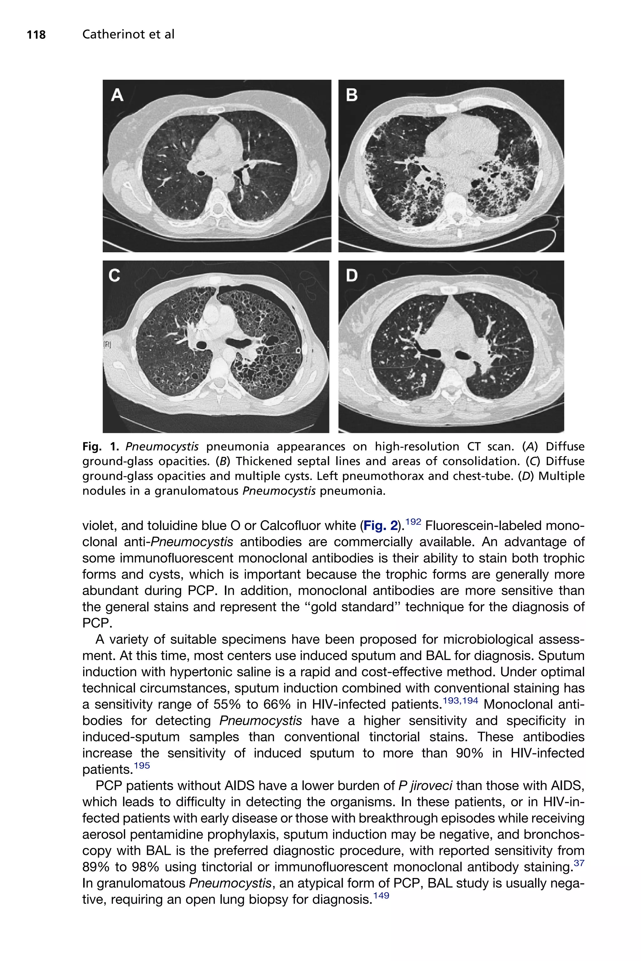
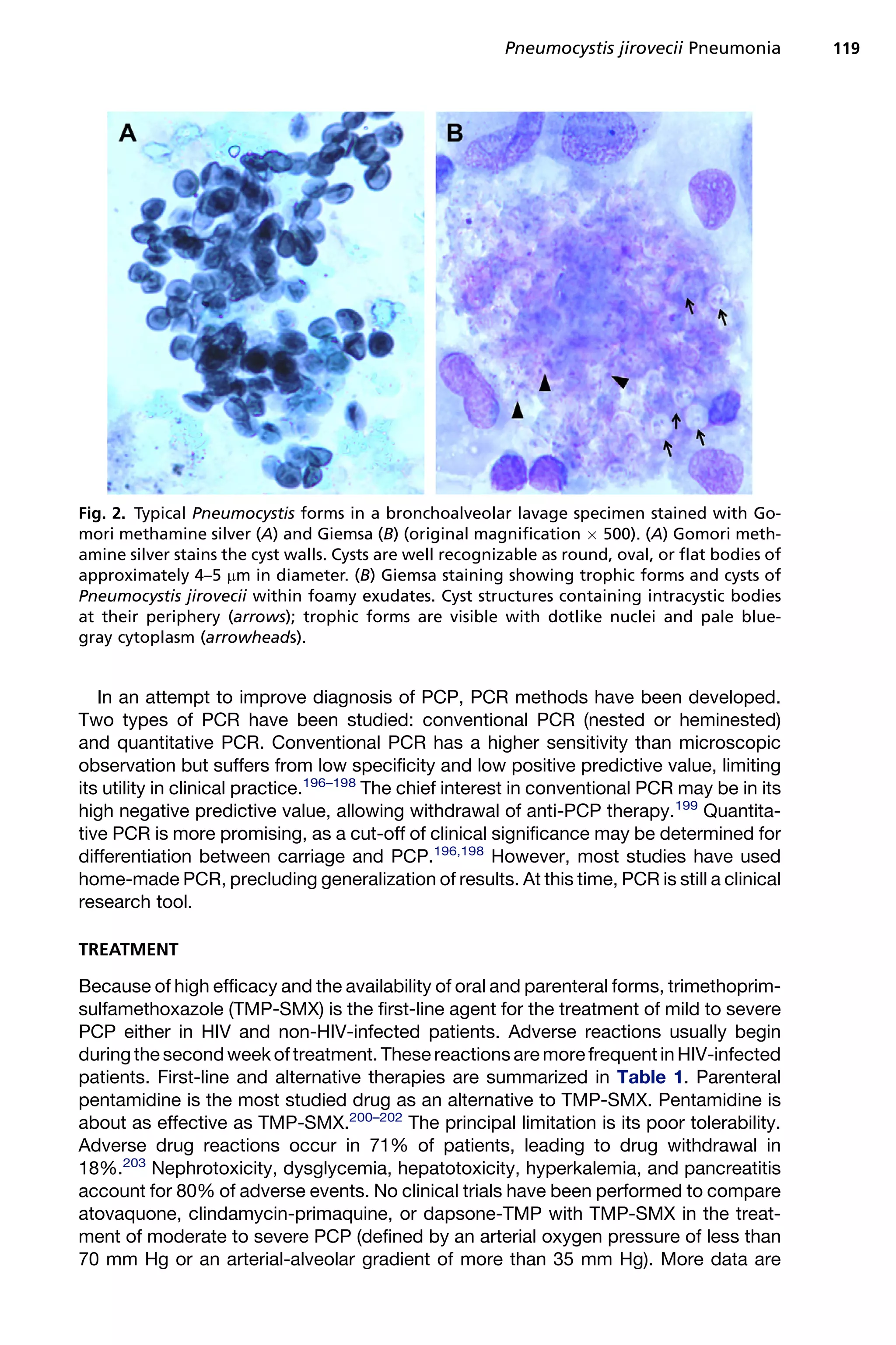
ribosomal RNA implies a close relationship with fungi. Exp Parasitol 1989;
68(4):450–61.
Pneumocystis jirovecii Pneumonia 125](https://image.slidesharecdn.com/pneumocystisjiroveciipneumonia-210225201621/75/Pneumocystis-jirovecii-pneumonia-19-2048.jpg)


![development of airspace enlargement, and pulmonary inflammation in mice.
Infect Immun 2008;76(8):3481–90.
112. Norris KA, Morris A, Patil S, et al. Pneumocystis colonization, airway inflamma-
tion, and pulmonary function decline in acquired immunodeficiency syndrome.
Immunol Res 2006;36(1–3):175–87.
113. Vargas SL, Ponce CA, Hughes WT, et al. Association of primary Pneumocystis
carinii infection and sudden infant death syndrome. Clin Infect Dis 1999;29(6):
1489–93.
114. Masur H, Ognibene FP, Yarchoan R, et al. CD4 counts as predictors of opportu-
nistic pneumonias in human immunodeficiency virus (HIV) infection. Ann Intern
Med 1989;111(3):223–31.
115. Phair J, Munoz A, Detels R, et al. The risk of Pneumocystis carinii pneumonia
among men infected with human immunodeficiency virus type 1. Multicenter
AIDS Cohort Study Group. N Engl J Med 1990;322(3):161–5.
116. D’Egidio GE, Kravcik S, Cooper CL, et al. Pneumocystis jiroveci pneumonia
prophylaxis is not required with a CD41 T-cell count 200 cells/microl when viral
replication is suppressed. AIDS 2007;21(13):1711–5.
117. Kovacs JA, Masur H. Evolving health effects of Pneumocystis: one hundred
years of progress in diagnosis and treatment. JAMA 2009;301(24):2578–85.
118. Walzer PD, Evans HE, Copas AJ, et al. Early predictors of mortality from Pneu-
mocystis jirovecii pneumonia in HIV-infected patients: 1985–2006. Clin Infect Dis
2008;46(4):625–33.
119. Pillonel J. [Surveillance de l’infection par le VIH/SIDA en France, 2006]. Bull
Epid
emiol Hebd 2007;46–47:386–93 [in French].
120. Mikaelsson L, Jacobsson G, Andersson R. Pneumocystis pneumonia—a retro-
spective study 1991–2001 in Gothenburg, Sweden. J Infect 2006;53(4):260–5.
121. Hui M, Kwok WT. Pneumocystis carinii pneumonia in Hong Kong: a 10 year
retrospective study. J Med Microbiol 2006;55(Pt 1):85–8.
122. Arend SM, Kroon FP, van’t Wout JW. Pneumocystis carinii pneumonia in patients
without AIDS, 1980 through 1993. An analysis of 78 cases. Arch Intern Med
1995;155(22):2436–41.
123. Russian DA, Levine SJ. Pneumocystis carinii pneumonia in patients without HIV
infection. Am J Med Sci 2001;321(1):56–65.
124. Overgaard UM, Helweg-Larsen J. Pneumocystis jiroveci pneumonia (PCP) in
HIV-1-negative patients: a retrospective study 2002-2004. Scand J Infect Dis
2007;39(6–7):589–95.
125. Sepkowitz KA, Brown AE, Telzak EE, et al. Pneumocystis carinii pneumonia
among patients without AIDS at a cancer hospital. JAMA 1992;267(6):832–7.
126. Leggiadro RJ, Winkelstein JA, Hughes WT. Prevalence of Pneumocystis
carinii pneumonitis in severe combined immunodeficiency. J Pediatr 1981;
99(1):96–8.
127. Walzer PD, Schultz MG, Western KA, et al. Pneumocystis carinii pneumonia and
primary immune deficiency diseases of infancy and childhood. J Pediatr 1973;
82(3):416–22.
128. Levy J, Espanol-Boren T, Thomas C, et al. Clinical spectrum of X-linked hyper-
IgM syndrome. J Pediatr 1997;131(1 Pt 1):47–54.
129. Winkelstein JA, Marino MC, Ochs H, et al. The X-linked hyper-IgM syndrome:
clinical and immunologic features of 79 patients. Medicine (Baltimore) 2003;
82(6):373–84.
130. Imai K, Morio T, Zhu Y, et al. Clinical course of patients with wasp gene muta-
tions. Blood 2004;103(2):456–64.
Pneumocystis jirovecii Pneumonia 131](https://image.slidesharecdn.com/pneumocystisjiroveciipneumonia-210225201621/75/Pneumocystis-jirovecii-pneumonia-25-2048.jpg)
![131. Elhasid R, Etzioni A. Major histocompatibility complex class II deficiency: a clin-
ical review. Blood Rev 1996;10(4):242–8.
132. Richman DD, Zamvil L, Remington JS. Recurrent Pneumocystis carinii pneumonia
in a child with hypogammaglobulinemia. Am J Dis Child 1973;125(1):102–3.
133. Dittrich AM, Schulze I, Magdorf K, et al. X-linked agammaglobulinaemia and
Pneumocystis carinii pneumonia—an unusual coincidence? Eur J Pediatr
2003;162(6):432–3.
134. Burke BA, Good RA. Pneumocystis carinii infection. Medicine (Baltimore) 1973;
52(1):23–51.
135. Lund FE, Hollifield M, Schuer K, et al. B cells are required for generation of
protective effector and memory CD4 cells in response to Pneumocystis lung
infection. J Immunol 2006;176(10):6147–54.
136. Alibrahim A, Lepore M, Lierl M, et al. Pneumocystis carinii pneumonia in an infant
withX-linkedagammaglobulinemia.J Allergy ClinImmunol1998;101(4Pt1):552–3.
137. Adinoff AD, Johnston RB Jr, Dolen J, et al. Chronic granulomatous disease and
Pneumocystis carinii pneumonia. Pediatrics 1982;69(1):133–4.
138. Rosenszweig SD, Holland SM. Phagocyte immunodeficiencies and their infec-
tions. J Allergy Clin Immunol 2004;113(4):620–6.
139. Kovacs JA, Hiemenz JW, Macher AM, et al. Pneumocystis carinii pneumonia:
a comparison between patients with the acquired immunodeficiency syndrome
and patients with other immunodeficiencies. Ann Intern Med 1984;100(5):663–71.
140. Yale SH, Limper AH. Pneumocystis carinii pneumonia in patients without
acquired immunodeficiency syndrome: associated illness and prior corticoste-
roid therapy. Mayo Clin Proc 1996;71(1):5–13.
141. Pagano L, Fianchi L, Mele L, et al. Pneumocystis carinii pneumonia in patients
with malignant haematological diseases: 10 years’ experience of infection in
gimema centres. Br J Haematol 2002;117(2):379–86.
142. Cheson BD. Infectious and immunosuppressive complications of purine analog
therapy. J Clin Oncol 1995;13(9):2431–48.
143. Byrd JC, Hargis JB, Kester KE, et al. Opportunistic pulmonary infections with flu-
darabine in previously treated patients with low-grade lymphoid malignancies:
a role for Pneumocystis carinii pneumonia prophylaxis. Am J Hematol 1995;
49(2):135–42.
144. Morra E, Nosari A, Montillo M. Infectious complications in chronic lymphocytic
leukaemia. Hematol Cell Ther 1999;41(4):145–51.
145. Roblot F, Imbert S, Godet C, et al. Risk factors analysis for Pneumocystis jiroveci
pneumonia (PCP) in patients with haematological malignancies and pneumonia.
Scand J Infect Dis 2004;36(11–12):848–54.
146. De Castro N, Pavie J, Lagrange-Xelot M, et al. [Pneumocystis jiroveci pneu-
monia in patients with cancer: is it unavoidable?]. Rev Mal Respir 2007;24(6):
741–50 [in French].
147. Martin SI, Marty FM, Fiumara K, et al. Infectious complications associated with
alemtuzumab use for lymphoproliferative disorders. Clin Infect Dis 2006;43(1):
16–24.
148. Lundin J, Kimby E, Bjorkholm M, et al. Phase II trial of subcutaneous anti-CD52
monoclonal antibody alemtuzumab (Campath-1H) as first-line treatment for
patients with B-cell chronic lymphocytic leukemia (B-CLL). Blood 2002;100(3):
768–73.
149. Otahbachi M, Nugent K, Buscemi D. Granulomatous Pneumocystis jiroveci
pneumonia in a patient with chronic lymphocytic leukemia: a literature review
and hypothesis on pathogenesis. Am J Med Sci 2007;333(2):131–5.
Catherinot et al
132](https://image.slidesharecdn.com/pneumocystisjiroveciipneumonia-210225201621/75/Pneumocystis-jirovecii-pneumonia-26-2048.jpg)
![150. Morrison VA. Update on prophylaxis and therapy of infection in patients
with chronic lymphocytic leukemia. Expert Rev Anticancer Ther 2001;1(1):
84–90.
151. Kolstad A, Holte H, Fossa A, et al. Pneumocystis jirovecii pneumonia in B-cell
lymphoma patients treated with the rituximab-CHOEP-14 regimen. Haematolog-
ica 2007;92(1):139–40.
152. Venhuizen AC, Hustinx WN, van Houte AJ, et al. Three cases of Pneumocystis
jirovecii pneumonia (PCP) during first-line treatment with rituximab in combina-
tion with CHOP-14 for aggressive B-cell non-Hodgkin’s lymphoma. Eur J Hae-
matol 2008;80(3):275–6.
153. Varthalitis I, Meunier F. Pneumocystis carinii pneumonia in cancer patients.
Cancer Treat Rev 1993;19(4):387–413.
154. Henson JW, Jalaj JK, Walker RW, et al. Pneumocystis carinii pneumonia in
patients with primary brain tumors. Arch Neurol 1991;48(4):406–9.
155. Kulke MH, Vance EA. Pneumocystis carinii pneumonia in patients receiving
chemotherapy for breast cancer. Clin Infect Dis 1997;25(2):215–8.
156. Godeau B, Coutant-Perronne V, Le Thi Huong D, et al. Pneumocystis carinii
pneumonia in the course of connective tissue disease: report of 34 cases.
J Rheumatol 1994;21(2):246–51.
157. Ognibene FP, Shelhamer JH, Hoffman GS, et al. Pneumocystis carinii pneu-
monia: a major complication of immunosuppressive therapy in patients with We-
gener’s granulomatosis. Am J Respir Crit Care Med 1995;151(3 Pt 1):795–9.
158. Kadoya A, Okada J, Iikuni Y, et al. Risk factors for Pneumocystis carinii pneu-
monia in patients with polymyositis/dermatomyositis or systemic lupus erythe-
matosus. J Rheumatol 1996;23(7):1186–8.
159. Alarcon GS. Infections in systemic connective tissue diseases: systemic lupus
erythematosus, scleroderma, and polymyositis/dermatomyositis. Infect Dis
Clin North Am 2006;20(4):849–75.
160. Marie E. [Infections au cours des polymyosites et des dermatomyosites]. Presse
Med 2009;38:303–16 [in French].
161. Bachelez H, Schremmer B, Cadranel J, et al. Fulminant Pneumocystis carinii pneu-
monia in 4 patients with dermatomyositis. Arch Intern Med 1997;157(13):1501–3.
162. Smith MB, Hanauer SB. Pneumocystis carinii pneumonia during cyclosporine
therapy for ulcerative colitis. N Engl J Med 1992;327:497–8.
163. Bernstein CN, Kolodny M, Block E, et al. Pneumocystis carinii pneumonia in
patients with ulcerative colitis treated with steroids. Am J Gastroenterol 1993;
88:574–7.
164. Kaur N, Mahl TC. Pneumocystis jiroveci (carinii) pneumonia after infliximab
therapy: a review of 84 cases. Dig Dis Sci 2007;52(6):1481–4.
165. Komano Y, Harigai M, Koike R, et al. Pneumocystis jiroveci pneumonia in
patients with rheumatoid arthritis treated with infliximab: a retrospective review
and case-control study of 21 patients. Arthritis Rheum 2009;61(3):305–12.
166. Gordon SM, LaRosa SP, Kalmadi S, et al. Should prophylaxis for Pneumocystis
carinii pneumonia in solid organ transplant recipients ever be discontinued? Clin
Infect Dis 1999;28(2):240–6.
167. Hardy AM, Wajszczuk CP, Suffredini AF, et al. Pneumocystis carinii pneumonia in
renal-transplant recipients treated with cyclosporine and steroids. J Infect Dis
1984;149(2):143–7.
168. Lufft V, Kliem V, Behrend M, et al. Incidence of Pneumocystis carinii pneumonia
after renal transplantation. Impact of immunosuppression. Transplantation 1996;
62(3):421–3.
Pneumocystis jirovecii Pneumonia 133](https://image.slidesharecdn.com/pneumocystisjiroveciipneumonia-210225201621/75/Pneumocystis-jirovecii-pneumonia-27-2048.jpg)


![Pneumocystis_jirovecii_pneumonia[1].pptx](https://cdn.slidesharecdn.com/ss_thumbnails/malamulo-pneumocystisjiroveciipneumonia1-241127111242-957cc6eb-thumbnail.jpg?width=640&height=640&fit=bounds)


