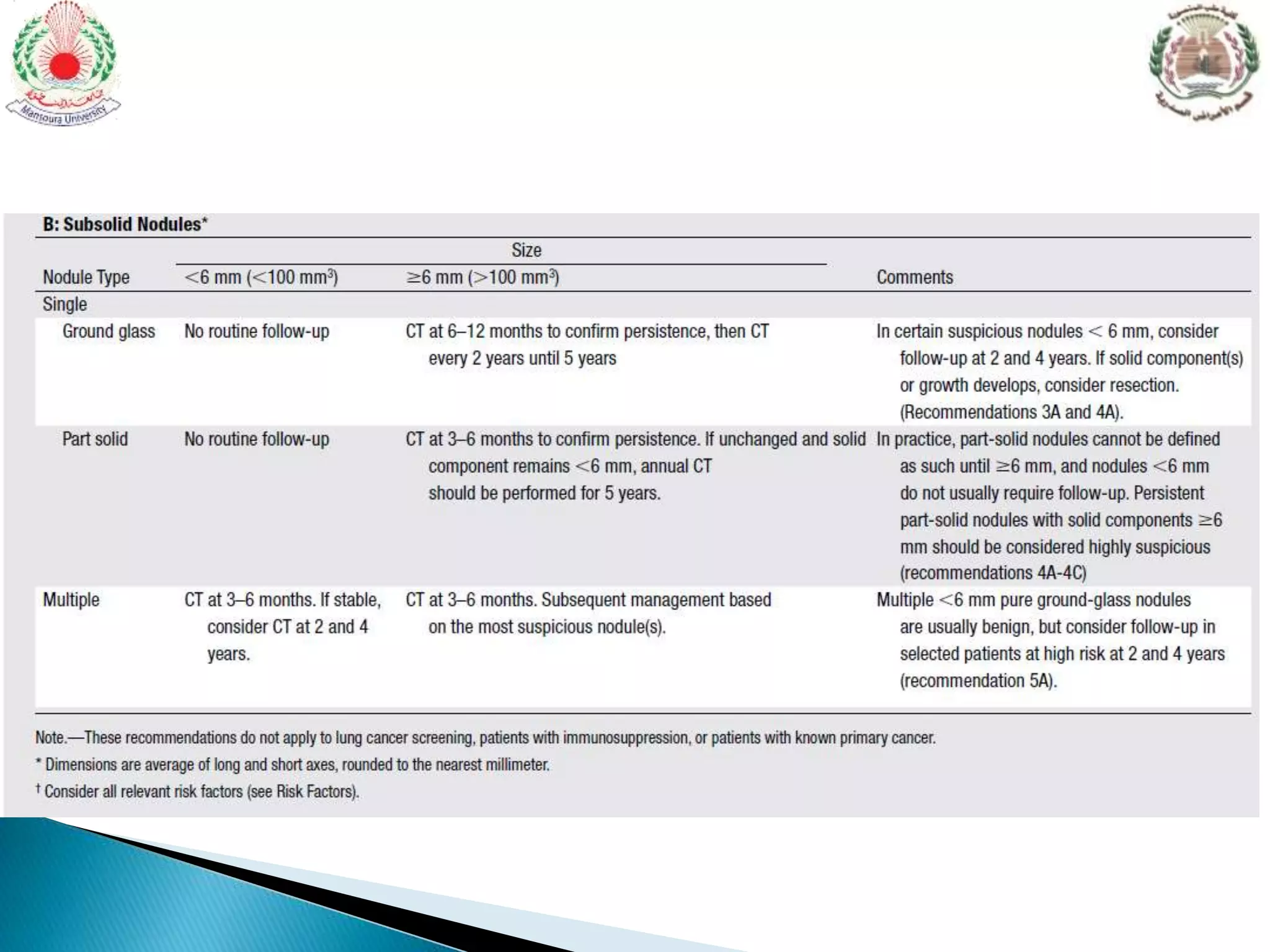
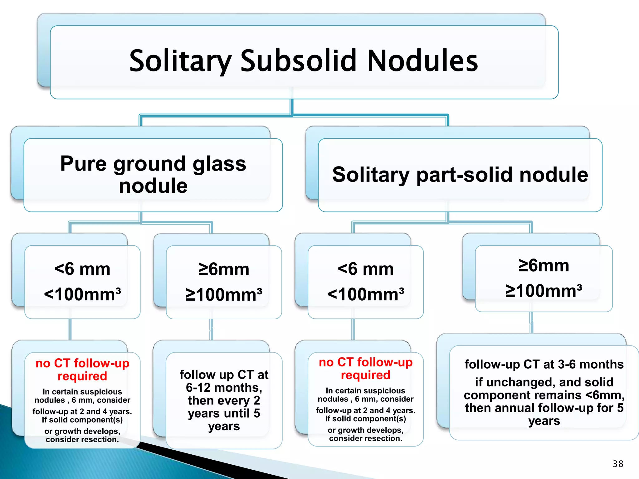
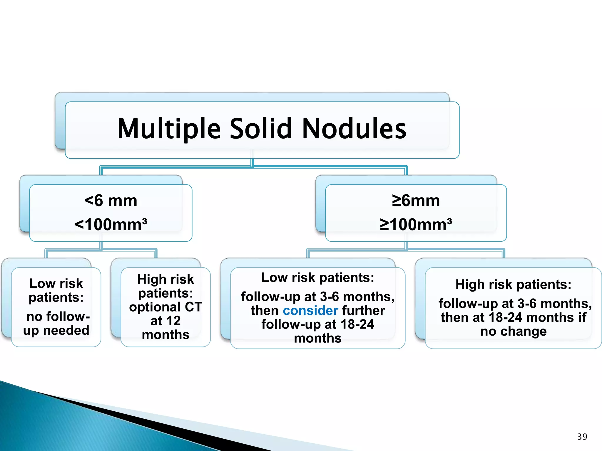
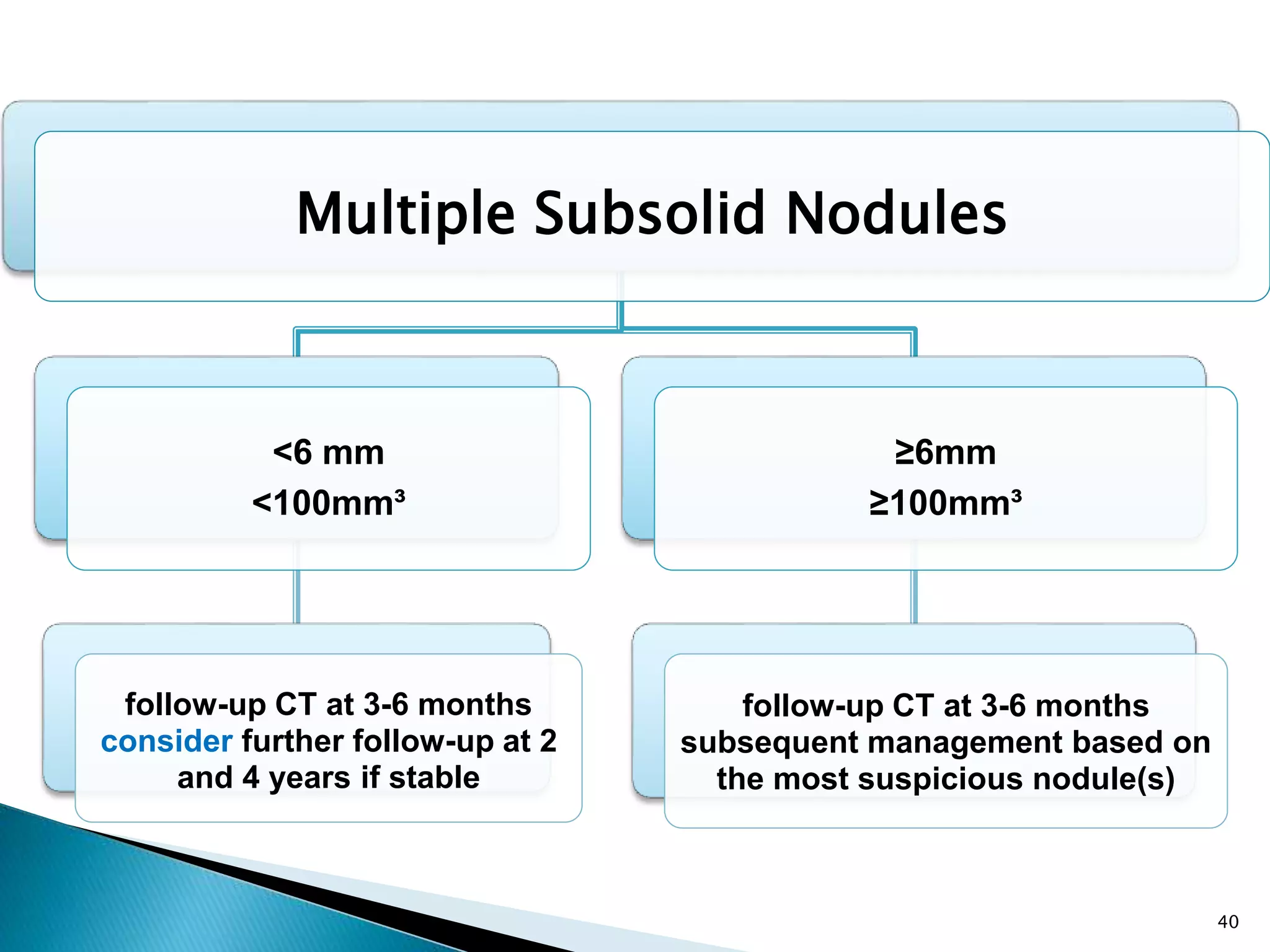
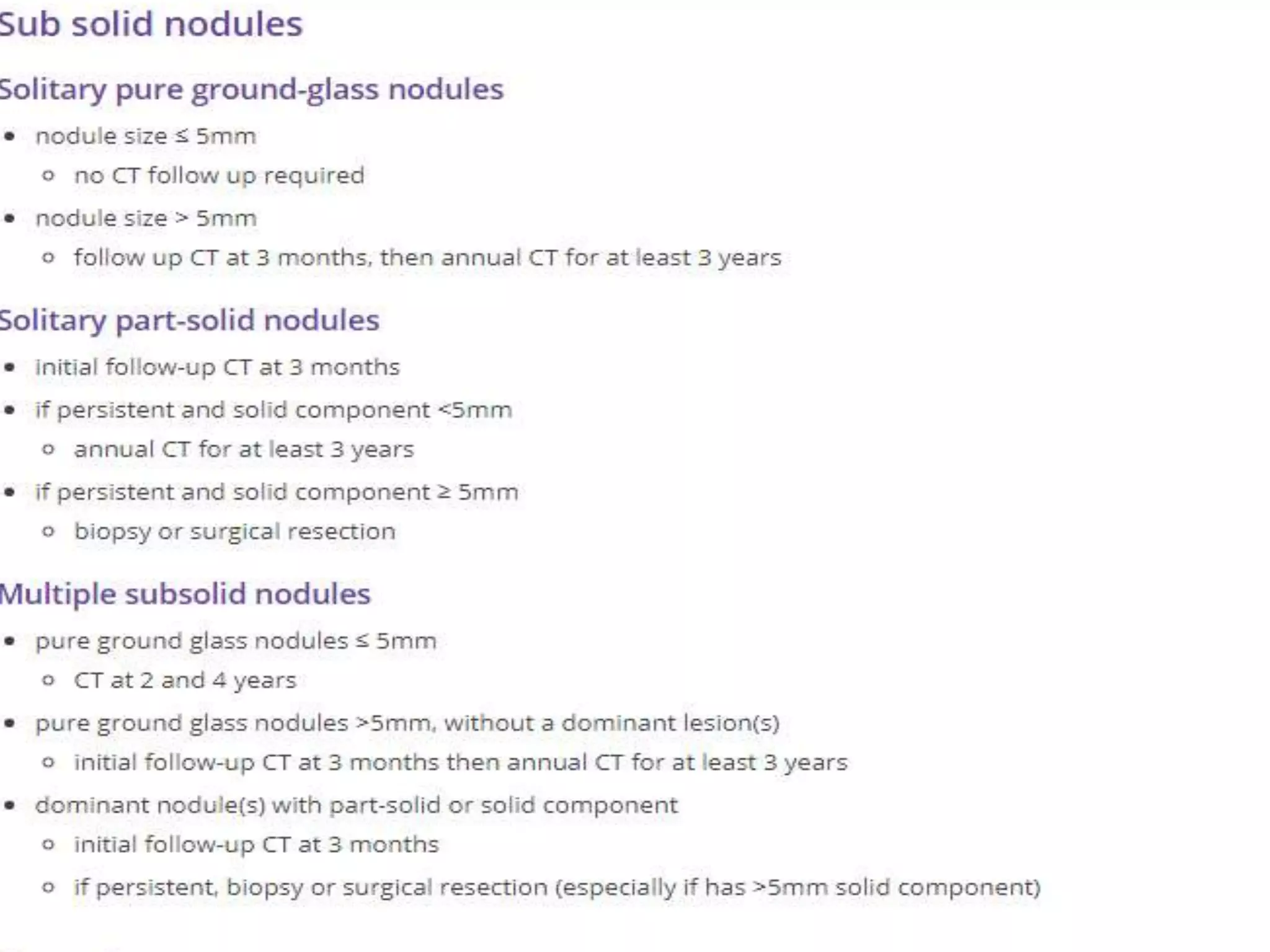
Guidelines for Management of Incidental
Pulmonary Nodules Detected on CT
Images
Fleischner Society 2017
 The Fleischner Society Guidelines for management of
solid nodules were published in 2005.
 Separate guidelines for subsolid nodules were issued in
2013.
 The guidelines for solid and subsolid nodules have been
combined in one simplified table, and specific
recommendations have been included for multiple
nodules.
 The purpose of these recommendations is to reduce the
number of unnecessary follow-up examinations while
providing greater discretion to the radiologist, clinician,
and patient to make management decisions.
 These guidelines represent the consensus of the
Fleischner Society, and as such, they incorporate the
opinions of a multidisciplinary international group of
thoracic radiologists, pulmonologists, surgeons,
pathologists, and other specialists.
 Definition: the solitary pulmonary nodule is a single,
well-circumscribed, radiographic opacity that
measures up to 3 cm in diameter is surrounded
completely by aerated lung and doesn`t touch hilum
or mediastinum. There is no associated atelectasis,
adenopathy, or pleural effusion.
 All CT scans of the thorax in adults should be reconstructed
and archived with contiguous thin sections (<1.5 mm, typically
1.0 mm) to enable accurate characterization and measurement
of small pulmonary nodules
Differential Diagnosis
of Solitary Pulmonary
Nodules
Malignant
Bronchogenic carcinoma
 Adenocarcinoma
 Squamous cell carcinoma
 Large cell carcinoma
 Small cell carcinoma
Metastatic lesions
 Breast Head and neck
 Melanoma Colon
 Kidney Sarcoma
 Germ cell tumor Others
Pulmonary carcinoid
Benign
Infectious granuloma
 Tuberculosis
 Histoplasmosis
 Coccidioidomycosis
 Atypical mycobacteria
 Cryptococcosis
 Blastomycosis
Other infections
 Bacterial abscess
 Dirofilaria immitis
 Echinococcus cyst( hydatid cyst).
 Ascariasis
 Pneumocystis carinii
 Aspergilloma
Benign
Benign neoplasms
 Hamartoma
 Lipoma
 Fibroma
Vascular
 Arteriovenous malformation
 Pulmonary varix
Developmental
 Bronchogenic cyst
Inflammatory
 Granulomatosis with polyangiitis (Wegener's)
 Rheumatoid nodule
Benign
others
 Amyloidoma
 Rounded atelectasis
 Intrapulmonary lymph nodes
 Hematoma
 Pulmonary infarct
 Pseudotumor (loculated fluid)
 Mucoid impaction
Risk Factors for Malignancy:
General Considerations
 Nodule Size and Morphology(calcification&edge)
 Nodule Location
 Nodule Multiplicity
 Nodule Growth Rate
 Emphysema and Fibrosis
 Age, Sex, Race, and Family History
 Tobacco and Other Inhaled Carcinogens
 Nodule size has a clear relationship with risk of
malignancy, as discussed previously, and it is a
dominant factor in management.
 In these guidelines, nodules are further divided
into solid, ground-glass, and part-solid
categories.
Nodule Size and Morphology(calcification&edge)
 Lung cancers occur more frequently in the upper
lobes, with a predilection for the right lung.
 In the PanCan trial, upper lobe nodule location was
confirmed as a risk factor, with an odds ratio of
approximately 2.0.
Nodule Location
 An analysis of patients with multiple nodules in the
NELSON trial showed increased risk of primary
cancer as the total nodule count increased from 1 to 4
but decreased risk in patients with 5 or more nodules.
 In the PanCan trial, multiplicity of nodules was
associated with a reduced risk of cancer when
compared with risk associated with one nodule.
Nodule Multiplicity
 Volume doubling times for solid cancers are well
established (one volume doubling corresponds to a
26% increase in diameter), with a large majority of
times being in the 100–400 day range (20-400).
 For subsolid cancerous nodules, which represent
primary adenocarcinomas, more indolent growth is
the rule, with average doubling times on the order of
3–5 years.
Nodule Growth Rate
 The presence of emphysema on a CT image is an
independent risk factor for lung cancer.
 NLST found that emphysema- predominant COPD
phenotype and increasing severity of centrilobular
emphysema were associated with increased risk of
malignancy.
 Pulmonary fibrosis, particularly idiopathic pulmonary
fibrosis, is also an independent risk factor, with a
hazard ratio of approximately 4.2 compared with
emphysema alone.
Emphysema and Fibrosis
 An increase in risk associated with advancing age.
 Family history of lung cancer is a risk factor for both
smokers and those who never smoked.
 No significant difference between both genders except
incidence of adenocarcinoma in nonsmokers is
increasing, with female nonsmokers being affected
significantly more often than male nonsmokers.
 Race is also a factor, with a significantly higher
incidence of lung cancer in black men.
Age, Sex, Race, and Family History
 Cigarette smoking has been established as the major
risk factor for lung cancer with10 to 35 fold increased
risk, exposure to secondhand smoke is a proven.
 Other inhaled carcinogens that are known risk factors
for lung cancer include exposure to asbestos,
uranium, or radon.
Tobacco and Other Inhaled Carcinogens
 we recommend that risk be assigned according to the
categories proposed by the American College of
Chest Physicians (ACCP). Low risk, which
corresponds to an estimated risk of cancer of less
than 5%.
 To estimate high risk, we recommend combining the
ACCP intermediate-risk (5%–65% risk) and high-risk
(> 65% risk) categories.
Solitary Solid Nodule
<6 mm
<100mm³
Low risk
patients:
no
follow-up
needed
High risk
patients:
optional
CT at 12
months
6-8mm
100-250 mm³
Low risk patients:
follow-up at 6-12
months, then
consider further
follow-up at 18-24
months
High risk patients:
initial follow-up CT at
6-12 months and then
at 18-24 months if no
change
> 8mm
>250 mm³
Low or High risk
consider follow-up CT
at 3 months, and/or
CT-PET, and/or biopsy
37
Solitary Subsolid Nodules
Pure ground glass
nodule
<6 mm
<100mm³
no CT follow-up
required
In certain suspicious
nodules , 6 mm, consider
follow-up at 2 and 4 years.
If solid component(s)
or growth develops,
consider resection.
≥6mm
≥100mm³
follow up CT at
6-12 months,
then every 2
years until 5
years
Solitary part-solid nodule
<6 mm
<100mm³
no CT follow-up
required
In certain suspicious
nodules , 6 mm, consider
follow-up at 2 and 4 years.
If solid component(s)
or growth develops,
consider resection.
≥6mm
≥100mm³
follow-up CT at 3-6 months
if unchanged, and solid
component remains <6mm,
then annual follow-up for 5
years
38
Multiple Solid Nodules
<6 mm
<100mm³
Low risk
patients:
no follow-
up needed
High risk
patients:
optional CT
at 12
months
≥6mm
≥100mm³
Low risk patients:
follow-up at 3-6 months,
then consider further
follow-up at 18-24
months
High risk patients:
follow-up at 3-6 months,
then at 18-24 months if
no change
39
Multiple Subsolid Nodules
<6 mm
<100mm³
follow-up CT at 3-6 months
consider further follow-up at 2
and 4 years if stable
≥6mm
≥100mm³
follow-up CT at 3-6 months
subsequent management based on
the most suspicious nodule(s)
40
Thank you
Old guidelines
Pulmonary nodules
Pulmonary nodules

Pulmonary nodules

  • 1.
    Guidelines for Managementof Incidental Pulmonary Nodules Detected on CT Images Fleischner Society 2017
  • 3.
     The FleischnerSociety Guidelines for management of solid nodules were published in 2005.  Separate guidelines for subsolid nodules were issued in 2013.  The guidelines for solid and subsolid nodules have been combined in one simplified table, and specific recommendations have been included for multiple nodules.
  • 4.
     The purposeof these recommendations is to reduce the number of unnecessary follow-up examinations while providing greater discretion to the radiologist, clinician, and patient to make management decisions.  These guidelines represent the consensus of the Fleischner Society, and as such, they incorporate the opinions of a multidisciplinary international group of thoracic radiologists, pulmonologists, surgeons, pathologists, and other specialists.
  • 5.
     Definition: thesolitary pulmonary nodule is a single, well-circumscribed, radiographic opacity that measures up to 3 cm in diameter is surrounded completely by aerated lung and doesn`t touch hilum or mediastinum. There is no associated atelectasis, adenopathy, or pleural effusion.  All CT scans of the thorax in adults should be reconstructed and archived with contiguous thin sections (<1.5 mm, typically 1.0 mm) to enable accurate characterization and measurement of small pulmonary nodules
  • 6.
  • 7.
    Malignant Bronchogenic carcinoma  Adenocarcinoma Squamous cell carcinoma  Large cell carcinoma  Small cell carcinoma Metastatic lesions  Breast Head and neck  Melanoma Colon  Kidney Sarcoma  Germ cell tumor Others Pulmonary carcinoid
  • 8.
    Benign Infectious granuloma  Tuberculosis Histoplasmosis  Coccidioidomycosis  Atypical mycobacteria  Cryptococcosis  Blastomycosis Other infections  Bacterial abscess  Dirofilaria immitis  Echinococcus cyst( hydatid cyst).  Ascariasis  Pneumocystis carinii  Aspergilloma
  • 9.
    Benign Benign neoplasms  Hamartoma Lipoma  Fibroma Vascular  Arteriovenous malformation  Pulmonary varix Developmental  Bronchogenic cyst Inflammatory  Granulomatosis with polyangiitis (Wegener's)  Rheumatoid nodule
  • 10.
    Benign others  Amyloidoma  Roundedatelectasis  Intrapulmonary lymph nodes  Hematoma  Pulmonary infarct  Pseudotumor (loculated fluid)  Mucoid impaction
  • 11.
    Risk Factors forMalignancy: General Considerations
  • 12.
     Nodule Sizeand Morphology(calcification&edge)  Nodule Location  Nodule Multiplicity  Nodule Growth Rate  Emphysema and Fibrosis  Age, Sex, Race, and Family History  Tobacco and Other Inhaled Carcinogens
  • 13.
     Nodule sizehas a clear relationship with risk of malignancy, as discussed previously, and it is a dominant factor in management.  In these guidelines, nodules are further divided into solid, ground-glass, and part-solid categories. Nodule Size and Morphology(calcification&edge)
  • 14.
    Nodule Size andMorphology(calcification&edge)
  • 15.
    Nodule Size andMorphology(calcification&edge)
  • 16.
    Nodule Size andMorphology(calcification&edge)
  • 17.
    Nodule Size andMorphology(calcification&edge)
  • 18.
     Lung cancersoccur more frequently in the upper lobes, with a predilection for the right lung.  In the PanCan trial, upper lobe nodule location was confirmed as a risk factor, with an odds ratio of approximately 2.0. Nodule Location
  • 19.
     An analysisof patients with multiple nodules in the NELSON trial showed increased risk of primary cancer as the total nodule count increased from 1 to 4 but decreased risk in patients with 5 or more nodules.  In the PanCan trial, multiplicity of nodules was associated with a reduced risk of cancer when compared with risk associated with one nodule. Nodule Multiplicity
  • 20.
     Volume doublingtimes for solid cancers are well established (one volume doubling corresponds to a 26% increase in diameter), with a large majority of times being in the 100–400 day range (20-400).  For subsolid cancerous nodules, which represent primary adenocarcinomas, more indolent growth is the rule, with average doubling times on the order of 3–5 years. Nodule Growth Rate
  • 21.
     The presenceof emphysema on a CT image is an independent risk factor for lung cancer.  NLST found that emphysema- predominant COPD phenotype and increasing severity of centrilobular emphysema were associated with increased risk of malignancy.  Pulmonary fibrosis, particularly idiopathic pulmonary fibrosis, is also an independent risk factor, with a hazard ratio of approximately 4.2 compared with emphysema alone. Emphysema and Fibrosis
  • 22.
     An increasein risk associated with advancing age.  Family history of lung cancer is a risk factor for both smokers and those who never smoked.  No significant difference between both genders except incidence of adenocarcinoma in nonsmokers is increasing, with female nonsmokers being affected significantly more often than male nonsmokers.  Race is also a factor, with a significantly higher incidence of lung cancer in black men. Age, Sex, Race, and Family History
  • 23.
     Cigarette smokinghas been established as the major risk factor for lung cancer with10 to 35 fold increased risk, exposure to secondhand smoke is a proven.  Other inhaled carcinogens that are known risk factors for lung cancer include exposure to asbestos, uranium, or radon. Tobacco and Other Inhaled Carcinogens
  • 34.
     we recommendthat risk be assigned according to the categories proposed by the American College of Chest Physicians (ACCP). Low risk, which corresponds to an estimated risk of cancer of less than 5%.  To estimate high risk, we recommend combining the ACCP intermediate-risk (5%–65% risk) and high-risk (> 65% risk) categories.
  • 37.
    Solitary Solid Nodule <6mm <100mm³ Low risk patients: no follow-up needed High risk patients: optional CT at 12 months 6-8mm 100-250 mm³ Low risk patients: follow-up at 6-12 months, then consider further follow-up at 18-24 months High risk patients: initial follow-up CT at 6-12 months and then at 18-24 months if no change > 8mm >250 mm³ Low or High risk consider follow-up CT at 3 months, and/or CT-PET, and/or biopsy 37
  • 38.
    Solitary Subsolid Nodules Pureground glass nodule <6 mm <100mm³ no CT follow-up required In certain suspicious nodules , 6 mm, consider follow-up at 2 and 4 years. If solid component(s) or growth develops, consider resection. ≥6mm ≥100mm³ follow up CT at 6-12 months, then every 2 years until 5 years Solitary part-solid nodule <6 mm <100mm³ no CT follow-up required In certain suspicious nodules , 6 mm, consider follow-up at 2 and 4 years. If solid component(s) or growth develops, consider resection. ≥6mm ≥100mm³ follow-up CT at 3-6 months if unchanged, and solid component remains <6mm, then annual follow-up for 5 years 38
  • 39.
    Multiple Solid Nodules <6mm <100mm³ Low risk patients: no follow- up needed High risk patients: optional CT at 12 months ≥6mm ≥100mm³ Low risk patients: follow-up at 3-6 months, then consider further follow-up at 18-24 months High risk patients: follow-up at 3-6 months, then at 18-24 months if no change 39
  • 40.
    Multiple Subsolid Nodules <6mm <100mm³ follow-up CT at 3-6 months consider further follow-up at 2 and 4 years if stable ≥6mm ≥100mm³ follow-up CT at 3-6 months subsequent management based on the most suspicious nodule(s) 40
  • 41.
  • 42.