Download to read offline
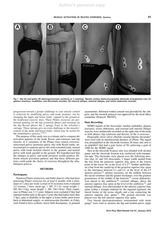
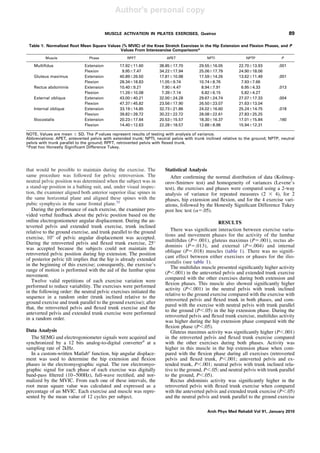

This document summarizes the results of a study that evaluated muscle activation patterns during four variations of Pilates core stability exercises performed in a quadruped position. The four exercises differed in pelvic and trunk positioning: 1) retroverted pelvis with flexed trunk, 2) anteverted pelvis with extended trunk, 3) neutral pelvis with inclined trunk, and 4) neutral pelvis with parallel trunk. Surface electromyography was used to measure activation of six core muscles during each exercise. The results showed that different pelvic and trunk positions produced varying activation patterns in the multifidus, gluteus maximus, and oblique muscles, but similar rectus abdominis activation.


![ONFH[AVN HIP] -TRIPLE REGIME -A NOVAL SURGICAL CONCEPT .pptx](https://cdn.slidesharecdn.com/ss_thumbnails/onfhavnhip2026koaconcalicutdrgokuldevdrmashraf-260210064517-213ec005-thumbnail.jpg?width=640&height=640&fit=bounds)


