Congenital CYNOTIC HEART DISEASE -1.
•
0 likes•38 views

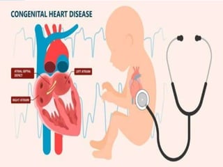
Congenital heart disease, also called a defect, refers to one or more problems with the heart structure that are present at birth. These abnormalities occur when the heart or blood vessels don't form correctly in utero. At least eight out of every 1000 infants born in the US each year have a heart defect.

Recommended
More Related Content
Similar to Congenital CYNOTIC HEART DISEASE -1.
Similar to Congenital CYNOTIC HEART DISEASE -1. (20)
More from DR .PALLAVI PATHANIA
More from DR .PALLAVI PATHANIA (20)
Recently uploaded
Recently uploaded (20)
Congenital CYNOTIC HEART DISEASE -1.
- 4. For thousands of years, the heart has been considered one of the most important organs in the body. Aristotle even believed that other organs existed just to cool it, including the brain and lungs (which we now know perform their own vital functions). Although it may not be exactly as Aristotle once thought, the heart does perform a role that is absolutely necessary for survival. The heart is the first organ to form and become functional, emphasizing the importance of transport of material to and from the developing infant. It originates about day 18 or 19 from the mesoderm and begins beating and pumping blood about day 21 or 22. It forms from the cardiogenic region near the head and is visible as a prominent heart bulge on the surface of the embryo. Originally, it consists of a pair of strands called cardiogenic cords that quickly form a hollow lumen and are referred to as endocardial tubes. These then fuse into a single heart tube and differentiate into the truncus arteriosus, bulbus cordis, primitive ventricle, primitive atrium, and sinus venosus, starting about day 22. The primitive heart begins to form an S shape within the pericardium between days 23 and 28.
- 5. The internal septa begin to form about day 28, separating the heart into the atria and ventricles, although the foramen ovale persists until shortly after birth. Between weeks five and eight, the atrioventricular valves form. The atrioventricular valves form. The semilunar valves form between weeks five and nine.
- 7. Schematic representation of the formation of the adult heart. With the onset of embryonic folding the left and right heart forming region (HFR). The yellow structures represent the endocardial cells (endo) that form the inner lining of the heart and in gray the formed primary myocardium. VP refers to the venous pole were the blood will enter the heart and AP to the arterial pole where the blood will leave the heart. For easy comparison of panels (a) through (d), a red line indicates the lateral border of the HFR and a blue line the medial border. With ongoing folding the HFR becomes positioned ventrally of the foregut (see also Figure 1). At the position where the lateral borders of the HFR meet, the linear heart tube is connected to the body wall via the dorsal mesocardium (dm) (panel d). The DM breaks due to which the heart is only attached to the body wall at the AP and VP (panel h–k). Whereas panels (a)–(d) show a dorsal view of the forming heart, panels (e)–(h) show a ventral view, illustrating the transition of linear heart tube to four-chambered heart. Only at the ventral site of the linear heart tube the differentiation of the embryonic ventricle (V) is locally initiated.
- 8. The forming working myocardium of the chambers is indicated in blue (panels f–k). Note in panel (g) that at the dorsal side of the heart tube, primary myocardium is retained, which is referred to as the inner curvature (IC). Flanking the forming ventricle, primary myocardium is retained which is referred to as the inflow tract (IFT) and outflow tract (OFT). With ongoing development, the linear heart tube loops to the right and chamber formation becomes evident (panel h). At this stage the right ventricle (RV) starts to form when primary myocardium of the OFT differentiates into chamber myocardium. Moreover, upstream of the left ventricle (LV), the primary myocardium of the IFT locally differentiates into the left atrium (LA) and right atrium (RA). In the meantime, newly differentiated cardiomyocytes are added to the lengthening heart forming the sinus venous (SV) myocardium. The heart is connected to the blood circulation at the VP via the left and right cardinal vein (cv) and at the AP via the pharyngeal arch arteries (paa). Panel (j) shows a representation of the 5 week old human heart showing the expanding (ballooning) atria and ventricles, as well as the remnants of the primary myocardium of the IFT, AVC (atrioventricular canal), IC and OFT. The forming primary atrial septum (pAS) and ventricular septum (VS) are identified. Within the LA the attachment to the body wall is identified as the mediastinal mesenchyme (mm) through which the cardinal vein (cv) and the future pulmonary vein (pv) drain into the heart. In the formed heart (panel k) the primary myocardium of the IFT and AVC has differentiated into the central conduction system, comprising the sinoatrial node (sn), the atrioventricular node (avn), the His bundle (His) and the bundle branches (bb). Within the right atrium the superior and inferior caval veins (scv and icv) drain in the RA and the pulmonary veins (pv) in the LA. Flanking the chambers, valves are formed of which only the mitral valve (mv) and the tricuspid valve (tv) are shown.
- 9. In the healthy normal post-natal heart, oxygen-rich blood enters the left atrium, is propagated to the left ventricle and then pumped via the aorta into the systemic circulation. The oxygen-deprived blood, returning from the body, enters the right atrium and is propelled by the right ventricle via the pulmonary trunk toward the lungs. The cardiac conduction system orchestrates the efficient contraction-relaxation cycle of the atria and ventricles. The electrical impulse resulting in cardiac contraction is triggered in the sinus node, which is located at the entrance of the superior caval vein into the right atrium. The electrical impulse spreads through both atria, but cannot directly activate the ventricles due to the electrical isolation of the atria from the ventricles by the annulus fibrosus (also called insulating plane or fibrous continuity). The electrical impulse is delayed in the atrioventricular (AV) node, and then quickly propagated through the His-bundle (AV-bundle), which penetrates the insulating annulus fibrosis plane, via the bundle branches and the peripheral conduction system (the Purkinje fibers) to the cardiomyocytes. The coordinated propagation of the electrical impulse ensures the synchronous contraction of the ventricles from the apex toward the aorta and pulmonary trunk.
- 10. PROCESS FORMATION AND GROWTH OF THE LINEAR HEART TUBE THE GROWTH AND LOOPING OF THE HEART TUBE FROM LINEAR TO FOUR CHAMBERED HEART DEVELOPMENT OF THE CONDUCTION SYSTEM THE SINUS NODE ATRIOVENTRICULAR NODE AND BUNDLE BRANCHES PERIPHERAL VENTRICULAR CONDUCTION SYSTEM SEPTATION
- 12. Schematic representation of septation. Panel (a) shows the same schematic drawing of the four chamber-forming heart as Figure 2(j), in which now the major cushions have been added within the atrioventricular canal (AVC) and outflow tract (OFT) and the left and right bloodstreams through the heart is indicated. The ventricular foramen is located between the distal edge of the ventricular septum (VS) and the inner curvature (IC). Noteworthy is that the left bloodstream passes the ventricular foramen during systole, while the right bloodstream already passes through the ventricular foramen during diastole. Panels (b)–(f) illustrate atrial septation and show the cutting edge (sagittal sections) at the level of the dashed line in panel (a). For easy comparison the cushions are numbered. The mesenchymal complex formed by the anterior (1) and posterior (2) atrioventricular cushion, the extension of the anterior cushion over the roof of the atrium (MC), and the extracardiac mesenchyme that protrudes into atrial lumen (DMP), surround the connection between the left (LA) and right atrium (RA). With expansion of this complex the primary atrial foramen (pAF) becomes smaller and eventually closes forming the primary atrial septum (pAS). Prior to closure of the pAF, the secondary atrial foramen (sAS) is formed. Within the RA the secondary atrial septum (sAS) folds down from the atrial wall into the lumen and covers the pAS partly and the pAF completely. The uncovered part of the pAS is recognized as the oval fossa (OF)
- 13. PROCESS CONTINUE ATRIAL SEPTATION VENTRICULAR SEPTATION SEPTATION OF THE OUTFLOW TRACT THE CARDIAC CONNECTIVE TISSUES EPICARDIUM AND ITS DERIVATIVES DEVELOPMENT OF THE VALVES REMODELING OF THE EMBRYONIC VALVES
- 23. Development of the heart begins in the third week with the formation of two endothelial strands called the angioblastic cords. These cords canalize forming two heart tubes, which fuse into single heart tube by the end of the third week due to lateral embryonic folding. By the fourth week, the developing heart receives blood from three pairs of veins: the vitelline veins, umbilical veins, and common cardinal veins. The vitelline veins carry poorly oxygenated blood from the yolk sac, and enter the sinus venosus; The umbilical veins carry oxygenated blood from the chorion, the primordial placenta; and the common cardinal veins carry poorly oxygenated blood from the rest of the embryo.
- 24. As the primordial liver develops in close association with the septum transversum, the hepatic cords join and surround epithelial-lined spaces, forming the primordial hepatic sinusoids. These primordial sinusoids become connected to the vitelline veins. Vitelline veins pass through the septum transversum and enter sinus venosus, also called as venous end of the heart. Left vitelline veins regress while right vitelline veins form the hepatic veins, and a network of vitelline veins around the duodenum form the portal vein.
- 25. As the development of liver progresses, umbilical veins lose connection with heart and empty into liver. The right umbilical vein and cranial part of the left umbilical vein degenerate during seventh week of gestation, leaving only the caudal part of the left umbilical vein. The caudal part of the left umbilical vein carries oxygenated blood to the embryo from the placenta. The umbilical vein is connected to the inferior vena cava (IVC) via the ductus venosus, a venous shunt that develops in the liver. This bypass directs most of the blood directly to the heart from placenta without passing through liver.
- 26. Umbilical vein (ventral view) The embryo is drained primarily by the cardinal veins, with the anterior cardinal vein draining the cranial part of the embryo and the posterior cardinal vein draining the caudal part. These two join to form the common cardinal vein, which enters the sinus venosus. By the eighth week, the anterior cardinal veins are connected by a vessel running obliquely between them. This oblique vessel allows for the shunting blood from the left anterior cardinal vein to the right. Once the caudal part of the left anterior cardinal vein degenerates, this oblique anastomotic vessel becomes the left brachiocephalic vein. The right anterior cardinal vein and right common cardinal vein eventually become the superior vena cava (SVC), and the posterior cardinal veins contribute to the common iliac veins and the azygos vein.
- 27. As the subcardinal and supracardinal veins form, they first supplement but soon replace the posterior cardinal veins. The subcardinal veins appear first, and eventually form parts of the left renal vein, suprarenal vein, gonadal vein, and inferior vena cava (IVC). Above the kidneys, anastomoses join the supracardinal veins, forming the azygos and hemiazygos veins. Below the kidneys, the right supracardinal vein contributes to IVC, while the left supracardinal vein degenerates. In the fourth and fifth weeks of development, the pharyngeal arches form. These are supplied by the pharyngeal arch arteries, which connect the aortic sac to the two dorsal aortae. The dorsal aortae extend the length of the embryo, eventually fusing in the caudal part of the embryo to form the lower thoracic/abdominal aorta. The rest of the right dorsal aorta degenerates, while the remainder of the left dorsal aorta becomes the primordial aorta.
- 28. The dorsal aortae give off the intersegmental arteries, which supply the somites and their derivatives. These intersegmental arteries become the vertebral arteries in the neck region, the intercostal arteries in the thorax, the lumbar arteries and common iliac arteries in the abdomen, and the lateral sacral arteries in the sacral region. The very caudal end of the dorsal aorta gives rise to the median sacral artery, and any other intersegmental arteries regress. The umbilical vesicle (i.e. yolk sac), allantois, and chorion are supplied by unpaired branches of the dorsal aorta. The umbilical vesicle is supplied by the vitelline arteries, and once part of the umbilical vesicle forms the primordial gut, this region is supplied by the vitelline arteries as well. The vitelline arteries give rise to the celiac artery, which supplies the foregut; the superior mesenteric artery, which supplies the midgut; and the inferior mesenteric artery, which supplies the hindgut
- 29. The two umbilical arteries, contained within the umbilical cord, carry poorly oxygenated blood from the embryo to the placenta. The proximal part of these arteries become the internal iliac and superior vesical arteries, while the distal parts regress and become the medial umbilical ligaments. Heart layers As the heart tubes fuse, the primordial myocardium begins to form from the splanchnic mesoderm around the pericardial cavity. This primordial myocardium becomes the middle, muscular layer of the heart. Separated from the primordial myocardium by gelatinous tissue called cardiac jelly, the heart begins to develop as a thin tube. This endothelial tube becomes the endocardium, the innermost layer of the heart. Epicardium, the outermost layer, originates from mesothelial cells from the outer surface of the sinus venosus.
- 30. Heart tube As the cranial part of the embryo folds, the heart tube elongates. As it elongates, the heart tube develops alternating constrictions and expansions, forming the bulbus cordis, ventricle, atrium, and sinus venosus. The bulbus cordis has multiple components, including the truncus arteriosus, conus arteriosus, and conus cordis. The truncus arteriosus is cranial to the aortic sac, to which it is connected, and gives off the pharyngeal arch arteries. Blood leaves the heart via the pharyngeal arch arteries, and returns to the sinus venosus of the heart via the umbilical, vitelline, and common cardinal veins. The bulbus cordis and ventricles grow at a faster rate than other parts of the developing heart, and because of this the heart bends and folds in on itself, forming the bulbo- ventricular loop. As this bending occurs, the atrium and sinus venosus move so that they are dorsal to the truncus arteriosus, bulbus cordis, and ventricle. During this time, the sinus venosus also develops lateral extensions, the left and right horns.
- 31. The heart is initially attached to the dorsal wall of the pericardial cavity by a mesentery called the dorsal mesocardium, but as the heart grows it begins to fill the pericardial cavity and the central part of the dorsal mesocardium degenerates. The loss of part of this mesentery allows a communication to form between the left and right sides of the pericardial cavity, the transverse pericardial sinus. Primitive circulation The sinus venosus receives blood from the common cardinal veins, umbilical veins and vitelline veins. The common cardinal veins carry blood from the embryo; the umbilical veins carry blood from the placenta; and the vitelline veins carry blood from the umbilical vesicle.
- 32. After entering the sinus venosus, blood flows through the sinuatrial valves into the primordial atrium. It then flows from the primordial atrium into the primordial ventricle via the atrioventricular (AV) canal. When the primordial ventricle contracts, it pumps blood into the bulbus cordis and through the truncus arteriosus, into the aortic sac. From there, blood enters the pharyngeal arch arteries, and then the dorsal aortae, which allows it to travel back to the embryo, placenta, and umbilical vesicle.
- 39. Partitioning of the developing heart : In the middle of the fourth week, the atrioventricular canal, primordial atrium and ventricle start to partition, and this process is completed by the end of week eight. It begins with the formation of the endocardial cushions, specialized extracellular matrix tissue related to myocardial tissue. At the end of the fourth week, these cushions appear on the ventral and dorsal walls of the AV canal and start to grow toward each other. They eventually fuse, separating the AV canal into left and right components, partially separating the atrium and ventricle and acting as AV valves. The primordial atrium becomes separated into the right and left atria by two septa, the septum primum and septum secundum. The septum primum appears first in the form of a thin membrane, growing out of the roof of the primordial atrium toward the endocardial cushions, leaving an opening between its edge and endocardial cushion. This opening is called the foramen primum, and it allows blood to continue to be shunted from the right atrium to the left. It progressively shrinks and eventually closes as the septum primum elongates and fuses with the endocardial cushions, forming the primordial AV septum. Before the foramen primum closes completely, however, apoptosis of cells in the middle of the septum primum forms perforations in the septum. These perforations form a new second opening, the foramen secundum, which allows oxygenated blood to continue to flow from the right atrium to the left even after the foramen primum has closed.
- 40. The muscular septum, the septum secundum, grows immediately adjacent to the septum primum, just to its right. It grows downward from the ventro- cranial wall of the atrium during the fifth and sixth weeks of development, gradually overlapping the foramen secundum in the septum primum. By overlapping the foramen secundum without fusing to the septum primum, an incomplete barrier between the atria is formed. At this point in development, the opening between the atria is called the foramen ovale, and it allows oxygenated blood to continue to flow from the right atrium, under the flap of the septum secundum, through the foramen secundum, and into the left atrium. This arrangement also prevents blood from flowing in the opposite direction, from the left atrium to the right atrium: the thin septum primum gets pressed up against the more firm and inflexible septum secundum, blocking blood from flowing through the foramen ovale. Although the cranial part of the septum primum slowly regresses, some parts of the septum primum remain attached to the endocardial cushions. These residual parts of the septum primum form the valve of the foramen ovale.
- 41. Valve of foramen ovale (lateral-left view) After a baby is born, the pressure in the left atrium increases significantly, becoming much higher than the pressure in the right atrium. This causes the septum primum to be pushed against the septum secundum and the valves of the foramen primum to fuse with the septum secundum, functionally closing the foramen ovale. When this occurs, the foramen ovale becomes the fossa ovalis and the two septae form a complete barrier between the atria. Sinus venosus: The sinuatrial orifice, the opening of the sinus venosus into the single primordial atrium, is initially located in the posterior wall of the primordial atrium. This changes, however, at the end of the fourth week, when the right sinual horn grows larger than the left. This unequal growth moves the sinuatrial orifice to the right, shifting it into what will become the adult right atrium. As the right sinual horn continues to grow, blood from the head and neck region of the embryo flows into it via the SVC, and blood from the placenta and the rest of the body of the embryo flows into it via the IVC.
- 42. As the heart continues to develop, the sinus venosus gets integrated into the wall of the right atrium as the smooth part of the internal surface of the right atrium, the sinus venarum. The rest of the internal surface of the right atrium and auricle has a thicker, trabeculated appearance; these parts of the adult atrium originate from the primordial atrium. The transition from the smooth to the rough internal surface of the right atrium is demarcated on the inside of the atrium by a ridge called the crista terminalis, which originates from the cranial part of the right sinuatrial valve, and on the outside by a groove called the sulcus terminalis. The caudal part of the right sinuatrial valve forms the valves of the IVC and coronary sinus. The left sinual horn develops into the coronary sinus; and the left sinuatrial valve eventually fuses with the septum secundum, becoming part of the interatrial septum.
- 43. Interatrial septum Primary pulmonary vein: The majority of the inner wall of the left atrium is smooth and is derived from the primordial pulmonary vein, which develops from the dorsal atrial wall just left of the septum primum. As the left atrium grows, the primordial pulmonary vein, as well as its main branches, become integrated into the atrial wall. This results in four pulmonary veins entering into the left atrium. The left auricle has the same origin as the right auricle: the primordial atrium. As such, its internal surface is trabeculated. Ventricles: The primordial ventricle begins its division into two ventricles with the growth of the median ridge, a muscular interventricular (IV) septum with a superior free edge that arises from the floor of the primordial ventricle, close to the apex of the heart. Dilation of the developing ventricles on either side of this septum is responsible for the initial increase in the septal height, with additional growth occurring due to the contribution of ventricular myocytes from both sides of the heart.
- 58. Between the upper free edge of this septum and the endocardial cushions, there remains an opening called the IV foramen. This foramen allows blood to continue to flow between the right and left ventricles until its closure at the end of the seventh week, when the left and right bulbar ridges fuse with the endocardial cushion, forming the membranous part of the IV septum. The bulbar ridges form in the fifth week as proliferations of mesenchymal neural crest cells in the walls of the bulbus cordis. The membranous part of the IV septum results when tissue from the right side of the endocardial cushion extends to the muscular part of the IV septum, ultimately merging with the aorticopulmonary septum and muscular IV septum. Once the IV foramen closes and the membranous part of the IV septum forms, the aorta becomes the sole outflow tract of the left ventricle, and the pulmonary trunk becomes the sole outflow tract of the right ventricle. As the ventricles continue to develop, cavitation results in the formation of muscular bundles. While some of these persist as trabeculae carneae (irregular columns of muscle on the inner surface of the ventricles), others form the papillary muscles and chordae tendinae (heart strings), which connect the papillary muscles to the AV valves.
- 59. Cardiac valves: The aortic and pulmonic semilunar valves each develop from three swellings of subendocardial tissue present around the opening of aorta and pulmonary trunk. They evolve into three thin cusps. Anterior cusp of mitral valve (cranial view): The tricuspid and mitral AV valves form from proliferations of tissue surrounding the AV canals. The tricuspid valve develops three cusps, whereas the mitral (i.e. bicuspid) valve develops two.
- 62. Conducting system: Heart conductive system Initially, the primordial atrium functions as the developing heart’s pacemaker; but the sinus venosus soon takes over this role. In the fifth week, the sinuatrial (SA) node develops in the right atrium near the opening of the SVC. After the sinus venosus is integrated into the heart, cells from its left wall can be found near the opening of the coronary sinus, at the base of the interatrial septum. With the addition of some cells from the AV region, the AV node and bundle are formed just above the endocardial cushions. Fibes originating from the AV bundle project from the atrium into the ventricle and divide into left and right bundle branches, which can be found throughout the ventricular myocardium. Although the SA node, AV node, and AV bundle eventually receive nervous innervation from outside the heart, the primordial conducting system develops before this occurs.
- 69. The majority of congenital developmental anomalies of the heart are present 6 weeks after conception.Congenital heart disease can be cyanotic or acyanotic. There are three types of congenital heart disease Grade 1 - Left to Right shunts Grade 2 - Right to Left shunts Grade 3 - Obsructive lesions Clinical features of LEFT to RIGHT shunts(acyanotic heart disease) Frequent chest infections are seen (6-8 attacks first year of life) Tendency for increased sweating that is related to their tendency for developing Congestive cardiac failure Precordial bulge is seen. Hyperkinetic precordium occur. Tricuspid /mitral mid diastolic murmur is heard. X-ray show plethoric lung field + cardiomegaly. Example are VSD,ASD, PDA,AV canal defect
- 73. Cyanotic Congenital Heart Defects Cyanotic heart defects are cardiac defects in which the blood pumped to the rest of the body contains less than normal amounts of oxygen. In other words, the heart pumps mixed oxygen-poor and oxygen-rich blood to the body. This can lead to cyanosis which is a bluish discoloration of the skin. Cyanotic heart defects typically contain right- to-left shunts, meaning deoxygenated blood from the right heart is shunted to the left heart. As a result, oxygen-poor blood is delivered to the body and can cause cyanosis.
- 81. ECG
- 90. Echocardiography Cardiac catheterization
- 91. The treatment of choice for most congenital heart diseases is surgery to repair the defect. There are many types of surgery, depending on the kind of birth defect. Surgery may be needed soon after birth, or it may be delayed for months or even years. Some surgeries may be staged as the child grows. Your child may need to take water pills (diuretics) and other heart medicines before or after surgery. Be sure to follow the correct dosage. Regular follow-up with the provider is important. Many children who have had heart surgery must take antibiotics before, and sometimes after having any dental work or other medical procedures. Make sure you have clear instructions from your child's heart provider. Ask your child's provider before getting any immunizations. Most children can follow the recommended guidelines for childhood vaccinations
- 95. 1. Truncus Arteriosus : Trick: Hold up 1 finger Truncus Arteriosus: One great vessel leaving the heart, instead of 2 The first cyanotic congenital heart defect is truncus arteriosus. You can hold up 1 finger to remember this. Truncus arteriosus is when one blood vessel leaves the heart instead of 2. You might remember from the anatomy of the heart lecture that normally there are 2 main arteries leaving the heart. The main pulmonary artery leaves the right side of the heart and delivers deoxygenated blood to the lungs. The aorta leaves the left side of the heart and delivers oxygenated blood to the rest of the body.
- 96. In the case of truncus arteriosus, the great vessel coming out of the heart fails to divide during development. This leaves a connection between the aorta and pulmonary artery. A ventricular septal defect (VSD) is typically present, and the blood from the right and left ventricle combine and exit the heart through one great vessel. A VSD is a hole in the wall between the right and left ventricle. As a result, oxygen-poor blood from the right heart and oxygen-rich blood from the left heart are delivered to the rest of the body. This can lead to potential cyanosis. So again, use 1 finger to remember truncus arteriosus and one great vessel leaving the heart.
- 98. Truncus Arteriosus: Hold up 1 finger to remember one great vessel leaves the heart, instead of two (normally pulmonary artery [1] and aorta [2] leave the heart).
- 106. Causes: In normal circulation, the pulmonary artery comes out of the right ventricle and the aorta comes out of the left ventricle, which are separate from each other. With truncus arteriosus, a single artery comes out of the ventricles. There is most often also a large hole between the 2 ventricles (ventricular septal defect). As a result, the blue (without oxygen) and red (oxygen-rich) blood mix. Some of this mixed blood goes to the lungs, and some goes to the rest of the body. Often, more blood than usual ends up going to the lungs. If this condition is not treated, two problems occur: Too much blood circulation in the lungs may cause extra fluid to build up in and around them. This makes it hard to breathe. If left untreated and more than normal blood flows to the lungs for a long time, the blood vessels to the lungs become permanently damaged. Over time, it becomes very hard for the heart to force blood through them. This is called pulmonary hypertension, which can be life threatening.
- 118. 2. Transposition of Great Arteries; Trick: Hold up 2 fingers and cross them Transposition of Great Arteries: Two great arteries leaving the heart are transposed The second cyanotic heart defect is transposition of great arteries. You can remember this by holding up 2 fingers and crossing them (to represent the transposition). Transposition of great arteries is when the 2 main arteries leaving the heart (main pulmonary artery and aorta) are transposed or reversed. Remember we said the main pulmonary artery normally leaves the right heart and goes to the lungs, and the aorta leaves the left heart and goes to the rest of the body. In transposition of great arteries, the pulmonary artery and aorta are reversed. Therefore, the main pulmonary artery arises from the left ventricle instead of the right, and the aorta arises from the right ventricle instead of the left.
- 119. Transposition of Great Arteries: Hold up 2 fingers and cross them to remember the pulmonary artery (1) and aorta (2) are transposed (reversed) as shown by the arrows.
- 120. The transposition of the aorta and pulmonary artery creates 2 separate circuits. Circuit 1: Deoxygenated blood from the right heart flows to the rest of the body (via the aorta) and back to the right side of the heart again. Normally deoxygenated blood would flow from the right heart to the lungs via the pulmonary artery. Circuit 2: Oxygenated blood from the left heart flows to the lungs (via the pulmonary artery) and back to the left side of the heart again. Normally oxygenated blood would flow from the left heart to the rest of the body via the aorta. In order to be compatible with life, there needs to be a connection between the 2 circuits to allow for mixing of oxygen-rich and poor blood. So there is typically a patent ductus arteriosus or a ventricular septal defect present. As you can imagine, this will lead to the delivery of oxygen-poor blood to the body and subsequent cyanosis. So again, use 2 fingers and cross them to remember transposition of great arteries and how the 2 great arteries are reversed or transposed.
- 132. 3. Tricuspid Atresia: Trick: Hold up 3 fingers Tricuspid Atresia: Tricuspid valve fails to form (Tri = 3) The third cyanotic heart defect is tricuspid atresia. Hold up 3 fingers to remember this defect. Tricuspid atresia is a congenital heart defect in which the tricuspid valve fails to form. Remember in our medical terminology lecture, we learned the prefix “tri-” means 3. So holding up 3 fingers will help you remember tricuspid atresia. You might remember from the anatomy of the heart lecture that the tricuspid valve is located between the right atrium and right ventricle. In the case of tricuspid atresia, the tricuspid valve fails to form. As a result, blood from the right atrium cannot enter the right ventricle. Instead, an atrial septal defect is present (a hole in the wall between the right and left atrium). This allows for deoxygenated blood in the right atrium to flow into the left atrium. As a result, oxygen-poor blood from the right heart mixes with the oxygen-rich blood in the left heart. This can lead to decreased oxygen levels in the blood delivered to the rest of the body, which can cause cyanosis.
- 133. Tricuspid Atresia: Hold up 3 fingers to remember the tricuspid valve (star) fails to form and blood is unable to flow from the right atrium to the right ventricle (red X)
- 134. There are different types of tricuspid atresia, but the right ventricle is typically underdeveloped and the presence of a ventricular septal defect allows blood from the left ventricle to enter the right ventricle. Remember the right ventricle is not receiving blood from the right atrium in this case, so it receives blood from the left ventricle instead. So again, use 3 fingers to remember tricuspid atresia and how the tricuspid valve fails to form.
- 140. Embryology
- 143. Types
- 179. 4. Tetralogy of Fallot: Trick: Hold up 4 fingers Tetralogy of Fallot: Tetrad of 4 cardiac defects (Tetra = 4) The fourth cyanotic heart defect is tetralogy of Fallot. Hold up 4 fingers to remember this, as tetralogy of Fallot is a tetrad of 4 cardiac defects. Remember in our medical terminology lecture, the prefix “tetra-” means 4. So holding up 4 fingers will help you remember tetralogy of Fallot is a tetrad. The tetrad includes: Pulmonary Stenosis Right Ventricular Hypertrophy (RVH) Overriding Aorta Ventricular Septal Defect (VSD) Pulmonary stenosis is narrowing of the pulmonary valve and main pulmonary artery. Right ventricular hypertrophy is thickening of the right ventricular wall. Overriding aorta refers to the enlarged aortic valve that seems to open from both ventricles and sits on top of the ventricular septal defect. Finally, the ventricular septal defect is a hole in the wall between the right and left ventricle.
- 180. The pulmonary stenosis, RVH, and VSD can alter pressure gradients and create a right-to-left shunt, allowing oxygen-poor blood in the right heart to flow to the left heart. This can lead to cyanosis. So again, use 4 fingers to remember tetralogy of Fallot and how there is a tetrad of 4 cardiac defects.
- 181. Tetralogy of Fallot: Hold up 4 fingers to remember the tetrad of cardiac defects including pulmonary stenosis (1), right ventricular hypertrophy (2), overriding aorta (3), and ventricular septal defect (4)
- 205. Total Anomalous Pulmonary Venous Return (TAPVR): Hold up 5 fingers to remember the 5 words in TAPVR. The pulmonary veins do not connect to the left atrium (X) like they normally should (star), instead they connect to the systemic venous system.
- 206. 5. Total Anomalous Pulmonary Venous Return (TAPVR) Trick: Hold up 5 fingers Total Anomalous Pulmonary Venous Return (5 words): Pulmonary veins connect to systemic venous system rather than the left atrium The fifth cyanotic heart defect is total anomalous pulmonary venous return (TAPVR). Hold up 5 fingers to remember this because there are 5 words that make up the defect. TAPVR is when the pulmonary veins connect to the systemic venous system rather than the left atrium. Normally the 4 pulmonary veins deliver oxygenated blood from the lungs to the left atrium. In the case of TAPVR, the pulmonary veins do not connect to the left atrium. They connect to the systemic venous system instead.
- 207. As a result, the oxygenated blood from the lungs mixes with the deoxygenated venous blood from the body, and the mixed blood flows back to the right atrium. Since the pulmonary veins are not delivering blood to the left atrium, there is usually an atrial septal defect present to allow blood to travel from the right atrium to the left atrium. Remember the right atrial blood in this case is mixed oxygen-rich and oxygen-poor blood coming from the rest of the body. So the left side of the heart is receiving blood with less than normal amounts of oxygen (compared to the oxygenated blood it would normally receive from the pulmonary veins). This can cause cyanosis. So again, use 5 fingers to remember total anomalous pulmonary venous return as the defect contains 5 words. This is when the pulmonary veins connect to the systemic venous system rather than the left atrium.