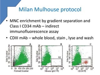
STEM CELL
ENUMERATION
Varun Kumar Singh
STEM CELL
• Pool of undifferentiated cells – all cells of bone
marrow – proliferation and differentiation
Hemangioblast
Vascular
endothelium
LTR-
HSC
STR-
HSC
CMP CLP
APOPT
OSIS
Phenotype
• CD34+ Thy-1lo CD38- Lin- HLA-DR- Rh123Dull
• CD117
• TPO
Indications
Therapeutic use of Stem cell
Hematologic Non- hematologic
Neoplastic Non – neoplastic Solid organ malignancies
ALL Aplastic anemia Inborn error of metabolism
AML PNH SCID
CML Thalassemia Wiskott- Aldrich syndrome
MF Sickle cell disease Congenital leukocyte
function disorder
CLL Fanconi’s anemia Malignant osteopenia
MDS Congenital PRCA
NHL
HL
Myeloma
Sources of stem cells
• Bone marrow
– Directly from marrow
– adequate number of MNCs
– Contains : RBC, WBC, MNCs, platelets, plasma, fat
• Cord blood
– From placenta – delivered/undelivered
• Peripheral blood –
– Less stem cells
– mobilized – chemotherapy/cytokine
– Apheresis ( leukapheresis )
• Collection target : recipient weight
– 2.5-5.0x10⁶ CD34+ cells/kg ( peripheral blood)
– 3.0x10⁷ Total nucleated cells/ kg (cord blood)
Quantification of stem cells
• Need
– Time of harvest and transplant
– to determine the volume of harvest (apheresis)
– To asses viability of PB and cord blood product
– viable CD34 count – critical parameter in
engraftment
Methods
• Determination of mononuclear cell counts
• Cell culture for colony forming units
• CD34 enumeration by flow cytometry
• New methods: -
– Microvolume fluorimetry
– Using automated hematology analyzer
Mononuclear cell counts
• Stem cells are mononuclear cells
• MNC count – indirect measure of number of stem
cells
• Dose – depends on weight of recipient – 2.5-5x10⁶
/kg
• Manual / automated cell counters
• Artefacts in automated counters : unlysed RBCs,
nRBCs, platelet clumps, fat – visual inspection of
smears
• Stored samples – post thaw- cell loss not determined
• Three categories :
– Nucleated RBCs
– Mature cells – neutrophils and bands
– Mononuclear cells – all lymphoid, monocytic and
myeloid precursors
• Advantages : cheap, readily available, short TAT,
good (negative ) predictor for collection efficacy
• Disadvantages : does not correlate well with
engraftment, yield variable owing to disease
status.
Cell culture for colony forming units
• Clonogenicity – cannot be demonstrated by MNC
or flow cytometry
• In vitro assay – stem cells and media ( agar,
methyl cellulose)
• PB/ cord blood/ BM – Ficoll-Paque gradient
centrifugation – separate the MNCs
• MNC suspended in solution – counted
• Aliquot the media and cell suspension – mixed –
incubated with growth factors
• Colony count :- colonies containing atleast 40
cells or more are counted, vary from lab to lab
• Colony morphology : -
• CFU/ml = no of colonies x dilution factor /
volume
• Advantages:- tests the capacity of stem cells to
divide – predict the engraftment potential
• Disadvantage: - time taking, variable results
from institutions, commercial kits are
available- not standardised.
Flowcytometric CD34+ enumeration
• Why CD34 – ubiquitous expression in stem cells,
large scale studies – proven – CD34+ enriched
product – successful transplant
• Optimal CD34 mAb
– Epitopes – 3 – sensitivity to neuraminidase and
glycoprotease
– Class I : sensitive to both – mAb – generate
inconsistent data
– Class II : sensitive only to glycoprotease
– Class III: insensitive to both
More consistent
data for CD34
counts
• Optimal fluorochrome :
• CD34 cells less in number – conjugate with a bright
fluorochrome
• PE (phycoerythrin) & FITC (fluorescein isothiocyanate)
• Class III conjugates of both, 8G12 (BD lifesciences) and
581(Beckman coulter) – can be used
• PE conjugate of Class II , QBend10( Beckman coulter)
• Avoid Class II conjugates of FITC – charge interference –
binds with less affinity
Dual platform vs. Single platform
Dual platform
• %CD34+ from
flowcytometer X TLC from
hematology analyser =
absolute CD34+ count
• Denominator = all nucleated
cells – includes nRBCs,
platelet clumps, cell debris,
& fat cells(bone marrow)
Single platform
• Fluorescent beads of known
concentration - absolute
CD34+ count from
flowcytometer
• Eliminates errors in
counting by cell counters
• Denominator issue
correction
• Reduces variation among
laboratories
Milan Mulhouse protocol
• MNC enrichment by gradient separation and
Class I CD34 mAb – indirect
immunofluorescence assay
• CDIII mAb – whole blood, stain , lyse and wash
SIHON protocol
• Used LDS751 – nucleic acid stain – identify
nucleated cells
• Counter stain CD34 with CD14 and CD66e –
exclude binding to myelomonocytic cells
ISHAGE protocol
• 4 sequential gating steps:
– All leukocytes CD45 (dim to bright)
– CD34+ cells from leukocytes
– Clustered CD45dim, SSClow HPC selected from
CD45+CD34+ events
– Events outside generic lymph-blast region –
excluded (apoptotic cells, platelet clumps)
• Origin protocol – isotype control , murine IgG1
• Later omitted in modified platform
ISHAGE
Housekeeping plots :
• all non malignant CD34+ cells
do not express CD45
• instrument optimisation and
improve gating
•R5 - in R1 – including all
lymphocytes - displayed on
FSC vs SSC (F) – minimum size
range for the lymph-blast
region
• set the FALS lower threshold
to include even the smallest of
lymphocytes – exclude non
specific stained cells, platelet
clumps and debris
• plot E ( CD45 vs CD34) adjust
the lower limit of the gates R1
and R2
Current day ISGAGE protocol
• Single platform
• Stain, lyse, no wash
• Incorporated vitality dye- 7AAD (7 amino
actinomycin D)
• Commercially available kits : BD trucount,
Stem Cell kit (BD), Stem-kit (Beckman Coulter)
• Beads – absolute counts
ProcountTM
With Trucount tube
Stem cell enumeration kitTM
• Meets ISHAGE guidelines
• Single tube assay
• Compatible with various sample types and
anticoagulants
• Single platform with BD trucount tube/liquid beads
• Contents:
– CD34PE/CD45FITC
– 7-AAD viability dye
– NH4Cl lysing solution
– Trucount tubes/ liquid beads
Comparison of Stem cell kit to Procount kit
SCE Kit Procount Kit
Meet ISHAGE Guidelines Yes No
No. of Tubes per Sample 1 2(requires isotype)
Viability Dye 7-AAD None
Incubation Time 30min 45min
Reagent Stability 20 months 5months
Specimen Type Peripheral Blood
Leukapheresis
Bone Marrow
Cord Blood
Peripheral Blood (PB)
Leukapheresis
Specimen Stability 24 hours for all samples 24hrs for PB
6 hrs for leukapheresis
Anticoagulant EDTA
Heparin
ACD-A
CPD
EDTA
Cytometer BD FACSCalibur (OS9 and OSX)
BD FACSCanto II
BD FACS Diva ( separate
BD FACSCalibur OS 9 only
Process
• Dilute samples : 40,000/ μL, PBS
• 3 tubes, test, high control (CD34 35cells/l), low control
CD35 10cells/l)
• Add 10 μL of CD45FITC and CD34PE to all tubes
• Add 100 μL controls to respective tubes
• Add 100 μL sample to test
• Add 20 μL &-AAD to test – incubate for 20min in dark
• Add 2ml NH4Cl (pharm lyse 1:10) to all 3 tubes –
incubate for 10min
• Add liquid beads 50 μL to the tubes
• Acquire immediately
• Stem cell control
– High/low
– Always run process controls prior to staining
samples
– Helps in instrument setup and performance
– Process control for
• Antibody staining for CD34+
• 7-AAD staining for non viable cells
• Red blood cell lysis
Workflow
• Start-up system : check for bead lot ID and
beads/pellet, set acquisition targets
– CD45 events : 75000
– Viable CD34 : 100
– Time : 900sec
• QC with setup beads
– Measures cytometer’s performace & consistency
• Optimize settings
– With either high stem control
(w/o 7AAD)
– Adjust threshold
– Adjust FSC gain
• Acquire controls and
Ensure that they pass
• Use high stem cell control (w/o 7AAD)
• Adjust compensation
• Vortex and acquire test sample
• Analysis
– Eliminate debris
– Identify viable cells
– Identify the leukocytes and lymphocytes
– Identify CD34+ cluster among viable cells
– Gate the beads
Sequential gating
BD FACS DIVA
Review gates
• Calculation :
Absolute count =
CD events x beads concentration x DF
Bead events x volume
• CD34+ cell count, and not change in total WBC
should determine the adequacy of stem cell
harvest
• Time : Total nucleated count – with in 6hrs,
CD34 count with in 12 hours of venepuncture
• Label samples accurately
• Storage: 40 C
• Commence harvest: CD34 count > 10 cells/ μL,
aim : final yield of > 2x10⁶ CD34 cells /kg
• Class II or Class III conjugated anti CD34 ab
• Anti pan CD45 conjugated FITC ab
• Minimise cell loss: stain , lyse , no wash is
recommended, fixative-free NH4Cl-based
erythrocyte lysis reagent
• Minimum of 100 CD34 events
• Sequential gating strategy : ISHAGE protocol
recommended
• CD34 count should be performed in duplicate,
mean CD34 count should be used.
• Single platform technology should be used for
absolute count employing reverse pipetting
• Isotype controls: not required unless using
Procount
• Process controls : daily (ideally), or at least once a
week
• Cytometer performance: monitor daily
Microvolume fluorimetry
• IMAGN 200 with STELLer CD34 assay ( Biometric Imaging,
BectonDickenson)
• Absolute CD34 on PB and PBSC
• Cy5 conjugated Class III mAb
• Isotype control
• No lyse method
• Excitation wavelength – 633nm – renders RBCs transparent
• Cells gated: fluorescence intensity, Cy5 peak intensity,
defined size range, uniform event staining
• Capillary tube : known volume - yield absolute cd34 value
• Sysmax XN series – immature cell population
• Cell wall – phospholipids – increases as the cells mature
• Separate chamber – surfactant+ diluting fluid+ nuclear dye
(fluorescent)
• Surfactant – dissolves the phospholipids- channels – nuclear
dye binds to the nucleus
• Argon laser – measure fluorescence
Take home message
• CD34 counts – vital
– Assessing time of harvest
– Yield of harvest
– Engraft potential
• Methodologies – MNCs, clonogenic assay,
flowcytometric enumeration, hemocytometic
enumeration.
• Single platform CD34 assay – least interlab
variation – standarised
• Newer methods – hemocytometer – cheaper
alternative with good correlation.
References
• Hsu YM, Cushing MM. Autologous Stem Cell Mobilization and Collection. Hematol
Oncol Clin North Am. 2016 Jun;30(3):573-89.
• Chapple P et al. Comparison of three methods of CD34+ cell enumeration in
peripheral blood: dual-platform ISHAGE protocol versus single-platform, versus
microvolume fluorimetry.Cytotherapy.2000;2(5):371-76.
• Gratama JW, Sutherland DR, Keeney M. Flow cytometric enumeration and
immunophenotyping of hematopoietic stem and progenitor cells. Semin Hematol.
2001 Apr;38(2):139-47. Review.
• Ngoma A, Saito S, Ohto H, Ikeda K, Yasuda H, Kawabata K, Kanno T, Kikuta A,
Mochizuki K, Nollet KE. CD34+ cell enumeration by flow cytometry: a comparison
of systems and methodologies. Arch Pathol Lab Med. 2011 Jul;135(7):909-14.
• Whitby A, Whitby L, Fletcher M, Reilly JT, Sutherland DR, Keeney M, Barnett D.
ISHAGE protocol: are we doing it correctly? Cytometry B Clin Cytom. 2012
Jan;82(1):9-17.
• Analyzing Samples for CD34 Enumeration Using the BD™ Stem Cell Enumeration
Kit, Ellen Meinelt, BD lifesciences.
Thank you !

Stem cell enumeration

  • 1.
  • 2.
    STEM CELL • Poolof undifferentiated cells – all cells of bone marrow – proliferation and differentiation Hemangioblast Vascular endothelium LTR- HSC STR- HSC CMP CLP APOPT OSIS
  • 3.
    Phenotype • CD34+ Thy-1loCD38- Lin- HLA-DR- Rh123Dull • CD117 • TPO
  • 4.
    Indications Therapeutic use ofStem cell Hematologic Non- hematologic Neoplastic Non – neoplastic Solid organ malignancies ALL Aplastic anemia Inborn error of metabolism AML PNH SCID CML Thalassemia Wiskott- Aldrich syndrome MF Sickle cell disease Congenital leukocyte function disorder CLL Fanconi’s anemia Malignant osteopenia MDS Congenital PRCA NHL HL Myeloma
  • 5.
    Sources of stemcells • Bone marrow – Directly from marrow – adequate number of MNCs – Contains : RBC, WBC, MNCs, platelets, plasma, fat • Cord blood – From placenta – delivered/undelivered • Peripheral blood – – Less stem cells – mobilized – chemotherapy/cytokine – Apheresis ( leukapheresis ) • Collection target : recipient weight – 2.5-5.0x10⁶ CD34+ cells/kg ( peripheral blood) – 3.0x10⁷ Total nucleated cells/ kg (cord blood)
  • 6.
    Quantification of stemcells • Need – Time of harvest and transplant – to determine the volume of harvest (apheresis) – To asses viability of PB and cord blood product – viable CD34 count – critical parameter in engraftment
  • 7.
    Methods • Determination ofmononuclear cell counts • Cell culture for colony forming units • CD34 enumeration by flow cytometry • New methods: - – Microvolume fluorimetry – Using automated hematology analyzer
  • 8.
    Mononuclear cell counts •Stem cells are mononuclear cells • MNC count – indirect measure of number of stem cells • Dose – depends on weight of recipient – 2.5-5x10⁶ /kg • Manual / automated cell counters • Artefacts in automated counters : unlysed RBCs, nRBCs, platelet clumps, fat – visual inspection of smears • Stored samples – post thaw- cell loss not determined
  • 9.
    • Three categories: – Nucleated RBCs – Mature cells – neutrophils and bands – Mononuclear cells – all lymphoid, monocytic and myeloid precursors • Advantages : cheap, readily available, short TAT, good (negative ) predictor for collection efficacy • Disadvantages : does not correlate well with engraftment, yield variable owing to disease status.
  • 10.
    Cell culture forcolony forming units • Clonogenicity – cannot be demonstrated by MNC or flow cytometry • In vitro assay – stem cells and media ( agar, methyl cellulose) • PB/ cord blood/ BM – Ficoll-Paque gradient centrifugation – separate the MNCs • MNC suspended in solution – counted • Aliquot the media and cell suspension – mixed – incubated with growth factors
  • 12.
    • Colony count:- colonies containing atleast 40 cells or more are counted, vary from lab to lab • Colony morphology : -
  • 13.
    • CFU/ml =no of colonies x dilution factor / volume • Advantages:- tests the capacity of stem cells to divide – predict the engraftment potential • Disadvantage: - time taking, variable results from institutions, commercial kits are available- not standardised.
  • 14.
    Flowcytometric CD34+ enumeration •Why CD34 – ubiquitous expression in stem cells, large scale studies – proven – CD34+ enriched product – successful transplant • Optimal CD34 mAb – Epitopes – 3 – sensitivity to neuraminidase and glycoprotease – Class I : sensitive to both – mAb – generate inconsistent data – Class II : sensitive only to glycoprotease – Class III: insensitive to both More consistent data for CD34 counts
  • 15.
    • Optimal fluorochrome: • CD34 cells less in number – conjugate with a bright fluorochrome • PE (phycoerythrin) & FITC (fluorescein isothiocyanate) • Class III conjugates of both, 8G12 (BD lifesciences) and 581(Beckman coulter) – can be used • PE conjugate of Class II , QBend10( Beckman coulter) • Avoid Class II conjugates of FITC – charge interference – binds with less affinity
  • 16.
    Dual platform vs.Single platform Dual platform • %CD34+ from flowcytometer X TLC from hematology analyser = absolute CD34+ count • Denominator = all nucleated cells – includes nRBCs, platelet clumps, cell debris, & fat cells(bone marrow) Single platform • Fluorescent beads of known concentration - absolute CD34+ count from flowcytometer • Eliminates errors in counting by cell counters • Denominator issue correction • Reduces variation among laboratories
  • 18.
    Milan Mulhouse protocol •MNC enrichment by gradient separation and Class I CD34 mAb – indirect immunofluorescence assay • CDIII mAb – whole blood, stain , lyse and wash
  • 19.
    SIHON protocol • UsedLDS751 – nucleic acid stain – identify nucleated cells • Counter stain CD34 with CD14 and CD66e – exclude binding to myelomonocytic cells
  • 20.
    ISHAGE protocol • 4sequential gating steps: – All leukocytes CD45 (dim to bright) – CD34+ cells from leukocytes – Clustered CD45dim, SSClow HPC selected from CD45+CD34+ events – Events outside generic lymph-blast region – excluded (apoptotic cells, platelet clumps) • Origin protocol – isotype control , murine IgG1 • Later omitted in modified platform
  • 21.
  • 22.
    Housekeeping plots : •all non malignant CD34+ cells do not express CD45 • instrument optimisation and improve gating •R5 - in R1 – including all lymphocytes - displayed on FSC vs SSC (F) – minimum size range for the lymph-blast region • set the FALS lower threshold to include even the smallest of lymphocytes – exclude non specific stained cells, platelet clumps and debris • plot E ( CD45 vs CD34) adjust the lower limit of the gates R1 and R2
  • 23.
    Current day ISGAGEprotocol • Single platform • Stain, lyse, no wash • Incorporated vitality dye- 7AAD (7 amino actinomycin D) • Commercially available kits : BD trucount, Stem Cell kit (BD), Stem-kit (Beckman Coulter) • Beads – absolute counts
  • 24.
  • 26.
  • 27.
    Stem cell enumerationkitTM • Meets ISHAGE guidelines • Single tube assay • Compatible with various sample types and anticoagulants • Single platform with BD trucount tube/liquid beads • Contents: – CD34PE/CD45FITC – 7-AAD viability dye – NH4Cl lysing solution – Trucount tubes/ liquid beads
  • 28.
    Comparison of Stemcell kit to Procount kit SCE Kit Procount Kit Meet ISHAGE Guidelines Yes No No. of Tubes per Sample 1 2(requires isotype) Viability Dye 7-AAD None Incubation Time 30min 45min Reagent Stability 20 months 5months Specimen Type Peripheral Blood Leukapheresis Bone Marrow Cord Blood Peripheral Blood (PB) Leukapheresis Specimen Stability 24 hours for all samples 24hrs for PB 6 hrs for leukapheresis Anticoagulant EDTA Heparin ACD-A CPD EDTA Cytometer BD FACSCalibur (OS9 and OSX) BD FACSCanto II BD FACS Diva ( separate BD FACSCalibur OS 9 only
  • 29.
    Process • Dilute samples: 40,000/ μL, PBS • 3 tubes, test, high control (CD34 35cells/l), low control CD35 10cells/l) • Add 10 μL of CD45FITC and CD34PE to all tubes • Add 100 μL controls to respective tubes • Add 100 μL sample to test • Add 20 μL &-AAD to test – incubate for 20min in dark • Add 2ml NH4Cl (pharm lyse 1:10) to all 3 tubes – incubate for 10min • Add liquid beads 50 μL to the tubes • Acquire immediately
  • 30.
    • Stem cellcontrol – High/low – Always run process controls prior to staining samples – Helps in instrument setup and performance – Process control for • Antibody staining for CD34+ • 7-AAD staining for non viable cells • Red blood cell lysis
  • 31.
    Workflow • Start-up system: check for bead lot ID and beads/pellet, set acquisition targets – CD45 events : 75000 – Viable CD34 : 100 – Time : 900sec • QC with setup beads – Measures cytometer’s performace & consistency
  • 32.
    • Optimize settings –With either high stem control (w/o 7AAD) – Adjust threshold – Adjust FSC gain • Acquire controls and Ensure that they pass
  • 33.
    • Use highstem cell control (w/o 7AAD) • Adjust compensation
  • 34.
    • Vortex andacquire test sample • Analysis – Eliminate debris – Identify viable cells – Identify the leukocytes and lymphocytes – Identify CD34+ cluster among viable cells – Gate the beads
  • 35.
  • 36.
  • 39.
    • Calculation : Absolutecount = CD events x beads concentration x DF Bead events x volume
  • 40.
    • CD34+ cellcount, and not change in total WBC should determine the adequacy of stem cell harvest • Time : Total nucleated count – with in 6hrs, CD34 count with in 12 hours of venepuncture • Label samples accurately • Storage: 40 C • Commence harvest: CD34 count > 10 cells/ μL, aim : final yield of > 2x10⁶ CD34 cells /kg
  • 41.
    • Class IIor Class III conjugated anti CD34 ab • Anti pan CD45 conjugated FITC ab • Minimise cell loss: stain , lyse , no wash is recommended, fixative-free NH4Cl-based erythrocyte lysis reagent • Minimum of 100 CD34 events • Sequential gating strategy : ISHAGE protocol recommended
  • 42.
    • CD34 countshould be performed in duplicate, mean CD34 count should be used. • Single platform technology should be used for absolute count employing reverse pipetting • Isotype controls: not required unless using Procount • Process controls : daily (ideally), or at least once a week • Cytometer performance: monitor daily
  • 43.
    Microvolume fluorimetry • IMAGN200 with STELLer CD34 assay ( Biometric Imaging, BectonDickenson) • Absolute CD34 on PB and PBSC • Cy5 conjugated Class III mAb • Isotype control • No lyse method • Excitation wavelength – 633nm – renders RBCs transparent • Cells gated: fluorescence intensity, Cy5 peak intensity, defined size range, uniform event staining • Capillary tube : known volume - yield absolute cd34 value
  • 44.
    • Sysmax XNseries – immature cell population • Cell wall – phospholipids – increases as the cells mature • Separate chamber – surfactant+ diluting fluid+ nuclear dye (fluorescent) • Surfactant – dissolves the phospholipids- channels – nuclear dye binds to the nucleus • Argon laser – measure fluorescence
  • 46.
    Take home message •CD34 counts – vital – Assessing time of harvest – Yield of harvest – Engraft potential • Methodologies – MNCs, clonogenic assay, flowcytometric enumeration, hemocytometic enumeration. • Single platform CD34 assay – least interlab variation – standarised • Newer methods – hemocytometer – cheaper alternative with good correlation.
  • 47.
    References • Hsu YM,Cushing MM. Autologous Stem Cell Mobilization and Collection. Hematol Oncol Clin North Am. 2016 Jun;30(3):573-89. • Chapple P et al. Comparison of three methods of CD34+ cell enumeration in peripheral blood: dual-platform ISHAGE protocol versus single-platform, versus microvolume fluorimetry.Cytotherapy.2000;2(5):371-76. • Gratama JW, Sutherland DR, Keeney M. Flow cytometric enumeration and immunophenotyping of hematopoietic stem and progenitor cells. Semin Hematol. 2001 Apr;38(2):139-47. Review. • Ngoma A, Saito S, Ohto H, Ikeda K, Yasuda H, Kawabata K, Kanno T, Kikuta A, Mochizuki K, Nollet KE. CD34+ cell enumeration by flow cytometry: a comparison of systems and methodologies. Arch Pathol Lab Med. 2011 Jul;135(7):909-14. • Whitby A, Whitby L, Fletcher M, Reilly JT, Sutherland DR, Keeney M, Barnett D. ISHAGE protocol: are we doing it correctly? Cytometry B Clin Cytom. 2012 Jan;82(1):9-17. • Analyzing Samples for CD34 Enumeration Using the BD™ Stem Cell Enumeration Kit, Ellen Meinelt, BD lifesciences. Thank you !

Editor's Notes

  • #4 CD34 – expressed by early HSC and progenitor cells , lost beyond the progenitor cells Thy 1 – t cell adhesion protein CD38 early myeloid marker Lin – absence of any linage specific marker
  • #16 The class II epitope is nested between clusters of negatively charged sialylated O-linked glycans (that contribute to class I epitopes) and, consequent to their conjugation with negatively charged FITC, class II FITC conjugates do not bind with high affinity.
  • #20 The denominator is nucleated cells (LDS-75 lbright). Owens and Loken*’ designed a three-color protocol based on counterstaining CD34 with 7-aminoactinomycin D (7-AAD) to exclude dead cells and CD14 to exclude monocytes. CD34 is analyzed versus SSC on living CD14- events, and compared with isotype control mAb versus SSC as in the SIHON protocol. The denominator is leukocytes based on FSC and SSC characteristics, irrespective of their viability.