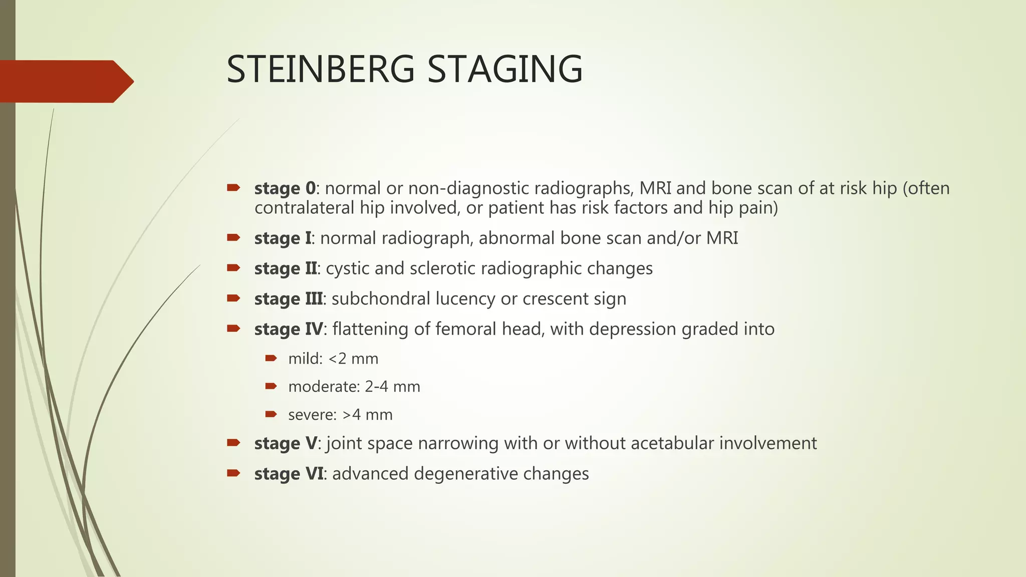
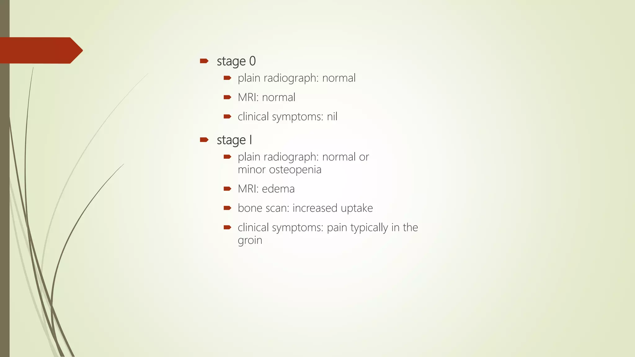
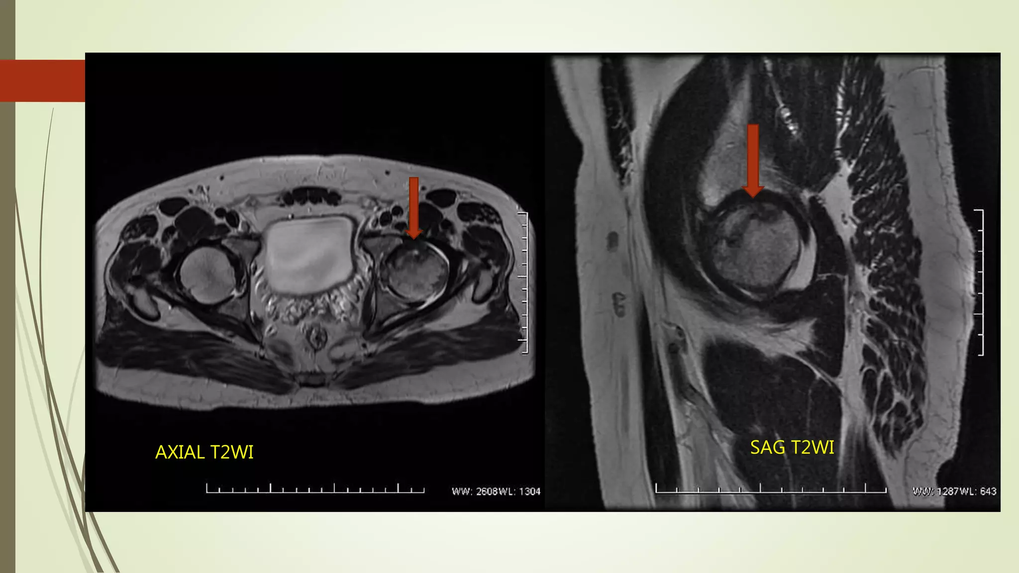
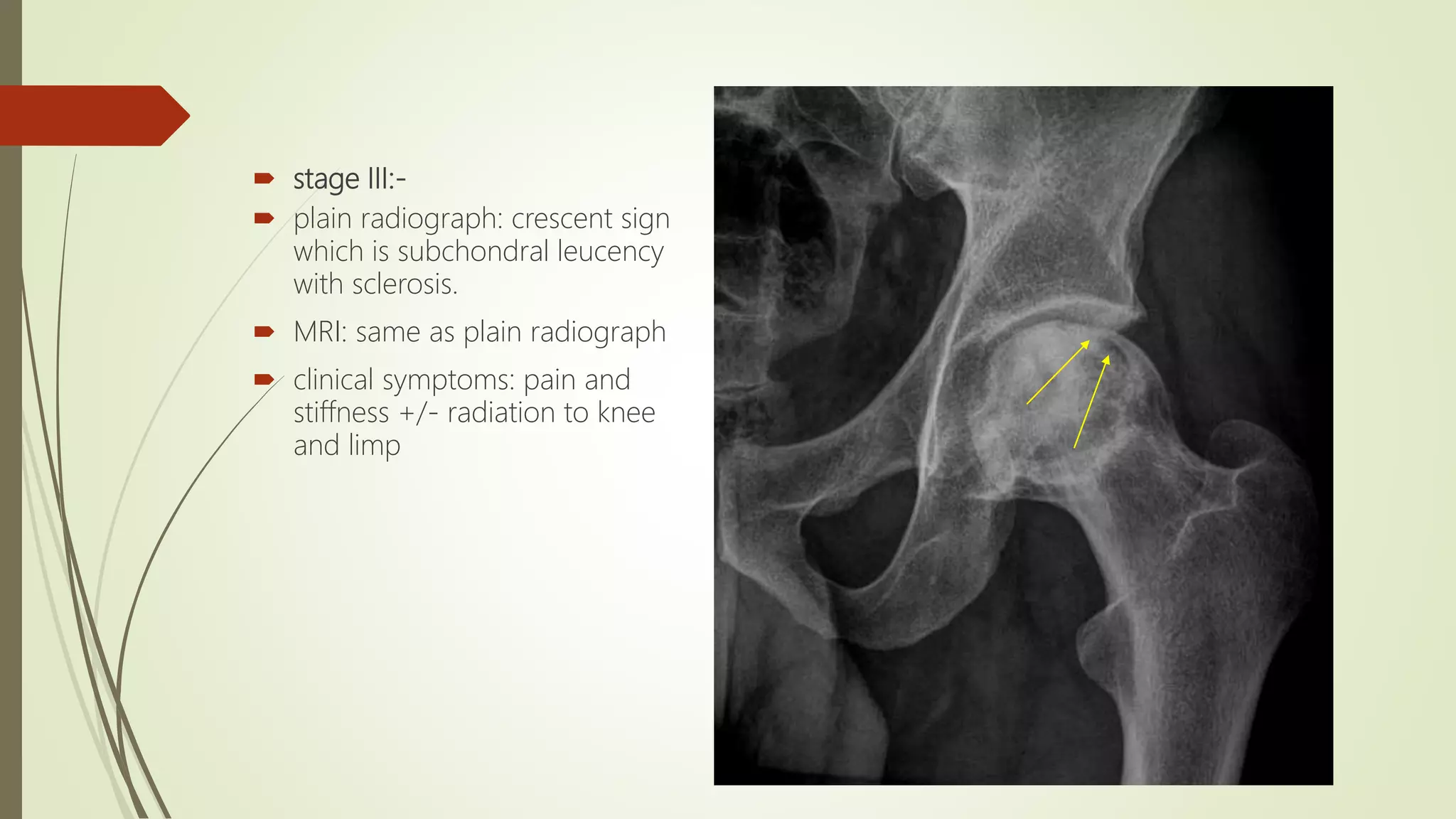
STAGES OF AVN
DR AVINASH DAHATRE
JR II RADIOLOGY
STEINBERG STAGING
 stage 0: normal or non-diagnostic radiographs, MRI and bone scan of at risk hip (often
contralateral hip involved, or patient has risk factors and hip pain)
 stage I: normal radiograph, abnormal bone scan and/or MRI
 stage II: cystic and sclerotic radiographic changes
 stage III: subchondral lucency or crescent sign
 stage IV: flattening of femoral head, with depression graded into
 mild: <2 mm
 moderate: 2-4 mm
 severe: >4 mm
 stage V: joint space narrowing with or without acetabular involvement
 stage VI: advanced degenerative changes
FICAT AND ARLET
STAGING OF AVN
 stage 0
 plain radiograph: normal
 MRI: normal
 clinical symptoms: nil
 stage I
 plain radiograph: normal or
minor osteopenia
 MRI: edema
 bone scan: increased uptake
 clinical symptoms: pain typically in the
groin
 stage II-
 plain radiograph: mixed osteopenia
and/or sclerosis and/or subchondral
cysts, without any subchondral
 MRI: geographic defect
 bone scan: increased uptake
 clinical symptoms: pain and stiffnes
COR T1WI
AXIAL T2WI SAG T2WI
 stage III:-
 plain radiograph: crescent sign
which is subchondral leucency
with sclerosis.
 MRI: same as plain radiograph
 clinical symptoms: pain and
stiffness +/- radiation to knee
and limp
SAG T1WI
 stage IV-
 plain radiograph: end-stage with
evidence of secondary
degenerative change.
 Flattening of femoral head
1. mild: <2 mm
2. moderate: 2-4 mm
3. severe: >4 mm
 MRI: same as plain radiograph
 clinical symptoms: pain and limp
COR T1WI
COR T2WI FATSAT
STAGES OF AVN

STAGES OF AVN

  • 1.
    STAGES OF AVN DRAVINASH DAHATRE JR II RADIOLOGY
  • 2.
    STEINBERG STAGING  stage0: normal or non-diagnostic radiographs, MRI and bone scan of at risk hip (often contralateral hip involved, or patient has risk factors and hip pain)  stage I: normal radiograph, abnormal bone scan and/or MRI  stage II: cystic and sclerotic radiographic changes  stage III: subchondral lucency or crescent sign  stage IV: flattening of femoral head, with depression graded into  mild: <2 mm  moderate: 2-4 mm  severe: >4 mm  stage V: joint space narrowing with or without acetabular involvement  stage VI: advanced degenerative changes
  • 3.
  • 4.
     stage 0 plain radiograph: normal  MRI: normal  clinical symptoms: nil  stage I  plain radiograph: normal or minor osteopenia  MRI: edema  bone scan: increased uptake  clinical symptoms: pain typically in the groin
  • 5.
     stage II- plain radiograph: mixed osteopenia and/or sclerosis and/or subchondral cysts, without any subchondral  MRI: geographic defect  bone scan: increased uptake  clinical symptoms: pain and stiffnes
  • 6.
  • 7.
  • 8.
     stage III:- plain radiograph: crescent sign which is subchondral leucency with sclerosis.  MRI: same as plain radiograph  clinical symptoms: pain and stiffness +/- radiation to knee and limp
  • 9.
  • 10.
     stage IV- plain radiograph: end-stage with evidence of secondary degenerative change.  Flattening of femoral head 1. mild: <2 mm 2. moderate: 2-4 mm 3. severe: >4 mm  MRI: same as plain radiograph  clinical symptoms: pain and limp
  • 11.
  • 12.