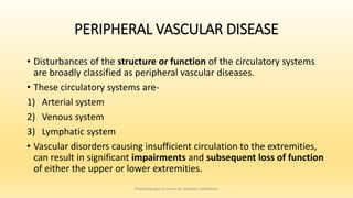
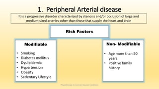
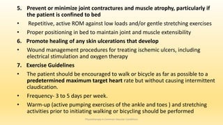
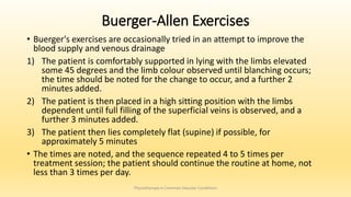
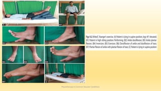
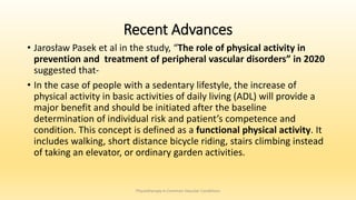
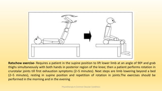
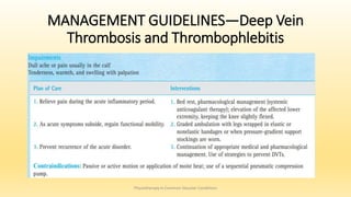
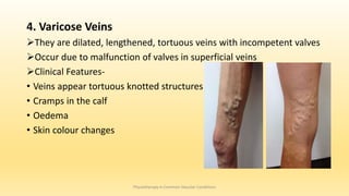
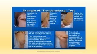
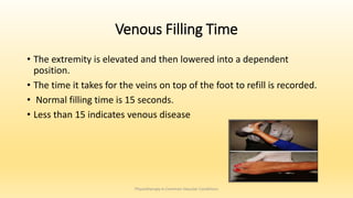
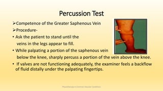
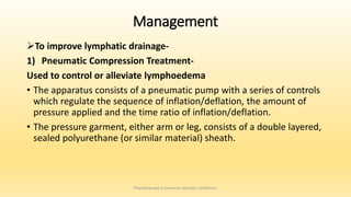
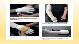
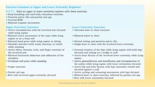
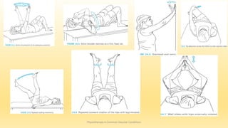
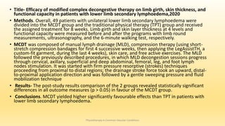
Vascular disease includes any condition that affects your circulatory system, or system of blood vessels. This ranges from diseases of your arteries, veins and lymph vessels to blood disorders that affect circulation.
Blood vessels are elastic-like tubes that carry blood to every part of your body. Blood vessels include:
Arteries that carry blood away from your heart.
Veins that return blood back to your heart.
Capillaries, your tiniest blood vessels, which link your small veins and arteries, deliver oxygen and nutrients to your tissues and take away their waste.
2. OBJECTIVES
At the end of this seminar, one will be able to-
• Define and discuss Peripheral arterial, venous and lymphatic disease
• Enumerate the risk factors of peripheral vascular disease
• List down the clinical manifestations of peripheral vascular disease
• Classify peripheral vascular disease
• Discuss the assessment and management of vascular, venous and
lymphatic disorders
Physiotherapy in Common Vascular Conditions
3. PERIPHERAL VASCULAR DISEASE
• Disturbances of the structure or function of the circulatory systems
are broadly classified as peripheral vascular diseases.
• These circulatory systems are-
1) Arterial system
2) Venous system
3) Lymphatic system
• Vascular disorders causing insufficient circulation to the extremities,
can result in significant impairments and subsequent loss of function
of either the upper or lower extremities.
Physiotherapy in Common Vascular Conditions
4. 1. Peripheral Arterial disease
Physiotherapy in Common Vascular Conditions
Risk Factors
Non- Modifiable
• Age more than 50
years
• Positive family
history
Modifiable
• Smoking
• Diabetes mellitus
• Dyslipidemia
• Hypertension
• Obesity
• Sedentary Lifestyle
It is a progressive disorder characterized by stenosis and/or occlusion of large and
medium sized arteries other than those that supply the heart and brain
5. 1) Atheroslerosis-
• Arteriosclerosis: thickening, hardening, and loss of elasticity of arterial
walls
• Atherosclerosis: the most common form of arteriosclerosis, associated
with damage to the endothelial lining of the vessels and the formation of
lipid deposits, eventually leading to plaque formation.
Physiotherapy in Common Vascular Conditions
Disorders of Arterial System
6. 2) Arteriosclerosis Obliterans
• It is characterized by chronic , progressive occlusion of peripheral
circulation, most often in large and medium sized arteries of lower
limb
• A peripheral manifestation of atherosclerosis characterized by
intermittent claudication, rest pain, and trophic changes. This is the
arterial disease most likely to lead to ulceration.
Physiotherapy in Common Vascular Conditions
7. 3) Acute Arterial Occlusion
• A thrombus, embolism, or trauma can cause acute loss of blood flow
to the peripheral arteries
• Immediate medical or surgical measures must be taken to maintain
the viability of the limb.
Physiotherapy in Common Vascular Conditions
8. 4) Thromboangitis Obliterans (Buerger’s Disease)
• Thromboangitis Obliterans is a chronic disease seen predominantly in young
males who smoke
• It involves an inflammatory reaction of the arteries to nicotine
• It results in vasoconstriction, decreased arterial circulation to the extremities,
ischemia, and eventual ulceration and necrosis of soft tissues.
Physiotherapy in Common Vascular Conditions
9. 5) Raynaud’s Disease-
• It is a arterial disorder that occurs more often in women than men.
• It is characterized by digital vasospasm, most often affecting the small
arteries and arterioles of the fingers and sometimes the toes.
• Vasospasm is brought on by exposure to cold, vibration, or stress. The
response is characterized by temporary pallor, then cyanosis and pain,
followed by numbness and a cold sensation of the digits.
Physiotherapy in Common Vascular Conditions
10. 6) Aneurysm
• The development of atheroma usually results in progressive stenosis
of the affected arteries.
• Sometimes, atheroma leads to a weakening of the vessel wall causing
it to dilate under arterial pressure.
• Localised dilatation of an artery is known as an aneurysm.
Physiotherapy in Common Vascular Conditions
11. 7) Thoracic outlet syndrome
• An abnormal rib which has developed on the 7th cervical vertebra or
a tough fibrous band attached to the 1st rib may press upon the
subclavian artery thereby stenosing it.
• An aneurysm commonly forms distal to the narrowing and, together
with pressure on the nerves of the brachial plexus, will cause tingling
and weakness in the forearm and Raynaud’s syndrome in the hand.
Physiotherapy in Common Vascular Conditions
12. Clinical Manifestations
1. Diminished or Absent Peripheral Pulses
2. Integumentary Changes
Skin discoloration, including pallor at rest or with exercise
Trophic changes include a shiny, waxy appearance and dryness of the
skin and loss of hair distal to the occlusion.
Skin temperature is decreased.
Ulcerations may develop, particularly at weight-bearing areas or over
bony prominences
3. Sensory Disturbances
Physiotherapy in Common Vascular Conditions
13. 4. Exercise Pain and Rest Pain
• Claudication (derived from the Latin word for limp) is defined as a
reproducible discomfort of a defined group of muscles that is induced
by exercise and relieved with rest.
• During the early stages of arterial disease, intermittent claudication is
characterized by a feeling of fatigue or weakness and, later, as
cramping or aching in the muscles used during exercise
• Critical limb Ischaemia- Chronic ischaemic pain occurring during rest,
lasting for more than 2 weeks, or as ischaemic lesions in the form of
foot ulceration or necrosis.
Physiotherapy in Common Vascular Conditions
15. Physiotherapy in Common Vascular Conditions
These collateral vessels maintain blood supply to the limb, but because they are of narrow calibre their resistance to
flow is very great and thus claudication still occurs. With the development of collaterals the distance the patient can
walk may improve markedly.
16. Classification of Peripheral Arterial Disease
1) Rutherford Classification
Physiotherapy in Common Vascular Conditions
18. Assessment of Arterial Disorders
• History
• Observation- Skin integrity and pigmentation
• Skin temperature
• Palpation and comparison of pulses in the involved and uninvolved upper
or lower extremities
• Tests for reactive hyperaemia
• Claudication time
• Ankle Brachial Index
• Transcutaneous oximetry
• Magnetic resonance angiography
• Arteriography
Physiotherapy in Common Vascular Conditions
19. 1) Rubor Dependency Test—Reactive Hyperemia
• Procedure- The legs are elevated for several minutes above the level
of the heart while the patient is lying supine.
• Pallor (blanching) of the skin occurs in the feet within 1 minute or less
if arterial circulation is poor.
• The time necessary for blanching to develop is noted. Then the legs
are placed in a dependent position, and the color of the feet is noted.
• Normally, a pinkish flush appears in the feet within several seconds
after the legs are placed in a dependent position. With occlusive
arterial disease, a bright bluish-red color, or rubor, of the distal legs
and feet is evident that is caused by reduced blood flow in the
capillaries. The rubor may take as long as 30 seconds to appear
Physiotherapy in Common Vascular Conditions
21. 2) Buerger’s test
• The vascular angle, which is also called Buerger's angle, is
the angle to which the leg has to be raised before it
becomes pale, whilst lying down.
• In a limb with a normal circulation the toes and sole of the
foot, stay pink, even when the limb is raised by 90
degrees.
• In an ischaemic leg, elevation to 15 degrees or 30 degrees
for 30 to 60 seconds may cause pallor.
• A vascular angle of less than 20 degrees indicates severe
ischaemia.
Physiotherapy in Common Vascular Conditions
22. 3. Claudication Time
• An objective assessment of exercise pain (intermittent claudication) is
performed to determine the amount of time a patient can exercise
before experiencing cramping and pain in the distal musculature.
• A commonly used test is to have the patient walk at a slow,
predetermined speed on a level treadmill (1 to 2 mph).
• The time that the patient is able to walk before the onset of pain or
before pain prohibits further walking is noted.
• This measurement should be undertaken to determine a baseline for
exercise tolerance before initiating a program to improve exercise
tolerance
Physiotherapy in Common Vascular Conditions
23. 4. Ankle Brachial Index
• The ankle-brachial index (ABI), also known as the ankle-arm index, is a
relatively easy, non-invasive test to assess for PAD
• This test can be performed by using an inexpensive handheld Doppler
probe and a sphygmomanometer.
• The ABI is calculated by dividing the highest ankle systolic pressure by
the highest brachial pressure.
Physiotherapy in Common Vascular Conditions
25. • Other methods-
• Transcutaneous Oximetry Transcutaneous -Provides information
about the oxygen saturation of blood by means of a photoelectric
device (a pulse oximeter).
• Arteriography - An invasive procedure that involves injecting a
radiopaque dye directly into an artery. The arteries are then
radiographically visualized to detect any restriction of movement of
the dye in arterial vessels indicating a partial or complete occlusion
• Magnetic Resonance Angiography-A noninvasive procedure, provides
radiographic visualization of arteries without the use of a contrast
medium
Physiotherapy in Common Vascular Conditions
26. MANAGEMENT
Medical Management-
• Anticoagulant
• Antiplatelets
• Lipid lowering agents
• Antihypertensives
• Antidiabetic drugs
• Analgesics
Risk factor modification-
• Smoking, tobacco cessation
• Dietary changes- such as limiting or avoiding salt, sucrose, and alcohol to lower
blood pressure and triglyceride and cholesterol levels.
• Weight reduction
• Lifestyle changes
Physiotherapy in Common Vascular Conditions
27. Management of Acute Arterial Occlusion
• Medical or surgical measures must be taken to reduce
ischemia and to restore circulation.
• Medical management includes bed rest and complete
systemic anticoagulation therapy.
• Complementary physical interventions to improve
peripheral blood flow while the patient is on bed rest may
include warming the limb by reflex heating of the torso or
opposite extremity or elevating the head of the bed slightly.
• Surgical interventions are thrombo-embolectomy or an
arterial bypass graft. If circulation cannot be significantly
improved or restored, gangrene develops within a very
short time, and amputation of the extremity is necessary.
Physiotherapy in Common Vascular Conditions
28. Contraindications-
• Exercise—passive or active
• Prolonged positioning during bed rest, which could cause pressure on
and potential breakdown of skin
• Local, direct heat on the involved extremity because of the potential
for a burn to the ischemic tissue
• Restrictive clothing that could compromise blood flow
Physiotherapy in Common Vascular Conditions
29. Management of Chronic Arterial Insufficiency
1. Patient Education
2. Medical or surgical management including medications; nutritional
counseling for weight control and to decrease salt, sucrose, cholesterol,
and caffeine intake; smoking cessation.
3. Relieve pain at rest- Advice to sleep with the legs in a dependent but
supported position over the edge of the bed or with the head of the bed
slightly elevated
4. Prevent skin ulcerations-
Proper care and protection of the skin, particularly the feet or hands.
Proper nail care.
Proper shoe selection and fit
Avoid restrictive clothing
Avoid exposure to extremes of temperature, both hot and cold
Physiotherapy in Common Vascular Conditions
30. 5. Prevent or minimize joint contractures and muscle atrophy, particularly if
the patient is confined to bed
• Repetitive, active ROM against low loads and/or gentle stretching exercises
• Proper positioning in bed to maintain joint and muscle extensibility
6. Promote healing of any skin ulcerations that develop
• Wound management procedures for treating ischemic ulcers, including
electrical stimulation and oxygen therapy
7. Exercise Guidelines
• The patient should be encouraged to walk or bicycle as far as possible to a
predetermined maximum target heart rate but without causing intermittent
claudication.
• Frequency- 3 to 5 days per week.
• Warm-up (active pumping exercises of the ankle and toes ) and stretching
activities prior to initiating walking or bicycling should be performed
Physiotherapy in Common Vascular Conditions
31. Buerger-Allen Exercises
• Buerger's exercises are occasionally tried in an attempt to improve the
blood supply and venous drainage
1) The patient is comfortably supported in lying with the limbs elevated
some 45 degrees and the limb colour observed until blanching occurs;
the time should be noted for the change to occur, and a further 2
minutes added.
2) The patient is then placed in a high sitting position with the limbs
dependent until full filling of the superficial veins is observed, and a
further 3 minutes added.
3) The patient then lies completely flat (supine) if possible, for
approximately 5 minutes
• The times are noted, and the sequence repeated 4 to 5 times per
treatment session; the patient should continue the routine at home, not
less than 3 times per day.
Physiotherapy in Common Vascular Conditions
33. Recent Advances
• Jarosław Pasek et al in the study, “The role of physical activity in
prevention and treatment of peripheral vascular disorders” in 2020
suggested that-
• In the case of people with a sedentary lifestyle, the increase of
physical activity in basic activities of daily living (ADL) will provide a
major benefit and should be initiated after the baseline
determination of individual risk and patient’s competence and
condition. This concept is defined as a functional physical activity. It
includes walking, short distance bicycle riding, stairs climbing instead
of taking an elevator, or ordinary garden activities.
Physiotherapy in Common Vascular Conditions
34. Physiotherapy in Common Vascular Conditions
Ratschow exercise- Requires a patient in the supine position to lift lower limb at an angle of 90º and grab
thighs simultaneously with both hands in posterior region of the knee; then a patient performs rotation in
crurotalar joints till first exhaustion symptoms (2–5 minutes). Next steps are limb lowering beyond a bed
(2–5 minutes), resting in supine position and repetition of rotation in joints.The exercises should be
performed in the morning and in the evening
35. • TITLE - THE EFFECT OF THE WARM WHIRLPOOL ON PERIPHERAL ARTERIAL INSUFFICIENCY IN LOWER
LIMB
• AUTHORS- Shorouk Ihab Ahmed; Hany Ezzat Obaya; Sally Said Abd Elhamed; Rana Hesham Elbanna
• AIM: To explore the warm whirlpool's influence on functions of the endothelium in type II diabetics with
peripheral arterial insufficiency in the lower distal limb.
• MATERIALS AND METHODS: In this study forty patients of both sex (aged from 40 to 55 years old) with
Insufficiency of the Peripheral Arteries as an outcome of Type II Diabetes Mellitus, were evenly split into 2
equal groups (A and B) randomly, group (A)managed to receive warm whirlpool and treadmill walking
sessions, group (B) received the treadmill walking sessions only. Data obtained from groups pre-and post-
treatment regarding nitric oxide blood analysis (NO), maximum walking time (MWT), and the intermittent
claudication questionnaire (ICQ) were statistically analyzed and compared.
• RESULTS: Nitric oxide in blood levels and maximum walking time increased significantly, while intermittent
claudication questionnaire in groups significantly decreased from pre- to post-treatment. Between groups,
there was a substantial increase in maximum walking time following therapy, while the intermittent
claudication questionnaire is significantly decreased; simultaneously, there was no significant difference in
nitric oxide between 2 groups after the management plan.
• CONCLUSION: The study concluded that a warm whirlpool is significantly effective for improving
endothelial function and decreasing symptoms in peripheral arterial diseases. As a result, incorporating
both warm whirlpool and treadmill walking exercise into a management program for lower limb peripheral
arterial insufficiency yielded remarkable outcomes.
Physiotherapy in Common Vascular Conditions
36. Traditional Treadmill Exercise
• Intensity of exercise: According to the maximal graded walking Test
• Frequency: 3 days/week.
• Duration: The time spent exercising in total (including rest periods) is
50 min/day. 5 minutes warming, 40 minutes active phase with rest
intervals, 5 minutes cooling down.
Programmed Warm Whirlpool
• Frequency- 3 times per week for 12 weeks
• Temperature: Mild warmth between 35.5°C and 37°C (96°F and 98°F)
• Leg immersion time: Immersion of the whole leg in a whirlpool for 20-
30 minutes every session
Physiotherapy in Common Vascular Conditions
37. Pain-Free Walking Exercise Therapy
• Piotr Mika et al. conducted 4 studies on - A 12-week intervention of treadmill training to
onset of pain
1. Experimental model of pain-free treadmill training in patients with claudication,2005
2. Red blood cell deformability in patients with claudication after pain-free treadmill training, 2006
3. The effect of pain-free treadmill training on fibrinogen, haematocrit, and lipid profile in patients
with claudication, 2011
4. Comparison of two treadmill training programs on walking ability and endothelial function in
intermittent claudication, 2013
• Studies 1–3 resulted in:
‒ Increase in pain-free walking distance
‒ Increase in peak walking distance
‒ No increases in inflammatory markers after exercise training
‒ No improvement in control group
Physiotherapy in Common Vascular Conditions
38. • Study 4 compared two treadmill walking protocols (12 weeks)
1. Traditional treadmill walking into moderate to severe discomfort
2. Treadmill walking only to the onset of claudication
Both groups had statistically significant improvement in walking
distance ‒ No statistical differences between groups
Physiotherapy in Common Vascular Conditions
Moderate Intensity Group
• Improved pain-free walking
distance 120% (121 meters)
• Improved peak walking distance
100% (393 meters)
Pain-Free Walking Group
• Improved pain-free walking
distance 93% (141 meters)
• Improved peak walking distance
98% (465 meters)
39. PAD Guideline Definitions (2016)
Supervised exercise program
• Program takes place in a hospital or outpatient facility.
• Program uses intermittent walking exercise as the treatment modality.
• Program can be standalone or within a cardiac rehabilitation program.
• Program is directly supervised by qualified healthcare provider(s).
• Training is performed for a minimum of 30–45 minutes/session; sessions
are performed at least 3 times/week for a minimum of 12 weeks.
• Training involves intermittent bouts of walking to moderate-to-maximum
claudication, alternating with periods of rest.
• Warm-up and cool-down periods precede and follow each session of
walking
Physiotherapy in Common Vascular Conditions
40. Intermittent Claudication
• Supervised exercise programs are more effective than non-supervised
programs in improving treadmill walking distances in patients with IC.
• The evidence suggests that programs focus on walking at an intensity
that elicits symptoms (score of 1 on the Claudication Pain Rating
Scale- figure 1) within 3 to 5 minutes, stopping if symptoms become
moderate (score of 2 on the Claudication Pain Rating Scale- figure 1),
resting until symptoms have resolved, and then resuming walking.
• The exercise program should be for 30 to 60 minutes of exercise and
rest cycles per session, 3 to 5 times per week, for a minimum of 3
months time period.
Physiotherapy in Common Vascular Conditions
42. UPPER VS LOWER EXTREMITY EXERCISE:
• The results of the randomized controlled trial conducted by Rena
Zwierska et al suggested that both upper- and lower- limb weight-
supported aerobic exercise training provide an adequate stimulus for
evoking improvements in walking performance in patients with PAD.
• Evidence from the this study suggests that the improvement in
walking performance after upper-limb training is due to increase in
the antioxidant potential leading to extension of painless and
maximum distance of claudication
• Certainly, it can also be expected that this form of training contributes
to strengthening the muscles of the upper limbs, possible blood
pressure normalization, and improvements in the circulatory system
as a whole.
Physiotherapy in Common Vascular Conditions
43. Venous Diseases
• Venous insufficiency occurs when the venous system is unable to
provide adequate antegrade blood flow back to the heart, i.e. venous
return, and fails to prevent retrograde flow into the extremities,
leading to peripheral edema
Physiotherapy in Common Vascular Conditions
44. Risk Factors
• Postoperative or post-fracture immobilization
• Prolonged bed rest
• Trauma to venous vessels
• Limb paralysis
• History of deep vein thrombosis or pulmonary embolism
• Advanced age
• Obesity
• Sedentary lifestyle
• Congestive heart failure
• Use of oral contraceptives
Physiotherapy in Common Vascular Conditions
45. Assessment
• History
• Limbs are examined, noting size, shape and the nature and extent of
any oedema.
• The peripheral pulses should be palpated, and the digital capillary
pressure tested by mild compression and release.
• The presence of varicose veins, signs of phlebitis, cellulitis or
thrombosis should be carefully assessed
• If ulcer is present, tracing of the ulcer outline is taken
• A visual assessment is made of the lesion, noting the tissue changes
in the floor of the ulcer and the surrounding skin.
Physiotherapy in Common Vascular Conditions
46. • It is important to make note of the cleanliness/infective state of the
ulcer, the presence of granulation tissue, and the formation of a
healing, spreading or indolent edge, in order that subsequent
progression or regression of the ulcer healing becomes obvious, and
the treatment can be varied according
• Ranges of movement are carefully measured at all lower limb joints,
and any reasons for lack of range are recorded.
• An estimate of muscle power is made and charted.
Physiotherapy in Common Vascular Conditions
47. Tests for Peripheral Venous Diseases
• Girth measurements of the upper or lower extremities
• Percussion test: compliance of the greater saphenous vein
• Homans’ sign
• Response to compression of the limb with a blood pressure cuff
• Doppler ultrasonography
• Venous duplex screening (non invasive)
• Venography- involves injecting radiopaque dye and radiographic
visualization of the venous system
Physiotherapy in Common Vascular Conditions
48. 1. Thrombophlebitis
Thrombophlebitis, a disorder typically affecting the lower extremities
is characterized by inflammation of inner walls of superficial veins
The thrombus becomes attached on the vessel wall but rarely forms
embolus
Clinical Features-
• Localised reddened warm area
• Hard cord like swelling along course of superficial vein
• Pain
Physiotherapy in Common Vascular Conditions
49. Test-
Application of a Blood Pressure Cuff Around the Calf
Procedure
• Inflate the cuff gradually until the patient experiences calf pain.
• A patient with acute thrombophlebitis usually cannot tolerate
pressures above 40 mm Hg.
Physiotherapy in Common Vascular Conditions
50. 2. Deep Vein Thrombosis
It is the blocking of deep vein by formation of thrombus
During the early stages of a DVT, only 25% to 50% of cases can be
identified by clinical manifestations, such as pain, swelling, or changes in
skin temperature and color.
Although edema in the vicinity of the clot may be present, it may be too
deep to palpate
Only measurement by ultrasonography, venous duplex screening, or
venography can confirm a DVT.
Physiotherapy in Common Vascular Conditions
51. Homans’ Sign Procedure.
• With the patient supine and the knee extended, passively dorsiflex
the ankle and gently squeeze the calf muscles.
• If pain occurs in the calf, Homans’ sign is positive, indicating the
possible presence of a DVT.
Physiotherapy in Common Vascular Conditions
52. Prevention of Deep Vein Thrombosis and
Thrombophlebitis
• Prophylactic use of anticoagulant therapy for the high-risk patient
• Initiation of ambulation as soon as possible after surgery
• Elevating the legs while lying supine and on a footstool when sitting
• No prolonged periods of sitting
• Active “pumping” exercises regularly throughout the day while lying
supine in bed
• Use of compression stockings to support the walls of the veins and
minimize venous pooling
• For patients on bed rest, use of a sequential pneumatic compression
unit
Physiotherapy in Common Vascular Conditions
54. • Aldrich and colleagues conducted a systematic review of the literature to
determine when a patient with DVT should be allowed to begin walking.
• Results of these studies suggest that early ambulation, begun within the
first 24 hours after initiating anticoagulant therapy, does not increase the
incidence of pulmonary embolism in patients without an existing
pulmonary embolism and who have adequate cardiopulmonary reserve.
• However, if a patient has a known pulmonary embolism, an ambulation
program must be initiated more cautiously. It is important to note that in
the studies reviewed all patients who participated in an early ambulation
program wore compression garments.
• The results also revealed that early ambulation is associated with more
rapid resolution of pain and swelling.
Physiotherapy in Common Vascular Conditions
55. 3. Pulmonary Embolism
When an embolus affects pulmonary
circulation, it is called a pulmonary embolism
Hallmark signs and symptoms are –
• Dyspnea
• Tachypnea
• Chest pain that intensifies with deep breathing
and coughing.
• Other signs and symptoms include- oedema,
anxiety, fever, diaphoresis, a cough,
hemoptysis
Physiotherapy in Common Vascular Conditions
56. Exercise therapy after acute pulmonary
embolism ( Recent Advances)
• Phase 1 was conducted while the subject was hospitalized and lasted up to
7 days or until they were ambulatory. It consisted of three 30-second bouts
for upper extremity cycling ergometry (approximately 5W at 75–85 rpm),
each followed by a 10-minute rest period performed three times a day.
• Phase 2 was initiated when patients became ambulatory or when they
were discharged, and consisted of weight-bearing aerobic exercise. They
were asked to walk 3 times a week, starting at 40–50% of their age
predicted HRR for 20 minutes, and progressing to 60–70% for 30 minutes
over the subsequent 3 weeks.
• Phase 3 was initiated 1 month after study-enrollment, concluded at 3
months, and consisted of 30 minutes of aerobic exercise at 60– 70% of HRR
followed by lower extremity stretching exercises 5 days a week
Physiotherapy in Common Vascular Conditions
57. 4. Varicose Veins
They are dilated, lengthened, tortuous veins with incompetent valves
Occur due to malfunction of valves in superficial veins
Clinical Features-
• Veins appear tortuous knotted structures
• Cramps in the calf
• Oedema
• Skin colour changes
Physiotherapy in Common Vascular Conditions
59. Venous Filling Time
• The extremity is elevated and then lowered into a dependent
position.
• The time it takes for the veins on top of the foot to refill is recorded.
• Normal filling time is 15 seconds.
• Less than 15 indicates venous disease
Physiotherapy in Common Vascular Conditions
60. Percussion Test
Competence of the Greater Saphenous Vein
Procedure-
• Ask the patient to stand until the
veins in the legs appear to fill.
• While palpating a portion of the saphenous vein
below the knee, sharply percuss a portion of the vein above the knee.
• If valves are not functioning adequately, the examiner feels a backflow
of fluid distally under the palpating fingertips.
Physiotherapy in Common Vascular Conditions
61. Girth Measurements
• Circumferential measurements of the involved and uninvolved limbs
are taken to determine the presence and extent of edema.
• Measurements are taken at anatomical landmarks or at
predetermined and consistent distances apart (e.g., 8 or 10 cm apart).
Physiotherapy in Common Vascular Conditions
62. 5. Chronic Venous Insufficiency
It is defined as inadequate venous return over a prolonged period of
time.
It may begin after a severe episode of DVT, may be associated with
varicose veins, or may be the result of trauma to the lower extremities or
blockage of the venous system by a neoplasm.
Clinical Features-
• Dependent, peripheral edema with
long periods of standing or sitting
• Dull aching in the affected extremity
• When edema persists, the skin becomes less
supple over time and takes on a brownish pigmentation.
Physiotherapy in Common Vascular Conditions
63. Physiotherapy in Common Vascular Conditions
Damaged or incompetent valves in the veins
Compromise venous return
Venous hypertension and Venous stasis in
the lower extremities
Chronic pooling of blood in the veins
Inadequate oxygenation and removal of
waste products leading to necrosis
Venous stasis ulcers
64. MANAGEMENT GUIDELINES—Chronic Venous
Insufficiency and Varicose Veins
1. Patient education and self-management skills for skin care, self-
massage for edema, and a home exercise program
2. Prevent lymphedema; minimize venous stasis.
• Pressure-gradient support stockings donned before getting out of
bed in the morning and worn every day.
• Walking, on a regular basis.
• Elevate the lower extremities after graded ambulation until the
heart rate returns to normal.
• Avoid prolonged periods of standing still and sitting with legs
dependent
Physiotherapy in Common Vascular Conditions
66. 3. Increase venous return and reduce lymphedema if already present.
• Intermittent mechanical compression pump
• Faradism under pressure
• Manual massage to drain edema.
• Relaxation and active ROM (pumping exercises) of the distal
muscles while involved limb is elevated.
4. Prevent skin abrasions, ulcerations, and wound infections.
• Proper skin care
Physiotherapy in Common Vascular Conditions
67. 5. Control of infection
• In the presence of acute or sub-acute infection, active physiotherapy
to the ulcer and surround is usually discontinued.
• In less acute condition, ultraviolet irradiation (UVR) may be used in
an attempt to sterilize the surface of the ulcer and improve the blood
supply.
• The lamp chosen should include UVC (i.e. less than 280 nm) in its
output in order to maximise the bactericidal effect.
• Chronic ulcer- Ultrasound therapy -pulsed, low output, at 3MHz to
the surrounding area to soften oedema and increase vascularity of
the skin and j subcutaneous tissue.
• Pulsed high frequency treatment may prove to be of value in this
stimulation and acceleration of healing
Physiotherapy in Common Vascular Conditions
68. 6. Increase muscle strength
• Muscle bulk and power may be considerably diminished because of
oedema, pain, joint stiffness, continued use of support bandaging,
faulty gait, and general lack of normal use.
• This also reduces the effectiveness of the muscle pump. An attempt
should be made to mobilize the stiff joints and improve muscle
strength while bearing in mind the patient’s ability to tolerate an
exercise program.
7. Mobilisation of the tissues in the floor and edges of the ulcer
• This helps to soften and increase the vascularity of the tissues, and,
possibly, hastens the more rapid, non-adherent healing of the ulcer.
Physiotherapy in Common Vascular Conditions
69. Non-contact low-frequency ultrasound therapy compared with UK standard of care
for venous leg ulcers: A single-centre, assessor-blinded, randomized controlled trial.
Int Wound J, 2015
Physiotherapy in Common Vascular Conditions
71. Disorders of Lymphatic Circulation
• Lymphedema is an excessive and persistent accumulation of
extravascular and extracellular fluid and proteins in tissue spaces.
• It occurs when lymph volume exceeds the capacity of the lymph
transport system, and it is associated with a disturbance of the water
and protein balance across the capillary membrane.
• An increased concentration of proteins draws larger amounts of water
into interstitial spaces, leading to lymphedema.
Physiotherapy in Common Vascular Conditions
72. • Primary Lymphedema- It is the result of insufficient development
and congenital malformation of the lymphatic system.
• It is further divided into-
1)Congenital- presents at birth and is sometimes known as Milroy’s
disease.
2)Praecox- (early) develops prior to 35 years of age.
3)Tarda- occurs in later life possibly as a result of deterioration in a
barely adequate lymph system.
Physiotherapy in Common Vascular Conditions
73. 2. Seconday Lymphoedema
• In these cases the normal lymphatics have been damaged.
• Causes include-
1. Surgical dissection of lymph nodes. Eg carcinoma of breast
2. Infection and Inflammation-
Inflammation of the lymph vessels (lymphangitis) or lymph nodes
(lymphadenitis) and enlargement of lymph nodes (lymphadenopathy)
can occur as the result of a systemic infection or local trauma.
3. Obstruction or fibrosis
(trauma, surgery, neoplasm)
4. Chronic venous insufficiency
Physiotherapy in Common Vascular Conditions
74. Clinical Manifestations of Lymphatic Disorders
• Lymphedema
• Sensory Disturbances
• Stiffness and Limited Range of Motion
• Decreased Resistance to Infection
Physiotherapy in Common Vascular Conditions
75. Examination and Evaluation of Lymphatic
Function
• Patient’s H/O- Note any history of infection, trauma, surgery, or radiation
therapy.
• Identify the occupation or daily activities of the patient and determine if
long periods of standing or sitting are required.
• Visual inspection and palpation of the skin. Skin changes (shiny, red
oedematous limb) and location of lymphoedema should be noted.
• Girth Measurements Circumferential measurements of the involved limb
should be taken and compared with the noninvolved limb if the problem is
unilateral
• Volumetric Measurements- An alternative method of measuring limb size is
to immerse the limb in a tank of water to a predetermined anatomical
landmark and measure the volume of water displaced
Physiotherapy in Common Vascular Conditions
76. Physiotherapy in Common Vascular Conditions
Volumetric Measurement of lower limb Volumetric Measurement of upper limb GirthMeasurement of lower limb
Stemmers Sign
77. Prevention of Lymphedema
• Avoid static, dependent positioning of the lower extremities
• When traveling long distances by car, stop periodically and walk around
• Elevate involved limb(s) and perform repetitive pumping exercises
frequently during the day.
• Avoid vigorous, repetitive activities with the involved limb and carrying
heavy loads
• Wear compressive garments while exercising.
• Avoid wearing clothing that restricts circulation. (avoid tight clothing)
• Monitor diet to maintain an ideal weight and minimize sodium intake.
• Avoid hot environments or use of local heat.
Physiotherapy in Common Vascular Conditions
78. Skin Care
• Keep the skin clean and supple
• Avoid infections; pay immediate attention to a skin abrasion or cut,
an insect bite, a blister, or a burn.
• Protect hands and feet; wear socks or hose, properly fitting shoes,
rubber gloves, oven mitts, etc.
• Avoid contact with harsh detergents and chemicals.
• Use caution when cutting nails. Women need to use an electric razor
when shaving legs or underarm area.
• Avoid hot baths, whirlpools, and saunas that elevate the body’s core
temperature.
Physiotherapy in Common Vascular Conditions
79. Management
To improve lymphatic drainage-
1) Pneumatic Compression Treatment-
Used to control or alleviate lymphoedema
• The apparatus consists of a pneumatic pump with a series of controls
which regulate the sequence of inflation/deflation, the amount of
pressure applied and the time ratio of inflation/deflation.
• The pressure garment, either arm or leg, consists of a double layered,
sealed polyurethane (or similar material) sheath.
Physiotherapy in Common Vascular Conditions
80. Physiotherapy in Common Vascular Conditions
Intermittent compression: In this method the sleeve inflates and
compresses at the chosen pressure (mmHg) and then deflates. The
whole limb is therefore compressed at the same time.
81. Physiotherapy in Common Vascular Conditions
Sequential compression: In this method, like a Ripple bed, the cells
inflate sequentially - as one set deflates so another set inflates
allowing for a ripple or sequential effect upon the tissues.
82. Method of treatment-
Before starting , limb measurements should be taken for comparison.
Preparation of patient- The limb to be treated should be completely
bared. Limb is then placed in a cotton gauze sleeve. The sleeve is
connected to plastic/rubber tubing to machine which is switched on.
Position for treatment- Limb is placed in some elevation , supported by
pillows.
Duration of treatment- In severe cases, 3 sessions of 3 hrs for 5-14
days. Out-patient cases, daily 1-1/2 hrs combining with home
treatment.
Timing of compression- If intermittent compression is given,
inflation/deflation ratio should be 3:1 with time of 1 min. so 45 secs
compression and 15 secs deflation. Start with low pressure and
increase to about 60 mmHg.
Physiotherapy in Common Vascular Conditions
83. 2. Manual lymphatic drainage
Manual lymphatic drainage involves slow, very light repetitive stroking
and circular massage movements done in a specific sequence with the
involved extremity elevated whenever possible.
Physiotherapy in Common Vascular Conditions
85. 3. Exercises
• Active ROM, stretching, and low-intensity resistance exercises are
integrated with manual drainage techniques.
• Exercises are performed while wearing a compressive garment or
bandages and in a specific sequence, often with the edematous
limb(s) elevated.
• A low-intensity cardiovascular/pulmonary endurance activity, such as
bicycling, often follows ROM and strengthening exercise
4. Elevation
• The involved limb is elevated during use of a sequential compression
pump, while sleeping or resting, or even during sedentary activities.
• The compressive bandages or garment are worn during periods of
elevation
Physiotherapy in Common Vascular Conditions
86. 5. Compressive bandages, garments, or pumps.
• No-stretch, nonelastic bandages or low-stretch elastic bandages or
garments are recommended because they provide relatively low
compressive forces on the edematous extremity at rest.
• In addition, they provide a higher working pressure with active
muscular contractions because of their less yielding nature than high-
stretch bandages.
6. Skin care and hygiene
• Lymphedema predisposes the patient to skin breakdown, infection,
and delayed wound healing.
• Meticulous attention to skin care and protection of the edematous
limb are essential elements of self-management of lymphedema.
Physiotherapy in Common Vascular Conditions
90. • Title- Efficacy of modified complex decongestive therapy on limb girth, skin thickness, and
functional capacity in patients with lower limb secondary lymphoedema,2020
• Methods. Overall, 49 patients with unilateral lower limb secondary lymphoedema were
divided into the MCDT group and the traditional physical therapy (TPT) group and received
the assigned treatment for 8 weeks. Limb girth and skin layer thickness at 4 levels and
functional capacity were measured before and after the programs with limb round
measurements, ultrasonography, and the 6-minute walking test, respectively.
• MCDT was composed of manual lymph drainage (MLD), compression therapy (using short-
stretch compression bandages for first 4 successive weeks, then applying the LegAssistTH, a
custom-fit garment, during the last 4 weeks), skin care, and free active exercises. The MLD
followed the previously described procedures, in which MLD decongestion sessions progress
through cervical, axillary, superficial and deep abdominal, femoral, leg, and foot lymph
nodes stimulation. It was started with firm pressure resorptive (strokes) techniques
proceeding from proximal to distal regions; the drainage stroke force took an upward, distal-
to-proximal application direction and was followed by a gentle sweeping pressure and fluid
mobilization technique
• Results- The post-study results comparison of the 2 groups revealed statistically significant
differences in all outcome measures (p > 0.05) in favour of the MCDT group.
• Conclusions. MCDT yielded higher significantly favourable effects than TPT in patients with
lower limb secondary lymphoedema.
Physiotherapy in Common Vascular Conditions
91. References
• Physical Rehabilitation- Susan O’ Sulllivan
• Therapeutic Exercises Carolyn Kisner and Colby
• Cash’s Textbook of Chest, Heart and Vascular Disorders for Physiotherapists
edited by PATRICIA A. DOWNIE FCSP
• American heart association guidelines
• Preeti Christian, Physiotherapy for intermittent claudication: a review article,2015
• Jaroslaw Pasek,The role of physical activity in prevention and treatment of
peripheral vascular disorders, 2020
• Anna Spannbauer, Intermittent Claudication in Physiotherapists’ Practice, 2019
• Fatma, Efficacy of modified complex decongestive therapy on limb girth, skin
thickness, and functional capacity in patients with lower limb secondary
lymphoedema,2020
• Rafael S. Cires-Drouet,Safety of exercise therapy after acute pulmonary
embolism,2020
Physiotherapy in Common Vascular Conditions