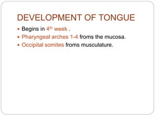
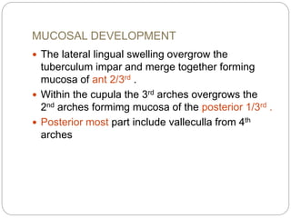
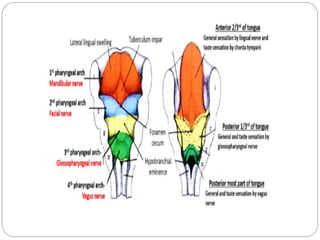
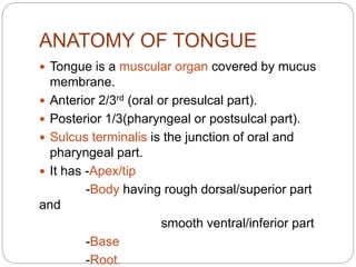
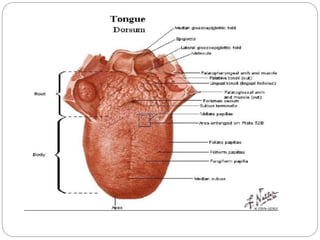
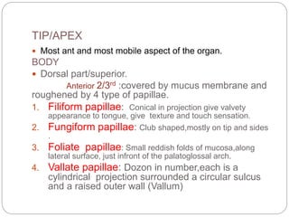
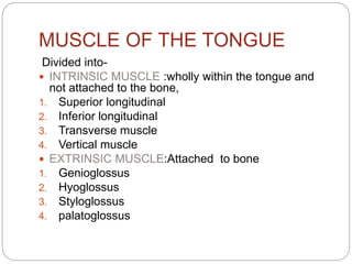
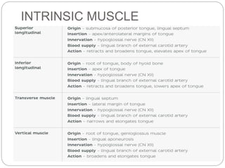
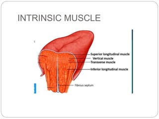
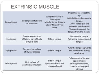
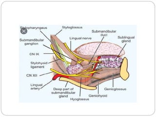
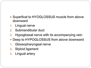
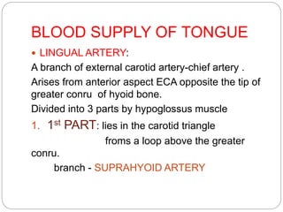
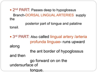
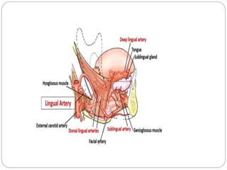
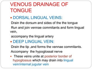
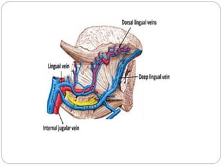
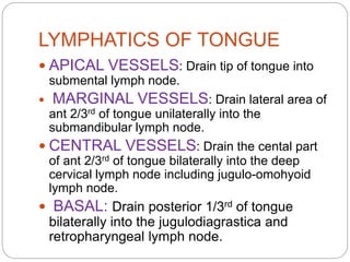
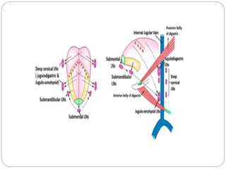
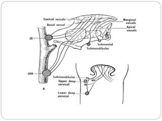
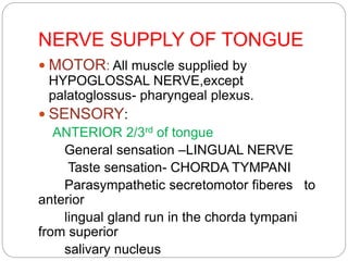
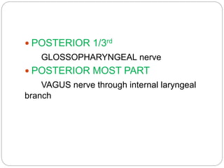
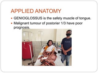
DR. CHIRANJIV BARUAH
PGT RADIATION
ONCOLOGY
ANATOMY OF TONGUE
DEVELOPMENT OF TONGUE
 Begins in 4th week .
 Pharyngeal arches 1-4 froms the mucosa.
 Occipital somites froms musculature.
MUCOSAL DEVELOPMENT
 The lateral lingual swelling overgrow the
tuberculum impar and merge together forming
mucosa of ant 2/3rd .
 Within the cupula the 3rd arches overgrows the
2nd arches formimg mucosa of the posterior 1/3rd .
 Posterior most part include valleculla from 4th
arches
Musculature development
 From the occipital somites which are the
segments of mesoderm.
 Somites migrate from the neck anteriorly to give
rise to muscles of the the tongue.
 Supplied by the hypoglossal nerve.
ANATOMY OF TONGUE
 Tongue is a muscular organ covered by mucus
membrane.
 Anterior 2/3rd (oral or presulcal part).
 Posterior 1/3(pharyngeal or postsulcal part).
 Sulcus terminalis is the junction of oral and
pharyngeal part.
 It has -Apex/tip
-Body having rough dorsal/superior part
and
smooth ventral/inferior part
-Base
-Root.
TIP/APEX
 Most ant and most mobile aspect of the organ.
BODY
 Dorsal part/superior.
Anterior 2/3rd :covered by mucus membrane and
roughened by 4 type of papillae.
1. Filiform papillae: Conical in projection give valvety
appearance to tongue, give texture and touch sensation.
2. Fungiform papillae: Club shaped,mostly on tip and sides
.
3. Foliate papillae: Small reddish folds of mucosa,along
lateral surface, just infront of the palatoglossal arch.
4. Vallate papillae: Dozon in number,each is a
cylindrical projection surrounded a circular sulcus
and a raised outer wall (Vallum)
 There are mucus and serious glands under the
tips and sides.
 On the under surface behind the tip there is a
rather large mixed gland on each side of midline
–Ant lingual gland.
 RANULA –Retention cyst of this gland.
Posterior 1/3rd
 Ant wall of oropharynx.
 Extends from sulcus terminalis and epiglotis.
 Foramen caecum: small depression at apex of
sulcus- upper end of the thyroglossal duct
 Smooth mucus membrane has a nadular
appearance constitute the lingual tonsil- part of
waldeyer”s ring.
 Between tongue and epiglotiss- median
glossoepiglottic fold.
 Lateral glossoepiglottic fold – extending from the
side of epiglottis to the wall of the pharynx.
 Inferior
surface/smooth
ventral part:
Lateral to lingual frenulum
we
have deep lingual vein
(visible),Lingual
artery/nerve
(not visible).
Farther lateral is ant fold
of
mucosa -fimbricated .
Some foliate papillae are
also
 Base :
postsulcal tongue attached to the floor of oral cavity.
populated by numerous lymphoid aggregate known
as
lingual tomsil.
 Root :
Part of post sulcal tongue attached to the floor of oral
cavity
MUSCLE OF THE TONGUE
Divided into-
 INTRINSIC MUSCLE :wholly within the tongue and
not attached to the bone,
1. Superior longitudinal
2. Inferior longitudinal
3. Transverse muscle
4. Vertical muscle
 EXTRINSIC MUSCLE:Attached to bone
1. Genioglossus
2. Hyoglossus
3. Styloglossus
4. palatoglossus
INTRINSIC MUSCLE
INTRINSIC MUSCLE
EXTRINSIC MUSCLE
 Superfical to HYOGLOSSUS muscle from above
downward
1. Lingual nerve
2. Submandibular duct
3. Hypoglossal nerve with its accompanying vein
 Deep to HYPOGLOSSUS from above downward
1. Glossopharyngeal nerve
2. Styloid ligament
3. Lingual artery
BLOOD SUPPLY OF TONGUE
 LINGUAL ARTERY:
A branch of external carotid artery-chief artery .
Arises from anterior aspect ECA opposite the tip of
greater conru of hyoid bone.
Divided into 3 parts by hypoglossus muscle
1. 1st PART: lies in the carotid triangle
froms a loop above the greater
conru.
branch - SUPRAHYOID ARTERY
 2nd PART: Passes deep to hypoglossus
Branch-DORSAL LINGUAL ARTERIES supply
the
posterior part of tongue and palatine
tonsil.
 3rd PART: Also called lingual artery /arteria
profunda linguae- runs upward
along
the ant border of hypoglossus
and then
go forward on on the
undersurface of
tongue.
VENOUS DRAINAGE OF
TONGUE
 DORSAL LINGUAL VEINS:
Drain the dorsum and sides of the the tongue
Run and join vennae commitants and form lingual
vein,
accompany the lingual artery
 DEEP LINGUAL VEIN:
Drain the tip ,and forms the vennae commitants.
Accompany the hypoglossal nerve
 These veins unite at posterior border of
hypoglossus which may drain into lingual
vein/internal jugular vein
LYMPHATICS OF TONGUE
 APICAL VESSELS: Drain tip of tongue into
submental lymph node.
 MARGINAL VESSELS: Drain lateral area of
ant 2/3rd of tongue unilaterally into the
submandibular lymph node.
 CENTRAL VESSELS: Drain the cental part
of ant 2/3rd of tongue bilaterally into the deep
cervical lymph node including jugulo-omohyoid
lymph node.
 BASAL: Drain posterior 1/3rd of tongue
bilaterally into the jugulodiagrastica and
retropharyngeal lymph node.
NERVE SUPPLY OF TONGUE
 MOTOR: All muscle supplied by
HYPOGLOSSAL NERVE,except
palatoglossus- pharyngeal plexus.
 SENSORY:
ANTERIOR 2/3rd of tongue
General sensation –LINGUAL NERVE
Taste sensation- CHORDA TYMPANI
Parasympathetic secretomotor fiberes to
anterior
lingual gland run in the chorda tympani
from superior
salivary nucleus
 POSTERIOR 1/3rd
GLOSSOPHARYNGEAL nerve
 POSTERIOR MOST PART
VAGUS nerve through internal laryngeal
branch
 GENIOGLOSSUS is the safety muscle of tongue.
 Malignant tumour of postorier 1/3 have poor
prognosis.
APPLIED ANATOMY
THANK YOU

Tongue

  • 1.
    DR. CHIRANJIV BARUAH PGTRADIATION ONCOLOGY ANATOMY OF TONGUE
  • 2.
    DEVELOPMENT OF TONGUE Begins in 4th week .  Pharyngeal arches 1-4 froms the mucosa.  Occipital somites froms musculature.
  • 3.
    MUCOSAL DEVELOPMENT  Thelateral lingual swelling overgrow the tuberculum impar and merge together forming mucosa of ant 2/3rd .  Within the cupula the 3rd arches overgrows the 2nd arches formimg mucosa of the posterior 1/3rd .  Posterior most part include valleculla from 4th arches
  • 5.
    Musculature development  Fromthe occipital somites which are the segments of mesoderm.  Somites migrate from the neck anteriorly to give rise to muscles of the the tongue.  Supplied by the hypoglossal nerve.
  • 6.
    ANATOMY OF TONGUE Tongue is a muscular organ covered by mucus membrane.  Anterior 2/3rd (oral or presulcal part).  Posterior 1/3(pharyngeal or postsulcal part).  Sulcus terminalis is the junction of oral and pharyngeal part.  It has -Apex/tip -Body having rough dorsal/superior part and smooth ventral/inferior part -Base -Root.
  • 8.
    TIP/APEX  Most antand most mobile aspect of the organ. BODY  Dorsal part/superior. Anterior 2/3rd :covered by mucus membrane and roughened by 4 type of papillae. 1. Filiform papillae: Conical in projection give valvety appearance to tongue, give texture and touch sensation. 2. Fungiform papillae: Club shaped,mostly on tip and sides . 3. Foliate papillae: Small reddish folds of mucosa,along lateral surface, just infront of the palatoglossal arch. 4. Vallate papillae: Dozon in number,each is a cylindrical projection surrounded a circular sulcus and a raised outer wall (Vallum)
  • 10.
     There aremucus and serious glands under the tips and sides.  On the under surface behind the tip there is a rather large mixed gland on each side of midline –Ant lingual gland.  RANULA –Retention cyst of this gland.
  • 11.
    Posterior 1/3rd  Antwall of oropharynx.  Extends from sulcus terminalis and epiglotis.  Foramen caecum: small depression at apex of sulcus- upper end of the thyroglossal duct  Smooth mucus membrane has a nadular appearance constitute the lingual tonsil- part of waldeyer”s ring.  Between tongue and epiglotiss- median glossoepiglottic fold.  Lateral glossoepiglottic fold – extending from the side of epiglottis to the wall of the pharynx.
  • 12.
     Inferior surface/smooth ventral part: Lateralto lingual frenulum we have deep lingual vein (visible),Lingual artery/nerve (not visible). Farther lateral is ant fold of mucosa -fimbricated . Some foliate papillae are also
  • 13.
     Base : postsulcaltongue attached to the floor of oral cavity. populated by numerous lymphoid aggregate known as lingual tomsil.  Root : Part of post sulcal tongue attached to the floor of oral cavity
  • 14.
    MUSCLE OF THETONGUE Divided into-  INTRINSIC MUSCLE :wholly within the tongue and not attached to the bone, 1. Superior longitudinal 2. Inferior longitudinal 3. Transverse muscle 4. Vertical muscle  EXTRINSIC MUSCLE:Attached to bone 1. Genioglossus 2. Hyoglossus 3. Styloglossus 4. palatoglossus
  • 16.
  • 17.
  • 18.
  • 20.
     Superfical toHYOGLOSSUS muscle from above downward 1. Lingual nerve 2. Submandibular duct 3. Hypoglossal nerve with its accompanying vein  Deep to HYPOGLOSSUS from above downward 1. Glossopharyngeal nerve 2. Styloid ligament 3. Lingual artery
  • 21.
    BLOOD SUPPLY OFTONGUE  LINGUAL ARTERY: A branch of external carotid artery-chief artery . Arises from anterior aspect ECA opposite the tip of greater conru of hyoid bone. Divided into 3 parts by hypoglossus muscle 1. 1st PART: lies in the carotid triangle froms a loop above the greater conru. branch - SUPRAHYOID ARTERY
  • 22.
     2nd PART:Passes deep to hypoglossus Branch-DORSAL LINGUAL ARTERIES supply the posterior part of tongue and palatine tonsil.  3rd PART: Also called lingual artery /arteria profunda linguae- runs upward along the ant border of hypoglossus and then go forward on on the undersurface of tongue.
  • 24.
    VENOUS DRAINAGE OF TONGUE DORSAL LINGUAL VEINS: Drain the dorsum and sides of the the tongue Run and join vennae commitants and form lingual vein, accompany the lingual artery  DEEP LINGUAL VEIN: Drain the tip ,and forms the vennae commitants. Accompany the hypoglossal nerve  These veins unite at posterior border of hypoglossus which may drain into lingual vein/internal jugular vein
  • 26.
    LYMPHATICS OF TONGUE APICAL VESSELS: Drain tip of tongue into submental lymph node.  MARGINAL VESSELS: Drain lateral area of ant 2/3rd of tongue unilaterally into the submandibular lymph node.  CENTRAL VESSELS: Drain the cental part of ant 2/3rd of tongue bilaterally into the deep cervical lymph node including jugulo-omohyoid lymph node.  BASAL: Drain posterior 1/3rd of tongue bilaterally into the jugulodiagrastica and retropharyngeal lymph node.
  • 29.
    NERVE SUPPLY OFTONGUE  MOTOR: All muscle supplied by HYPOGLOSSAL NERVE,except palatoglossus- pharyngeal plexus.  SENSORY: ANTERIOR 2/3rd of tongue General sensation –LINGUAL NERVE Taste sensation- CHORDA TYMPANI Parasympathetic secretomotor fiberes to anterior lingual gland run in the chorda tympani from superior salivary nucleus
  • 30.
     POSTERIOR 1/3rd GLOSSOPHARYNGEALnerve  POSTERIOR MOST PART VAGUS nerve through internal laryngeal branch
  • 32.
     GENIOGLOSSUS isthe safety muscle of tongue.  Malignant tumour of postorier 1/3 have poor prognosis. APPLIED ANATOMY
  • 33.