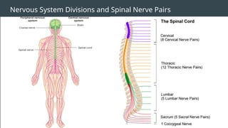
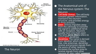
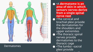
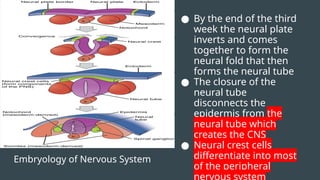
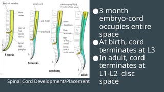
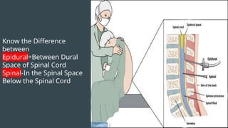
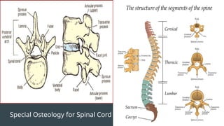
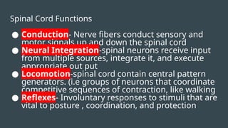
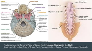
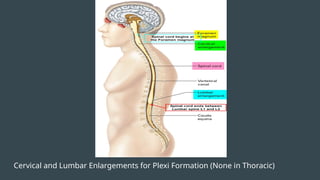
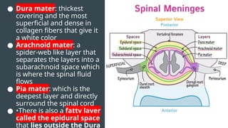
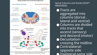
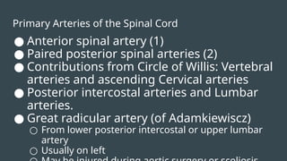
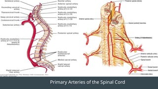
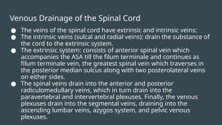
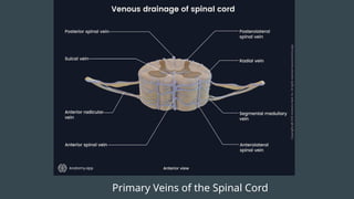
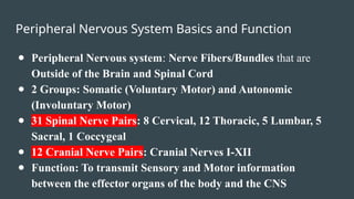
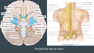
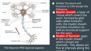
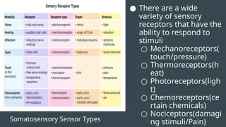
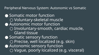
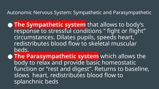
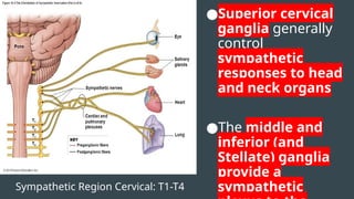
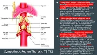
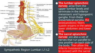
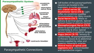
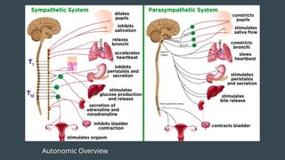
Nervous System
Nervous System Basics
● Central Nervous System: Made up of Brain and Spinal
Cord
● Peripheral Nervous system: Nerve Fibers/Bundles that
are Outside of the Brain and Spinal Cord
● 31 Spinal Nerve Pairs: 8 Cervical, 12 Thoracic, 5
Lumbar, 5 Sacral, 1 Coccygeal
● 12 Cranial Nerve Pairs: Cranial Nerves I-XII
Nervous System Divisions and Spinal Nerve Pairs
The Neuron
● The Anatomical unit of
the Nervous system: The
Neuron
● Cell body (Soma): The cell body
contains genetic information,
maintains the neuron’s
structure, and provides energy
to drive activities.
● Axon: Directs impulses away
from the cell body to transmit
the signal to additional nerve
cells or receptors. Neurons
usually have one main axon
● The term nerve fibers refers to
axons NOT dendrites
● Dendrites: The name is derived
from ‘dendron’ which means
tree. Like antennae, dendrites
receive and process signals from
the axons of other neurons.
● Increases the surface area to
receive signals from other
Dermatomes
● •A dermatome is an
area of skin in which
sensory nerves derive
from a single spinal
nerve root
● •The cervical and
brachial plexi provide
the dermatomes for
the shoulders and
upper extremites
● •The thoracic spinal
nerves provide
dermatomes to the
thoracic cage
● •The lumbo –sacral
plexi provide
Embryology of Nervous System
● By the end of the third
week the neural plate
inverts and comes
together to form the
neural fold that then
forms the neural tube
● The closure of the
neural tube
disconnects the
epidermis from the
neural tube which
creates the CNS
● Neural crest cells
differentiate into most
of the peripheral
Spinal Cord Development/Placement
●3 month
embryo-cord
occupies entire
space
●At birth, cord
terminates at L3
●In adult, cord
terminates at
L1-L2 disc
space
Know the Difference
between
Epidural=Between Dural
Space of Spinal Cord
Spinal-In the Spinal Space
Below the Spinal Cord
Special Osteology for Spinal Cord
Spinal Cord Functions
● Conduction- Nerve fibers conduct sensory and
motor signals up and down the spinal cord
● Neural Integration-spinal neurons receive input
from multiple sources, integrate it, and execute
appropriate out put
● Locomotion-spinal cord contain central pattern
generators. (i.e groups of neurons that coordinate
competitive sequences of contraction, like walking
● Reflexes- Involuntary responses to stimuli that are
vital to posture , coordination, and protection
Anatomic Superior Terminal Point of Spinal Cord: Foramen Magnum in the Skull
Anatomic Inferior End Point of Spinal Cord: Conus medullaris, Cauda Equina, Fillamentum Terminale
Cervical and Lumbar Enlargements for Plexi Formation (None in Thoracic)
● Dura mater: thickest
covering and the most
superficial and dense in
collagen fibers that give it
a white color
● Arachnoid mater: a
spider-web like layer that
separates the layers into a
subarachnoid space which
is where the spinal fluid
flows
● Pia mater: which is the
deepest layer and directly
surround the spinal cord
● •There is also a fatty layer
called the epidural space
that lies outside the Dura
Spinal Columns and Nuclei (DON’T
MEMORIZE)
● Tracts are
aggregated into
columns (dorsal ,
lateral and ventral)
● Columns are divided
into tracts that
ascend (sensory)
and descend (motor)
● Decusitation-
crossing the midline
● Contralateral-
opposite side
Primary Arteries of the Spinal Cord
● Anterior spinal artery (1)
● Paired posterior spinal arteries (2)
● Contributions from Circle of Willis: Vertebral
arteries and ascending Cervical arteries
● Posterior intercostal arteries and Lumbar
arteries.
● Great radicular artery (of Adamkiewiscz)
○ From lower posterior intercostal or upper lumbar
artery
○ Usually on left
Primary Arteries of the Spinal Cord
Venous Drainage of the Spinal Cord
● The veins of the spinal cord have extrinsic and intrinsic veins:
● The intrinsic veins (sulcal and radial veins): drain the substance of
the cord to the extrinsic system.
● The extrinsic system: consists of anterior spinal vein which
accompanies the ASA till the filum terminale and continues as
filum terminale vein, the greatest spinal vein which traverses in
the posterior median sulcus along with two posterolateral veins
on either sides.
● The spinal veins drain into the anterior and posterior
radiculomedullary veins, which in turn drain into the
paravertebral and intervertebral plexuses. Finally, the venous
plexuses drain into the segmental veins, draining into the
ascending lumbar veins, azygos system, and pelvic venous
plexuses.
Primary Veins of the Spinal Cord
Peripheral Nervous System Basics and Function
● Peripheral Nervous system: Nerve Fibers/Bundles that are
Outside of the Brain and Spinal Cord
● 2 Groups: Somatic (Voluntary Motor) and Autonomic
(Involuntary Motor)
● 31 Spinal Nerve Pairs: 8 Cervical, 12 Thoracic, 5 Lumbar, 5
Sacral, 1 Coccygeal
● 12 Cranial Nerve Pairs: Cranial Nerves I-XII
● Function: To transmit Sensory and Motor information
between the effector organs of the body and the CNS
Peripheral Nerve Pairs
The Neuron-PNS Special aspects
● Similar Structure and
Function to CNS except the
following:
● Myelin sheath: a layer of
tissue which covers the
axon. Formed by glial
cells called Schwann
cells, the myelin sheath
provides insulation as
well as structural support
for the axon.
● Nodes of Ranvier: gaps
in the myelin sheath
which contain ion
channels. This allows ion
flux at intervals along the
Peripheral Nerve Anatomy
● Each peripheral nerve is a
bundle of neural axons,
encased in a sheet of
connective tissue and
aggregated in groups called
fascicles
● Peripheral nerve fascicles are
the equivalent of spinal tracts
in the CNS
● AXONAL INJURY
● Following transection or other
serious injury, that portion of
the axon distal to the injury
will undergo degeneration,
known as anterograde or
Wallerian degeneration.
● The axon stump proximal to the
injury will sprout and regrow at
the rate of 1-2 mm/day. If the
regenerating nerve fibers re-
enter the endoneural tube,
Remember SAME DAVE for PNS root Functions
●S=
sensory
●A=afferent
●M= motor
●E=efferent
D= dorsal
A= afferent
V= ventral
E= efferent
Peripheral Nervous System Afferent vs. Efferent
● Afferent nerve fibers transmit data from receptors in the
periphery (touch, temperature, vibration, proprioception)
to the central nervous system. It gives us the ability to
sense and perceive our surroundings. PERCEPTION
● Efferent nerve fibers transmit both conscious and
unconscious instructions from the central nervous
system to motor cells (including vasomotor and
secretomotor cells) in the periphery. This allows us to
physically (voluntarily or involuntarily) respond to the
perception input. RESPONSE
Somatosensory System
●The somatosensory
system is the part of
the sensory system
concerned with the
conscious perception
of touch, pressure,
pain, temperature,
position, movement,
and vibration, which
arise from the
muscles, joints, skin,
and fascia.
●This is where the signal
(sense) in perceived
and processed
Somatosensory Sensor Types
● There are a wide
variety of sensory
receptors that have the
ability to respond to
stimuli
○ Mechanoreceptors(
touch/pressure)
○ Thermoreceptors(h
eat)
○ Photoreceptors(ligh
t)
○ Chemoreceptors(ce
rtain chemicals)
○ Nociceptors(damagi
ng stimuli/Pain)
Peripheral Nervous System: Autonomic vs Somatic
● Somatic motor function
○ Voluntary-skeletal muscle
● Autonomic motor function
○ Involuntary-smooth, cardiac muscle,
Gland tissue
● Somatic sensory function
○ Precise, well localized (e.g. skin)
● Autonomic sensory function
○ Vague, poorly localized (e.g. visceral)
Autonomic Nervous System: Sympathetic and Parasympathetic
● The Sympathetic system that allows to body’s
response to stressful conditions “ fight or flight”
circumstances. Dilates pupils, speeds heart,
redistributes blood flow to skeletal muscular
beds.
● The Parasympathetic system which allows the
body to relax and provide basic homeostatic
function or “rest and digest”. Returns to baseline,
slows heart, redistributes blood flow to
splanchnic beds
Sympathetic Region Cervical: T1-T4
●Superior cervical
ganglia generally
control
sympathetic
responses to head
and neck organs
●The middle and
inferior (and
Stellate) ganglia
provide a
sympathetic
Sympathetic Region Thoracic: T5-T12
● T5-T9 ganglia greater splanchnic nerve: helps
with the motility of the foregut and provides
sympathetic innervation to the adrenal
medulla. Specifically, it supplies the stomach
duodenum, liver, gallbladder, pancreas,
adrenal medulla, and the spleen. Nerve
descends from thorax, pierces the crus of the
diaphragm and synapses primarily in the
celiac ganglion
● T10-T11 ganglia lesser splanchnic nerve:
courses lateral and in parallel to the greater
splanchnic nerves, over the anterior surface of
the spine and enters the abdomen in the
same manner - by traversing the ipsilateral
diaphragmatic crus. The lesser splanchnic
nerves terminate by synapsing within the
aorticorenal or superior mesenteric ganglia.
The corresponding postsynaptic fibers then
modulate the activity of the midgut
● T12 ganglion least splanchnic nerve: travel
lateral and parallel to the lesser splanchnic
nerves, over the anterior surface of the spine,
close to its lateral margin. It enters the
abdomen by passing beneath the medial
arcuate ligament and then joins the aorto-
renal ganglia, contributing to the sympathetic
innervation for the renal plexus. The fibers of
this nerve are sometimes joined by the lesser
splanchnic nerve. In this case, the lesser
splanchnic nerve gives off small branches to
Sympathetic Region Lumbar: L1-L2
● The lumbar splanchnic
nerves: arise from the
upper lumbar levels and
terminate in the inferior
mesenteric and hypogastric
ganglia. From these
prevertebral ganglia, the
postganglionic fibers
supply organs in the pelvis,
lower abdomen and lower
limb.
● The sacral splanchnic
nerves: are also a set of
nerves that run parallel to
one another on each side of
the body. They allow the
inferior hypogastric plexus
to connect with the sacral
Parasympathetic Connections
● Cell bodies of the parasympathetic
neurons are located in the
brainstem, in nuclei associated
with cranial nerves III,
Oculomotor nerve (CN 3): Light
reflex via constrictor pupillae
Accommodation via ciliary muscle
● Facial Nerve (CN 7): Motor to
salivary & lacrimal glands. Special
sensory (taste) to anterior tongue
● Glossopharyngeal Nerve (CN 9):
Motor to parotid gland. Sensory to
carotid body & sinus. Special
sensory (taste) to posterior tongue
● Vagus Nerve CN 10): Sensory to
carotid body. All parasympathetics
to thoracic viscera & to abdominal
viscera supplied by celiac and
superior mesenteric arteries
● Ventral horns of spinal cord
segments S2-4: control bladder
and reproductive organs
Autonomic Overview

Anatomy study guide nervous system pa school

  • 1.
  • 2.
    Nervous System Basics ●Central Nervous System: Made up of Brain and Spinal Cord ● Peripheral Nervous system: Nerve Fibers/Bundles that are Outside of the Brain and Spinal Cord ● 31 Spinal Nerve Pairs: 8 Cervical, 12 Thoracic, 5 Lumbar, 5 Sacral, 1 Coccygeal ● 12 Cranial Nerve Pairs: Cranial Nerves I-XII
  • 3.
    Nervous System Divisionsand Spinal Nerve Pairs
  • 4.
    The Neuron ● TheAnatomical unit of the Nervous system: The Neuron ● Cell body (Soma): The cell body contains genetic information, maintains the neuron’s structure, and provides energy to drive activities. ● Axon: Directs impulses away from the cell body to transmit the signal to additional nerve cells or receptors. Neurons usually have one main axon ● The term nerve fibers refers to axons NOT dendrites ● Dendrites: The name is derived from ‘dendron’ which means tree. Like antennae, dendrites receive and process signals from the axons of other neurons. ● Increases the surface area to receive signals from other
  • 5.
    Dermatomes ● •A dermatomeis an area of skin in which sensory nerves derive from a single spinal nerve root ● •The cervical and brachial plexi provide the dermatomes for the shoulders and upper extremites ● •The thoracic spinal nerves provide dermatomes to the thoracic cage ● •The lumbo –sacral plexi provide
  • 6.
    Embryology of NervousSystem ● By the end of the third week the neural plate inverts and comes together to form the neural fold that then forms the neural tube ● The closure of the neural tube disconnects the epidermis from the neural tube which creates the CNS ● Neural crest cells differentiate into most of the peripheral
  • 7.
    Spinal Cord Development/Placement ●3month embryo-cord occupies entire space ●At birth, cord terminates at L3 ●In adult, cord terminates at L1-L2 disc space
  • 8.
    Know the Difference between Epidural=BetweenDural Space of Spinal Cord Spinal-In the Spinal Space Below the Spinal Cord
  • 9.
  • 10.
    Spinal Cord Functions ●Conduction- Nerve fibers conduct sensory and motor signals up and down the spinal cord ● Neural Integration-spinal neurons receive input from multiple sources, integrate it, and execute appropriate out put ● Locomotion-spinal cord contain central pattern generators. (i.e groups of neurons that coordinate competitive sequences of contraction, like walking ● Reflexes- Involuntary responses to stimuli that are vital to posture , coordination, and protection
  • 11.
    Anatomic Superior TerminalPoint of Spinal Cord: Foramen Magnum in the Skull Anatomic Inferior End Point of Spinal Cord: Conus medullaris, Cauda Equina, Fillamentum Terminale
  • 12.
    Cervical and LumbarEnlargements for Plexi Formation (None in Thoracic)
  • 13.
    ● Dura mater:thickest covering and the most superficial and dense in collagen fibers that give it a white color ● Arachnoid mater: a spider-web like layer that separates the layers into a subarachnoid space which is where the spinal fluid flows ● Pia mater: which is the deepest layer and directly surround the spinal cord ● •There is also a fatty layer called the epidural space that lies outside the Dura
  • 14.
    Spinal Columns andNuclei (DON’T MEMORIZE) ● Tracts are aggregated into columns (dorsal , lateral and ventral) ● Columns are divided into tracts that ascend (sensory) and descend (motor) ● Decusitation- crossing the midline ● Contralateral- opposite side
  • 15.
    Primary Arteries ofthe Spinal Cord ● Anterior spinal artery (1) ● Paired posterior spinal arteries (2) ● Contributions from Circle of Willis: Vertebral arteries and ascending Cervical arteries ● Posterior intercostal arteries and Lumbar arteries. ● Great radicular artery (of Adamkiewiscz) ○ From lower posterior intercostal or upper lumbar artery ○ Usually on left
  • 16.
    Primary Arteries ofthe Spinal Cord
  • 17.
    Venous Drainage ofthe Spinal Cord ● The veins of the spinal cord have extrinsic and intrinsic veins: ● The intrinsic veins (sulcal and radial veins): drain the substance of the cord to the extrinsic system. ● The extrinsic system: consists of anterior spinal vein which accompanies the ASA till the filum terminale and continues as filum terminale vein, the greatest spinal vein which traverses in the posterior median sulcus along with two posterolateral veins on either sides. ● The spinal veins drain into the anterior and posterior radiculomedullary veins, which in turn drain into the paravertebral and intervertebral plexuses. Finally, the venous plexuses drain into the segmental veins, draining into the ascending lumbar veins, azygos system, and pelvic venous plexuses.
  • 18.
    Primary Veins ofthe Spinal Cord
  • 19.
    Peripheral Nervous SystemBasics and Function ● Peripheral Nervous system: Nerve Fibers/Bundles that are Outside of the Brain and Spinal Cord ● 2 Groups: Somatic (Voluntary Motor) and Autonomic (Involuntary Motor) ● 31 Spinal Nerve Pairs: 8 Cervical, 12 Thoracic, 5 Lumbar, 5 Sacral, 1 Coccygeal ● 12 Cranial Nerve Pairs: Cranial Nerves I-XII ● Function: To transmit Sensory and Motor information between the effector organs of the body and the CNS
  • 20.
  • 21.
    The Neuron-PNS Specialaspects ● Similar Structure and Function to CNS except the following: ● Myelin sheath: a layer of tissue which covers the axon. Formed by glial cells called Schwann cells, the myelin sheath provides insulation as well as structural support for the axon. ● Nodes of Ranvier: gaps in the myelin sheath which contain ion channels. This allows ion flux at intervals along the
  • 22.
    Peripheral Nerve Anatomy ●Each peripheral nerve is a bundle of neural axons, encased in a sheet of connective tissue and aggregated in groups called fascicles ● Peripheral nerve fascicles are the equivalent of spinal tracts in the CNS ● AXONAL INJURY ● Following transection or other serious injury, that portion of the axon distal to the injury will undergo degeneration, known as anterograde or Wallerian degeneration. ● The axon stump proximal to the injury will sprout and regrow at the rate of 1-2 mm/day. If the regenerating nerve fibers re- enter the endoneural tube,
  • 23.
    Remember SAME DAVEfor PNS root Functions ●S= sensory ●A=afferent ●M= motor ●E=efferent D= dorsal A= afferent V= ventral E= efferent
  • 24.
    Peripheral Nervous SystemAfferent vs. Efferent ● Afferent nerve fibers transmit data from receptors in the periphery (touch, temperature, vibration, proprioception) to the central nervous system. It gives us the ability to sense and perceive our surroundings. PERCEPTION ● Efferent nerve fibers transmit both conscious and unconscious instructions from the central nervous system to motor cells (including vasomotor and secretomotor cells) in the periphery. This allows us to physically (voluntarily or involuntarily) respond to the perception input. RESPONSE
  • 25.
    Somatosensory System ●The somatosensory systemis the part of the sensory system concerned with the conscious perception of touch, pressure, pain, temperature, position, movement, and vibration, which arise from the muscles, joints, skin, and fascia. ●This is where the signal (sense) in perceived and processed
  • 26.
    Somatosensory Sensor Types ●There are a wide variety of sensory receptors that have the ability to respond to stimuli ○ Mechanoreceptors( touch/pressure) ○ Thermoreceptors(h eat) ○ Photoreceptors(ligh t) ○ Chemoreceptors(ce rtain chemicals) ○ Nociceptors(damagi ng stimuli/Pain)
  • 27.
    Peripheral Nervous System:Autonomic vs Somatic ● Somatic motor function ○ Voluntary-skeletal muscle ● Autonomic motor function ○ Involuntary-smooth, cardiac muscle, Gland tissue ● Somatic sensory function ○ Precise, well localized (e.g. skin) ● Autonomic sensory function ○ Vague, poorly localized (e.g. visceral)
  • 28.
    Autonomic Nervous System:Sympathetic and Parasympathetic ● The Sympathetic system that allows to body’s response to stressful conditions “ fight or flight” circumstances. Dilates pupils, speeds heart, redistributes blood flow to skeletal muscular beds. ● The Parasympathetic system which allows the body to relax and provide basic homeostatic function or “rest and digest”. Returns to baseline, slows heart, redistributes blood flow to splanchnic beds
  • 29.
    Sympathetic Region Cervical:T1-T4 ●Superior cervical ganglia generally control sympathetic responses to head and neck organs ●The middle and inferior (and Stellate) ganglia provide a sympathetic
  • 30.
    Sympathetic Region Thoracic:T5-T12 ● T5-T9 ganglia greater splanchnic nerve: helps with the motility of the foregut and provides sympathetic innervation to the adrenal medulla. Specifically, it supplies the stomach duodenum, liver, gallbladder, pancreas, adrenal medulla, and the spleen. Nerve descends from thorax, pierces the crus of the diaphragm and synapses primarily in the celiac ganglion ● T10-T11 ganglia lesser splanchnic nerve: courses lateral and in parallel to the greater splanchnic nerves, over the anterior surface of the spine and enters the abdomen in the same manner - by traversing the ipsilateral diaphragmatic crus. The lesser splanchnic nerves terminate by synapsing within the aorticorenal or superior mesenteric ganglia. The corresponding postsynaptic fibers then modulate the activity of the midgut ● T12 ganglion least splanchnic nerve: travel lateral and parallel to the lesser splanchnic nerves, over the anterior surface of the spine, close to its lateral margin. It enters the abdomen by passing beneath the medial arcuate ligament and then joins the aorto- renal ganglia, contributing to the sympathetic innervation for the renal plexus. The fibers of this nerve are sometimes joined by the lesser splanchnic nerve. In this case, the lesser splanchnic nerve gives off small branches to
  • 31.
    Sympathetic Region Lumbar:L1-L2 ● The lumbar splanchnic nerves: arise from the upper lumbar levels and terminate in the inferior mesenteric and hypogastric ganglia. From these prevertebral ganglia, the postganglionic fibers supply organs in the pelvis, lower abdomen and lower limb. ● The sacral splanchnic nerves: are also a set of nerves that run parallel to one another on each side of the body. They allow the inferior hypogastric plexus to connect with the sacral
  • 32.
    Parasympathetic Connections ● Cellbodies of the parasympathetic neurons are located in the brainstem, in nuclei associated with cranial nerves III, Oculomotor nerve (CN 3): Light reflex via constrictor pupillae Accommodation via ciliary muscle ● Facial Nerve (CN 7): Motor to salivary & lacrimal glands. Special sensory (taste) to anterior tongue ● Glossopharyngeal Nerve (CN 9): Motor to parotid gland. Sensory to carotid body & sinus. Special sensory (taste) to posterior tongue ● Vagus Nerve CN 10): Sensory to carotid body. All parasympathetics to thoracic viscera & to abdominal viscera supplied by celiac and superior mesenteric arteries ● Ventral horns of spinal cord segments S2-4: control bladder and reproductive organs
  • 33.