Report
Share

Recommended
Differential diagnosis of chest pain
This document discusses the differential diagnosis of chest pain by listing common cardiac, respiratory, gastrointestinal, and musculoskeletal causes. Key cardiac causes mentioned are angina, myocardial infarction, mitral valve prolapse, and pericarditis. Common respiratory causes listed are pleurisy, pneumothorax, and pulmonary embolism. Gastrointestinal causes such as reflux esophagitis and diffuse esophageal spasm are also summarized. The document provides brief descriptions of symptoms, signs, and investigations for each of these differential diagnoses of chest pain.
Pericarditis
Pericarditis is the inflammation of the pericardium, a thin, two-layered sac that surrounds your heart. The layers have a small amount of fluid between them to prevent friction when the heart beats. When the layers are inflamed, it can result in chest pain.
How to approach a patient with chest pain
History, Examination and Investigations of patient with chest pain and the differentials you can come up with.
Cardiac Versus Non Cardiac
There is no definitive way to differentiate between cardiac and non-cardiac chest pain without further evaluation. Severe chest pain should always be taken seriously and evaluated in an emergency department. Cardiac chest pain is caused by reduced blood flow and oxygen to the heart, which can lead to a heart attack without treatment. Non-cardiac chest pain can be due to structures in the chest like the esophagus or lungs. A medical history and physical exam can provide clues but further testing is often needed to determine the underlying cause of chest pain.
Chest Pain- Differential Diagnosis
This document discusses the differential diagnosis of chest pain by describing various cardiac and non-cardiac causes. It outlines key factors in the history and physical exam that can help determine if chest pain is typical angina, atypical angina, or non-anginal. Common cardiac causes discussed include acute coronary syndromes, aortic dissection, and pericarditis. Common non-cardiac causes discussed include pulmonary embolism, pneumonia, gastrointestinal issues like pancreatitis and peptic ulcer disease. Diagnostic tests for different conditions are also mentioned.
Approach to chest pain
This document provides guidance on evaluating and treating chest pain. It outlines many potential causes of chest pain including cardiac, pulmonary, gastrointestinal and musculoskeletal issues. For serious potential causes like myocardial ischemia, pericarditis or aortic dissection, the document describes relevant history, examination findings and initial investigations. It emphasizes doing an ECG, reviewing risk factors and considering stress testing to evaluate for cardiac ischemia as a common cause of chest pain. The document also reviews palpitations as an unrelated symptom and potential cardiac and non-cardiac causes.
Chest pain differential diagnosis
1. Acute coronary syndrome can present with substernal chest pressure, pain radiating to the shoulders, or pain with exertion. ECG may show ST elevations or new left bundle branch block indicating AMI. Troponin and CK-MB elevations are needed to diagnose AMI.
2. Aortic dissection often presents with sudden, severe chest pain and may be suggested on ECG by discrepancies in blood pressure between arms or signs of ischemia. Chest X-ray may show a widened mediastinum.
3. Pulmonary embolism presentations can vary widely but often include dyspnea. ECG may show signs of right heart strain. Most chest X-rays are normal but some show
Differential Dx Chest Pain
The document discusses the differential diagnosis of chest pain. It notes that the chest x-ray and history are the most important tests to evaluate non-cardiac chest pain. The history requires a meticulous examination of all relevant details. Many life-threatening and non-life threatening potential causes of chest pain are outlined, including cardiac, pulmonary, gastrointestinal, musculoskeletal, psychiatric, and neurological conditions. Thoracic outlet syndromes are given a detailed differential diagnosis. The evaluation of chest pain is emphasized to be complex, requiring a defined diagnosis beyond simply ruling out heart attack.
Chest pain
Chest pain can have many potential causes. A thorough history and physical exam are important to help determine the likely diagnosis and guide appropriate testing. Key aspects of the history include characteristics of the pain such as location, radiation, onset and nature. The physical exam focuses on identifying potential causes of the pain or associated symptoms. Initial tests may include an ECG, blood tests, chest x-ray and echocardiogram to help differentiate causes such as heart disease, pulmonary embolism, pneumonia or musculoskeletal issues. Further tests are guided by the initial findings.
Approach to chest pain
This document provides guidance on evaluating chest pain and discusses the approach to diagnosing aortic dissection. It emphasizes maintaining a high index of suspicion for aortic dissection as the symptoms can mimic other conditions. Aortic dissection often presents with sudden, severe chest or back pain and may migrate. Examination findings like pulse/blood pressure differences between limbs can help but have low sensitivity. The document reviews risk factors and recommends promptly ordering tests like CT scans to diagnose this dangerous condition given the high mortality if left untreated.
Approach chest pain & acs
The document provides information on evaluating and managing patients presenting with chest pain and acute coronary syndrome. It discusses how to take a clinical history to determine the nature and cause of chest pain. Differential diagnoses are provided for cardiac and non-cardiac causes. Acute coronary syndrome is described as encompassing unstable angina, NSTEMI, and STEMI, which present with chest pain at rest or minimal exertion due to plaque rupture or erosion in coronary arteries. Immediate management in the first 12 hours and long-term management are outlined.
Approach to a patient with chest pain
This document provides information on evaluating and diagnosing chest pain. It discusses:
- The importance of history taking in determining the cause of chest pain.
- Common life-threatening causes of chest pain like myocardial infarction, aortic dissection, pulmonary embolism, and tension pneumothorax.
- How to assess chest pain through physical examination, ECG, cardiac enzymes, imaging studies, and other investigations.
- Distinguishing cardiac from non-cardiac chest pain and differentiating conditions like angina, unstable angina, and STEMI based on features.
Chest Pain
This document discusses chest pain, including its definition, location, potential causes, assessment process (using PQRST), and initial management. Chest pain can be caused by life-threatening conditions like a heart attack, pulmonary embolism, or aortic dissection. The assessment of a patient presenting with chest pain involves taking a medical history and examining the quality, location, radiation, timing and associated symptoms of the pain, as well as performing physical assessments like vital signs, electrocardiogram, chest x-ray and blood tests to check for potential cardiac issues. The initial management focuses on stabilizing the patient, administering oxygen, monitoring vitals, performing an electrocardiogram and blood tests, providing pain relief, and determining
L 6.approach to chest pain
This document provides guidance on evaluating and diagnosing chest pain. It begins by defining chest pain and noting that it is a common reason patients present for medical care. A list of 10 questions for history taking on chest pain is then provided related to onset, location, character, duration, radiation, aggravating/relieving factors, tenderness, associated symptoms and severity. Causes of chest pain are discussed, distinguishing ischemic causes like angina and myocardial infarction from non-ischemic causes. Characteristics of cardiac pain like gradual onset and associated sweating or nausea are outlined. Differential diagnoses including aortic dissection, pulmonary embolism, pericarditis and esophageal spasm are also reviewed. An approach to diagnosis
Chest pain
This document provides guidance on evaluating and diagnosing the cause of chest pain. It notes that the majority of chest pain is non-cardiac in origin but can be disabling. A thorough history, physical exam, ECG and imaging can identify most causes, including life-threatening ones like myocardial infarction that require immediate treatment. The description of chest pain symptoms and related factors can provide clues to potential etiologies such as cardiac, pulmonary, gastrointestinal or musculoskeletal origin.
Chest pain emergencies
This document discusses the approach, differential diagnosis, initial tests, diagnostic tests, temporary treatment, and definitive management for three cases of chest pain. Case 1 involves a 48-year-old male with sudden severe chest pain. Case 2 is a 72-year-old female with chest discomfort and breathlessness. Case 3 is a 37-year-old female with central chest pain and history of breast cancer. Common differential diagnoses for chest pain include acute myocardial infarction, aortic dissection, pulmonary embolism, pneumonia, and perforated viscus. The document outlines the clinical clues, initial tests like ECG and CXR, additional diagnostic tests, and treatment strategies for each case and potential diagnosis.
Chest pain 2009 ppt
This document discusses differentiating between cardiac and non-cardiac causes of chest pain. Cardiac causes include ischemic issues like myocardial infarction and non-ischemic problems like pericarditis. Non-cardiac causes range from gastrointestinal issues, pulmonary problems, musculoskeletal pain and even shingles. It is important to thoroughly assess patients experiencing chest pain to determine location, quality and accompanying symptoms to correctly identify potential life-threatening issues and provide appropriate intervention.
Classification, FetaL Circulation and TOF.pptx
This document discusses cyanotic cardiac defects, fetal circulation, and tetralogy of Fallot. It classifies cyanotic diseases based on reduced or increased pulmonary blood flow. Fetal circulation is described. The key features of tetralogy of Fallot are outlined as severe right ventricle outflow obstruction, large ventricular septal defect, overriding aorta, and right ventricular hypertrophy. Complications include anoxic spells, neurological issues, and congestive heart failure if left untreated. Treatment options are discussed as total surgical correction or palliative shunt procedures.
12 cardio-infectious
This document discusses several cardiovascular disorders related to disturbances in oxygen transport, including structural heart valve disorders like mitral valve prolapse, mitral regurgitation, and mitral stenosis. It also covers infective endocarditis, rheumatic heart disease, myocarditis, pericarditis, aortic aneurysm, and vascular disorders like Buerger's disease, Raynaud's disease, and venous thrombosis. For each condition, it provides information on clinical manifestations, diagnosis, and treatment approaches.
Palpitation
This document discusses palpitations, which refer to abnormal awareness of one's heartbeat. Palpitations can be caused by rapid, slow, or irregular heart rhythms and may result from primary cardiac diseases or systemic conditions affecting the heart. Common causes include anxiety, hyperthyroidism, caffeine, smoking, sinus tachycardia, supraventricular tachycardia, ventricular tachycardia, atrial fibrillation, extrasystoles, and Wolff-Parkinson-White syndrome. A thorough history and electrocardiogram can help diagnose the underlying rhythm abnormality.
17 pericardial disease
The document summarizes different types of pericardial diseases. It describes the normal anatomy and functions of the pericardium. It then discusses various pericardial conditions such as acute pericarditis, pericardial effusion, constrictive pericarditis and their causes, symptoms, diagnostic criteria and treatments. Acute pericarditis is usually caused by viral or bacterial infections and presents with chest pain and pericardial friction rub. Constrictive pericarditis occurs after acute pericarditis and causes decreased diastolic filling through pericardial thickening and fibrosis.
Cardiology 1.1. Chest pain - by Dr. Farjad Ikram
Introduction to one of the most common symptoms that can represent a wide range of diseases, from benign to life-threatening, covering number of systems including gastrointestinal, cardiovascular, pulmonary, musculoskeletal and psychiatric. Includes a brief explanation of anti-anginal therapy.
Template design credits - http://www.slidescarnival.com
Evaluation of syncope in adults
This document discusses the evaluation of syncope in adults. Syncope is defined as a brief, self-limited loss of consciousness due to decreased blood flow to the brain. The causes of syncope can be categorized as neurally-mediated, orthostatic, cardiac, or structural/cardiopulmonary. A thorough history, physical exam, and diagnostic testing are needed to determine the underlying cause and guide treatment. The history provides clues to distinguish syncope from other conditions and identify risk factors, while the physical exam focuses on vital signs and signs of end-organ damage or dysfunction.
Cardiac tamponade
Pericardial effusion and cardiac tamponade are conditions caused by an abnormal accumulation of fluid in the pericardial space. Cardiac tamponade occurs when excess fluid buildup leads to reduced ventricular filling and hemodynamic compromise. Symptoms include breathlessness, chest pain, and hypotension. Diagnosis is made through ECG, echocardiogram, and x-ray. Treatment depends on severity but may include medications, drainage of fluid via pericardiocentesis, or surgery. Cardiac tamponade requires urgent pericardiocentesis to drain fluid and prevent further hemodynamic compromise.
Rhuematic heart disease
Rheumatic heart disease is a chronic condition caused by rheumatic fever that results in scarring and deformity of the heart valves. It occurs due to acute rheumatic fever and recurrent streptococcal infections, which can cause inflammation of the heart muscles and valves, leading to thickened and stenotic valves with regurgitation. Diagnosis involves echocardiogram, chest x-ray, and ECG showing prolonged PR interval or delayed AV conduction. Management includes medical treatment, nursing care like assessments, and health promotion to prevent recurrent infections.
Syncope Presentation
Syncope, or transient loss of consciousness, can be caused by various cardiac and non-cardiac conditions. A thorough history, physical exam, ECG and diagnostic testing are needed to evaluate the cause. Patients found to have cardiac syncope, abnormal vital signs, ECG changes or structural heart disease have a higher risk of adverse outcomes and should be admitted. Risk stratification tools like the San Francisco Syncope Rule and Oesil Risk Score can help determine which low-risk patients can be safely discharged. High-risk features predicting serious underlying rhythm issues include age over 45, history of heart disease or abnormal ECG.
TACHYCARDIA IN ECG ,CAUSES AND MANAGEMENT OF TACHYARRHYTHMIAS
Tachycardia can be caused by physiological factors like infancy, childhood, emotion, exertion, pregnancy, or pathological factors such as arrhythmias, heart disease, myocardial infarction, anxiety, heart failure, shock, low blood volume, drugs, chronic lung disease, blood clots, or pain. Some specific arrhythmias mentioned include AVNRT, AVRT, AT, SANRT, AF, and VT.
Final pericardial effusion
This document provides an overview of pericardial diseases. It begins with the anatomy and functions of the pericardium. It then discusses various pericardial diseases like acute pericarditis, pericardial effusion, and cardiac tamponade. For acute pericarditis, it describes the key symptoms of chest pain, pericardial friction rub, and ECG changes. It also outlines the diagnostic criteria and treatment approaches for pericardial effusion and cardiac tamponade, including the use of echocardiography, medications, and pericardiocentesis.
aorticregurgitation anuradha mam.docx
Aortic regurgitation is a condition where the aortic valve leaks, allowing blood to flow back into the left ventricle during diastole. It can be caused by conditions affecting the aortic valve or root. Over time, the left ventricle undergoes remodeling to compensate for the increased volume load. Clinical features include a diastolic murmur, widened pulse pressure, and later symptoms of heart failure. Treatment depends on severity but may involve surgery to repair or replace the leaky valve.
ARTERIAL PULSES.pptx
Clinical examination in cardiolgy starts with the examination of arterial pulse.
In this PowerPoint presentation we have discussed all about this important topic
More Related Content
What's hot
Chest pain
Chest pain can have many potential causes. A thorough history and physical exam are important to help determine the likely diagnosis and guide appropriate testing. Key aspects of the history include characteristics of the pain such as location, radiation, onset and nature. The physical exam focuses on identifying potential causes of the pain or associated symptoms. Initial tests may include an ECG, blood tests, chest x-ray and echocardiogram to help differentiate causes such as heart disease, pulmonary embolism, pneumonia or musculoskeletal issues. Further tests are guided by the initial findings.
Approach to chest pain
This document provides guidance on evaluating chest pain and discusses the approach to diagnosing aortic dissection. It emphasizes maintaining a high index of suspicion for aortic dissection as the symptoms can mimic other conditions. Aortic dissection often presents with sudden, severe chest or back pain and may migrate. Examination findings like pulse/blood pressure differences between limbs can help but have low sensitivity. The document reviews risk factors and recommends promptly ordering tests like CT scans to diagnose this dangerous condition given the high mortality if left untreated.
Approach chest pain & acs
The document provides information on evaluating and managing patients presenting with chest pain and acute coronary syndrome. It discusses how to take a clinical history to determine the nature and cause of chest pain. Differential diagnoses are provided for cardiac and non-cardiac causes. Acute coronary syndrome is described as encompassing unstable angina, NSTEMI, and STEMI, which present with chest pain at rest or minimal exertion due to plaque rupture or erosion in coronary arteries. Immediate management in the first 12 hours and long-term management are outlined.
Approach to a patient with chest pain
This document provides information on evaluating and diagnosing chest pain. It discusses:
- The importance of history taking in determining the cause of chest pain.
- Common life-threatening causes of chest pain like myocardial infarction, aortic dissection, pulmonary embolism, and tension pneumothorax.
- How to assess chest pain through physical examination, ECG, cardiac enzymes, imaging studies, and other investigations.
- Distinguishing cardiac from non-cardiac chest pain and differentiating conditions like angina, unstable angina, and STEMI based on features.
Chest Pain
This document discusses chest pain, including its definition, location, potential causes, assessment process (using PQRST), and initial management. Chest pain can be caused by life-threatening conditions like a heart attack, pulmonary embolism, or aortic dissection. The assessment of a patient presenting with chest pain involves taking a medical history and examining the quality, location, radiation, timing and associated symptoms of the pain, as well as performing physical assessments like vital signs, electrocardiogram, chest x-ray and blood tests to check for potential cardiac issues. The initial management focuses on stabilizing the patient, administering oxygen, monitoring vitals, performing an electrocardiogram and blood tests, providing pain relief, and determining
L 6.approach to chest pain
This document provides guidance on evaluating and diagnosing chest pain. It begins by defining chest pain and noting that it is a common reason patients present for medical care. A list of 10 questions for history taking on chest pain is then provided related to onset, location, character, duration, radiation, aggravating/relieving factors, tenderness, associated symptoms and severity. Causes of chest pain are discussed, distinguishing ischemic causes like angina and myocardial infarction from non-ischemic causes. Characteristics of cardiac pain like gradual onset and associated sweating or nausea are outlined. Differential diagnoses including aortic dissection, pulmonary embolism, pericarditis and esophageal spasm are also reviewed. An approach to diagnosis
Chest pain
This document provides guidance on evaluating and diagnosing the cause of chest pain. It notes that the majority of chest pain is non-cardiac in origin but can be disabling. A thorough history, physical exam, ECG and imaging can identify most causes, including life-threatening ones like myocardial infarction that require immediate treatment. The description of chest pain symptoms and related factors can provide clues to potential etiologies such as cardiac, pulmonary, gastrointestinal or musculoskeletal origin.
Chest pain emergencies
This document discusses the approach, differential diagnosis, initial tests, diagnostic tests, temporary treatment, and definitive management for three cases of chest pain. Case 1 involves a 48-year-old male with sudden severe chest pain. Case 2 is a 72-year-old female with chest discomfort and breathlessness. Case 3 is a 37-year-old female with central chest pain and history of breast cancer. Common differential diagnoses for chest pain include acute myocardial infarction, aortic dissection, pulmonary embolism, pneumonia, and perforated viscus. The document outlines the clinical clues, initial tests like ECG and CXR, additional diagnostic tests, and treatment strategies for each case and potential diagnosis.
Chest pain 2009 ppt
This document discusses differentiating between cardiac and non-cardiac causes of chest pain. Cardiac causes include ischemic issues like myocardial infarction and non-ischemic problems like pericarditis. Non-cardiac causes range from gastrointestinal issues, pulmonary problems, musculoskeletal pain and even shingles. It is important to thoroughly assess patients experiencing chest pain to determine location, quality and accompanying symptoms to correctly identify potential life-threatening issues and provide appropriate intervention.
What's hot (9)
Similar to Perikarditis
Classification, FetaL Circulation and TOF.pptx
This document discusses cyanotic cardiac defects, fetal circulation, and tetralogy of Fallot. It classifies cyanotic diseases based on reduced or increased pulmonary blood flow. Fetal circulation is described. The key features of tetralogy of Fallot are outlined as severe right ventricle outflow obstruction, large ventricular septal defect, overriding aorta, and right ventricular hypertrophy. Complications include anoxic spells, neurological issues, and congestive heart failure if left untreated. Treatment options are discussed as total surgical correction or palliative shunt procedures.
12 cardio-infectious
This document discusses several cardiovascular disorders related to disturbances in oxygen transport, including structural heart valve disorders like mitral valve prolapse, mitral regurgitation, and mitral stenosis. It also covers infective endocarditis, rheumatic heart disease, myocarditis, pericarditis, aortic aneurysm, and vascular disorders like Buerger's disease, Raynaud's disease, and venous thrombosis. For each condition, it provides information on clinical manifestations, diagnosis, and treatment approaches.
Palpitation
This document discusses palpitations, which refer to abnormal awareness of one's heartbeat. Palpitations can be caused by rapid, slow, or irregular heart rhythms and may result from primary cardiac diseases or systemic conditions affecting the heart. Common causes include anxiety, hyperthyroidism, caffeine, smoking, sinus tachycardia, supraventricular tachycardia, ventricular tachycardia, atrial fibrillation, extrasystoles, and Wolff-Parkinson-White syndrome. A thorough history and electrocardiogram can help diagnose the underlying rhythm abnormality.
17 pericardial disease
The document summarizes different types of pericardial diseases. It describes the normal anatomy and functions of the pericardium. It then discusses various pericardial conditions such as acute pericarditis, pericardial effusion, constrictive pericarditis and their causes, symptoms, diagnostic criteria and treatments. Acute pericarditis is usually caused by viral or bacterial infections and presents with chest pain and pericardial friction rub. Constrictive pericarditis occurs after acute pericarditis and causes decreased diastolic filling through pericardial thickening and fibrosis.
Cardiology 1.1. Chest pain - by Dr. Farjad Ikram
Introduction to one of the most common symptoms that can represent a wide range of diseases, from benign to life-threatening, covering number of systems including gastrointestinal, cardiovascular, pulmonary, musculoskeletal and psychiatric. Includes a brief explanation of anti-anginal therapy.
Template design credits - http://www.slidescarnival.com
Evaluation of syncope in adults
This document discusses the evaluation of syncope in adults. Syncope is defined as a brief, self-limited loss of consciousness due to decreased blood flow to the brain. The causes of syncope can be categorized as neurally-mediated, orthostatic, cardiac, or structural/cardiopulmonary. A thorough history, physical exam, and diagnostic testing are needed to determine the underlying cause and guide treatment. The history provides clues to distinguish syncope from other conditions and identify risk factors, while the physical exam focuses on vital signs and signs of end-organ damage or dysfunction.
Cardiac tamponade
Pericardial effusion and cardiac tamponade are conditions caused by an abnormal accumulation of fluid in the pericardial space. Cardiac tamponade occurs when excess fluid buildup leads to reduced ventricular filling and hemodynamic compromise. Symptoms include breathlessness, chest pain, and hypotension. Diagnosis is made through ECG, echocardiogram, and x-ray. Treatment depends on severity but may include medications, drainage of fluid via pericardiocentesis, or surgery. Cardiac tamponade requires urgent pericardiocentesis to drain fluid and prevent further hemodynamic compromise.
Rhuematic heart disease
Rheumatic heart disease is a chronic condition caused by rheumatic fever that results in scarring and deformity of the heart valves. It occurs due to acute rheumatic fever and recurrent streptococcal infections, which can cause inflammation of the heart muscles and valves, leading to thickened and stenotic valves with regurgitation. Diagnosis involves echocardiogram, chest x-ray, and ECG showing prolonged PR interval or delayed AV conduction. Management includes medical treatment, nursing care like assessments, and health promotion to prevent recurrent infections.
Syncope Presentation
Syncope, or transient loss of consciousness, can be caused by various cardiac and non-cardiac conditions. A thorough history, physical exam, ECG and diagnostic testing are needed to evaluate the cause. Patients found to have cardiac syncope, abnormal vital signs, ECG changes or structural heart disease have a higher risk of adverse outcomes and should be admitted. Risk stratification tools like the San Francisco Syncope Rule and Oesil Risk Score can help determine which low-risk patients can be safely discharged. High-risk features predicting serious underlying rhythm issues include age over 45, history of heart disease or abnormal ECG.
TACHYCARDIA IN ECG ,CAUSES AND MANAGEMENT OF TACHYARRHYTHMIAS
Tachycardia can be caused by physiological factors like infancy, childhood, emotion, exertion, pregnancy, or pathological factors such as arrhythmias, heart disease, myocardial infarction, anxiety, heart failure, shock, low blood volume, drugs, chronic lung disease, blood clots, or pain. Some specific arrhythmias mentioned include AVNRT, AVRT, AT, SANRT, AF, and VT.
Final pericardial effusion
This document provides an overview of pericardial diseases. It begins with the anatomy and functions of the pericardium. It then discusses various pericardial diseases like acute pericarditis, pericardial effusion, and cardiac tamponade. For acute pericarditis, it describes the key symptoms of chest pain, pericardial friction rub, and ECG changes. It also outlines the diagnostic criteria and treatment approaches for pericardial effusion and cardiac tamponade, including the use of echocardiography, medications, and pericardiocentesis.
aorticregurgitation anuradha mam.docx
Aortic regurgitation is a condition where the aortic valve leaks, allowing blood to flow back into the left ventricle during diastole. It can be caused by conditions affecting the aortic valve or root. Over time, the left ventricle undergoes remodeling to compensate for the increased volume load. Clinical features include a diastolic murmur, widened pulse pressure, and later symptoms of heart failure. Treatment depends on severity but may involve surgery to repair or replace the leaky valve.
ARTERIAL PULSES.pptx
Clinical examination in cardiolgy starts with the examination of arterial pulse.
In this PowerPoint presentation we have discussed all about this important topic
ACUTE ABDOMENAL PAIN.pptx
This presentation is about the differential diagnosis of abdominal pain and how to manage the different diseases, presenting as acute abdominal pain.
Pericarditis
Pericarditis is inflammation of the pericardium that commonly occurs between ages 20-50. It is usually caused by viral infections but can also be due to other infections, immune reactions, medications, or unknown causes. Diagnosis requires at least two of four criteria - chest pain, pericardial friction rub, ECG changes, or pericardial effusion on echo. Treatment focuses on relieving pain, treating the underlying cause, and preventing complications like cardiac tamponade through NSAIDs, colchicine, steroids, or pericardiocentesis.
Chest pain structured approach
by Dr. Anas Babiker, Consulant Cardiologist, Alzaytouna Hospital
& Academic Secretary of the Sudan Heart Society
Acute renal failure (2)
This document discusses acute renal failure (ARF), also known as acute kidney injury (AKI). It defines ARF, discusses its epidemiology and causes. The main causes of ARF are pre-renal (decreased blood flow/volume), renal (damage within the kidneys), and post-renal (obstruction of urine flow). The most common form of intrinsic ARF is acute tubular necrosis, often due to ischemia or nephrotoxins. Diagnosis involves lab tests of kidney function and urine analysis. Treatment focuses on identifying and reversing the underlying cause, maintaining fluid/electrolyte balance, and potentially initiating renal replacement therapy like dialysis.
Coronary heart disease
Coronary heart disease is a condition caused by an inadequate blood supply to the heart muscle. It occurs when there is an imbalance between the heart's oxygen supply and demand. The main coronary arteries supply blood to the heart and can become narrowed or blocked by atherosclerosis.
Risk factors include age, male sex, family history, smoking, high cholesterol, hypertension, diabetes and obesity. Symptoms range from stable angina to acute coronary syndromes like heart attack. Diagnosis involves evaluating the medical history, symptoms, electrocardiogram and cardiac enzyme levels. Treatment depends on the type and severity of coronary heart disease.
DISEASES OF PERICARDIUM BY DRV AILU SHASHANK
The document discusses diseases of the pericardium, including acute pericarditis, constrictive pericarditis, pericardial effusion, and cardiac tamponade. It describes the anatomy and functions of the pericardium, symptoms and signs of different pericardial diseases, diagnostic tests including ECG, echo, CT and treatment approaches.
Arrhythmias 1DR NIKUNJ R SHEKHADA (MBBS,MS GEN SURG DNB CTS SR)
This document summarizes cardiac arrhythmias and the normal cardiac conduction system. It describes how arrhythmias occur due to problems in the sinus node, atrial cells, AV junction, or ventricular cells. Common arrhythmias discussed include sinus bradycardia, sinus tachycardia, atrial fibrillation, atrial flutter, supraventricular tachycardia, heart block, and bundle branch block. Causes, characteristics, and treatment approaches are provided for each type of arrhythmia. The normal cardiac conduction pathway and components of the ECG are also outlined.
Similar to Perikarditis (20)
TACHYCARDIA IN ECG ,CAUSES AND MANAGEMENT OF TACHYARRHYTHMIAS
TACHYCARDIA IN ECG ,CAUSES AND MANAGEMENT OF TACHYARRHYTHMIAS
Arrhythmias 1DR NIKUNJ R SHEKHADA (MBBS,MS GEN SURG DNB CTS SR)
Arrhythmias 1DR NIKUNJ R SHEKHADA (MBBS,MS GEN SURG DNB CTS SR)
More from JelenaNastasic
More from JelenaNastasic (8)
Recently uploaded
Pollock and Snow "DEIA in the Scholarly Landscape, Session One: Setting Expec...
Pollock and Snow "DEIA in the Scholarly Landscape, Session One: Setting Expec...National Information Standards Organization (NISO)
This presentation was provided by Steph Pollock of The American Psychological Association’s Journals Program, and Damita Snow, of The American Society of Civil Engineers (ASCE), for the initial session of NISO's 2024 Training Series "DEIA in the Scholarly Landscape." Session One: 'Setting Expectations: a DEIA Primer,' was held June 6, 2024.What is Digital Literacy? A guest blog from Andy McLaughlin, University of Ab...
What is Digital Literacy? A guest blog from Andy McLaughlin, University of Aberdeen
BBR 2024 Summer Sessions Interview Training
Qualitative research interview training by Professor Katrina Pritchard and Dr Helen Williams
How to Manage Your Lost Opportunities in Odoo 17 CRM
Odoo 17 CRM allows us to track why we lose sales opportunities with "Lost Reasons." This helps analyze our sales process and identify areas for improvement. Here's how to configure lost reasons in Odoo 17 CRM
DRUGS AND ITS classification slide share
Any substance (other than food) that is used to prevent, diagnose, treat, or relieve symptoms of a
disease or abnormal condition
Exploiting Artificial Intelligence for Empowering Researchers and Faculty, In...
Exploiting Artificial Intelligence for Empowering Researchers and Faculty, In...Dr. Vinod Kumar Kanvaria
Exploiting Artificial Intelligence for Empowering Researchers and Faculty,
International FDP on Fundamentals of Research in Social Sciences
at Integral University, Lucknow, 06.06.2024
By Dr. Vinod Kumar KanvariaPengantar Penggunaan Flutter - Dart programming language1.pptx
Pengantar Penggunaan Flutter - Dart programming language1.pptx
The simplified electron and muon model, Oscillating Spacetime: The Foundation...
Discover the Simplified Electron and Muon Model: A New Wave-Based Approach to Understanding Particles delves into a groundbreaking theory that presents electrons and muons as rotating soliton waves within oscillating spacetime. Geared towards students, researchers, and science buffs, this book breaks down complex ideas into simple explanations. It covers topics such as electron waves, temporal dynamics, and the implications of this model on particle physics. With clear illustrations and easy-to-follow explanations, readers will gain a new outlook on the universe's fundamental nature.
Digital Artifact 1 - 10VCD Environments Unit
Digital Artifact 1 - 10VCD Environments Unit - NGV Pavilion Concept Design
ANATOMY AND BIOMECHANICS OF HIP JOINT.pdf
it describes the bony anatomy including the femoral head , acetabulum, labrum . also discusses the capsule , ligaments . muscle that act on the hip joint and the range of motion are outlined. factors affecting hip joint stability and weight transmission through the joint are summarized.
Hindi varnamala | hindi alphabet PPT.pdf
हिंदी वर्णमाला पीपीटी, hindi alphabet PPT presentation, hindi varnamala PPT, Hindi Varnamala pdf, हिंदी स्वर, हिंदी व्यंजन, sikhiye hindi varnmala, dr. mulla adam ali, hindi language and literature, hindi alphabet with drawing, hindi alphabet pdf, hindi varnamala for childrens, hindi language, hindi varnamala practice for kids, https://www.drmullaadamali.com
The History of Stoke Newington Street Names
Presented at the Stoke Newington Literary Festival on 9th June 2024
www.StokeNewingtonHistory.com
Advanced Java[Extra Concepts, Not Difficult].docx
This is part 2 of my Java Learning Journey. This contains Hashing, ArrayList, LinkedList, Date and Time Classes, Calendar Class and more.
Recently uploaded (20)
Pollock and Snow "DEIA in the Scholarly Landscape, Session One: Setting Expec...
Pollock and Snow "DEIA in the Scholarly Landscape, Session One: Setting Expec...
What is Digital Literacy? A guest blog from Andy McLaughlin, University of Ab...
What is Digital Literacy? A guest blog from Andy McLaughlin, University of Ab...
Liberal Approach to the Study of Indian Politics.pdf
Liberal Approach to the Study of Indian Politics.pdf
How to Manage Your Lost Opportunities in Odoo 17 CRM
How to Manage Your Lost Opportunities in Odoo 17 CRM
Exploiting Artificial Intelligence for Empowering Researchers and Faculty, In...
Exploiting Artificial Intelligence for Empowering Researchers and Faculty, In...
Pengantar Penggunaan Flutter - Dart programming language1.pptx
Pengantar Penggunaan Flutter - Dart programming language1.pptx
The simplified electron and muon model, Oscillating Spacetime: The Foundation...
The simplified electron and muon model, Oscillating Spacetime: The Foundation...
Film vocab for eal 3 students: Australia the movie
Film vocab for eal 3 students: Australia the movie
Digital Artefact 1 - Tiny Home Environmental Design
Digital Artefact 1 - Tiny Home Environmental Design
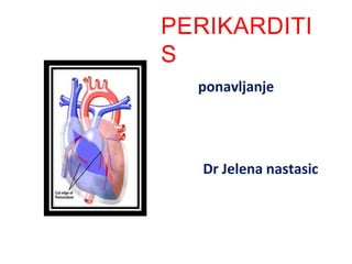
Perikarditis
- 2. DEFINICIJA •Zapaljenje srcane ovojnice(perikarda) •Moze biti:akutni i hronicni.
- 3. Gradjasrca
- 5. •AKUTNI PERIKARDITIS •Uzrok:bakterije,virusi,razni alergeni •Kl.slika:povisena T,jeza,drhtavica,bol iza grudne kosti •Dijagnoza: Lecenje:AB •auskultacija(trenje) simptomatsko •RTG •Punkcija(gnojav sadrzaj)
- 6. • HRONICNI PERIKARDITIS • Uzrok:kao posledica akutnog • Kl.slika:perikard postaje fibrozno zadebljan,srascuje za srce,u nega se taloze soli Ca i stvaraju oklop oko srca.Simptomi:brzo zamaranje,dispneja,ascites,povecana jetra Dijagnoza: • auskultacija(oslabljeni tonovi,perikardno trenje) • RTG-kalcifikacije • EKG-niska voltaza • Biopsija perikarda,angiokardiografija • Lecenje: • Hirursko(perikardiektomija)
- 7. EKG Electrocardiogram in acute pericarditis showing diffuse upsloping ST segment elevations seen best here in leads II, III, aVF, and V2 to V6. There is also subtle PR segment deviation (positive in aVR, negative in most other leads). ST segment elevation is due to a ventricular current of injury associated with epicardial inflammation; similarly, the PR segment changes are due to an atrial current of injury which, in Pericarditis, typically displaces the PR segment upward in lead aVR and downward in most other leads.
- 8. Pericardial Effusion Cardiomegaly due to a massive pericardial effusion. At least 200 mL of pericardial fluid must accumulate before the cardiac silhouette enlarges.