PEDIATRIC RESPIRATORY EMERGENCIES By Duangruethai  Tunprom, MD. 3 rd  years emergency medical resident PMK hospital
OUTLINE Upper airway obstruction & infection Lower airway obstruction Disease of the lung
ASTHMA History Physical examination Diagnostic strategies Pluse oximetry & ABG Peak expiratory flow rate CXR
MANAGEMENT APPROACH BASED ON CONTROL ADULTS AND CHILDREN OLDER THAN 5 YEARS
 
ESTIMATED EQUIPMENT DAILY DOSES OF INHALED GLUCOCORTICOSTEROIDS FOR ADULTS AND CHILDREN OLDER THAN 5 YEARS
 
SEVERITY OF ASTHMA EXACERBATIONS 1
SEVERITY OF ASTHMA EXACERBATIONS 2
SEVERITY OF ASTHMA EXACERBATIONS 3
DIFFERENTIAL DIAGNOSIS OF ASTHMA Condition Distinguishing characteristics Infection Anatomic /Congenital Acquired
DIFFERENTIAL DIAGNOSIS OF ASTHMA Condition Distinguishing characteristics Infection Bronchiolitis Laryngotracheobronchitis(croup) Pneumonia Tuberculosis Bronchiolitis obliterans Infant, preceding URI, sesonal, no history of atopy,  no family history of asthma Inspiratory stridor, barking cough, fever, response to humidified air Focal wheezing, rhonchi, rales, grunting, fever Diffuse adenopathy, weight loss, prolong fever Prolnged cough and/or chest pain,inhalational exposure to toxin Anatomic /Congenital Acquired
DIFFERENTIAL DIAGNOSIS OF ASTHMA Condition Distinguishing characteristics Infection Anatomic /Congenital GERD Cystic fibrosis Congestive heart failure Tracheoesophageal fistula Mediastinal mass Vascular ring Frequent emesis, weight loss, aspiration Diarrhea, weight loss, chronic cough, salty sweat Rales, murmur, gallop, hepatosplenomegaly, cardiomegaly and/or pulmonary vascular congestion on CXR Chocking, coughing, cyanosis with feeds Chest pain, mediastinal density on CXR Stridor, cyanosis, apnea, high piched brassy cough, dysphagia Acquired
DIFFERENTIAL DIAGNOSIS OF ASTHMA Condition Distinguishing characteristics Infection Anatomic /Congenital Acquired Foreign body aspiration Anaphylaxis History of choking, toddler, asymmetrical pulmonary examination, unilateral hyperinflation on CXR Abrupt onset, urticarial rash, angioedema, history of allergies
ED MANAGEMENT OF ACUTE ASTHMA
ED MANAGEMENT OF ACUTE ASTHMA Mental status Heart rate Oxygen saturation Degree of wheezing Inspiratory:expiratory ratio Accessory muscle use PEFR
ED MANAGEMENT OF ACUTE ASTHMA SABA by NEB or MDI-S Up to 3 SABA doses in 1 st  hour Oral corticosteroids if incomplete response or if already taking at home
ED MANAGEMENT OF ACUTE ASTHMA D/C  SABA q 4 hrs for 3-4 days Continue corticosteriod 3-10 days,if begun in ED Continue controller medications Initiate ICS if persistent asthma
ED MANAGEMENT OF ACUTE ASTHMA SABA  and IB  by NEB  in 1 st  hour Oral corticosteroids  If emesis,  consider  parenteral corticosteriods
ED MANAGEMENT OF ACUTE ASTHMA Continue SABA q 30-45 mins or continuously Observe for 60-90 mins after SABA end If sustained response: D/C If minimal or no improvement : admit
ED MANAGEMENT OF ACUTE ASTHMA Continue  SABA & IB by NEB —consider heliox Consider SQ or IM epinephrine or terbutaline IV corticosteroids  Magnesium sulfate IV SABA infusion
ED MANAGEMENT OF ACUTE ASTHMA Admit to PICU : Continued severe respiratory distress Altered mental status Need for IV SABA Need for mechanical ventilation Admit others to hospital ward
RECOMMENED DOSES OF MEDICATIONS FOR ACUTE ASTHMA Drug Dose Albuterol 0.15 mg/kg/dose (0.03ml/kg/dose,max 1.0ml) < 10 kg  2.5  mg (0.5ml) 11-19kg  3.75 mg (0.75ml) > 20 kg  5  mg (1.0ml) Continuous albuterol 1.0 mg/kg/hr by NB (max 20 mg/hr)  < 10 kg  10 mg/hr (2 ml/hr) 10-20 kg  15 mg/hr (3 ml/hr) > 20 kg  20 mg/hr (4 ml/hr) Albuterol by MDI Dose is not well established < 10 kg  2-4 puffs 11-19kg  4-6 puffs > 20 kg  6-8 puffs Levalbuterol Half the recommended albuterol doses Ipratropium bromide < 20 kg  250 µg/dose > 20 kg  500 µg/dose
RECOMMENED DOSES OF MEDICATIONS FOR ACUTE ASTHMA(CONT.) Drug Dose L-epinephrine (1:1000) or terbutaline (1.0 mg/mg) 0.01ml/kg/dose SC or IM (max 0.4 ml) May be repeated every 10-15 min IV terbutaline 10 µg/kg bolus over 10 min,then 0.1-0.3 µg/kg/min infusion Every 30 min,may increase infusion by 0.3 µg/kg/min to a max of 5 µg/kg/min Prednisolone 2 mg/kg (max 60mg), in ED 1 mg/kg/dose bid, home therapy Dexamethasone 0.6mg/kg PO, 2 dose 24 hrs apart IV methylprednisolone 1-2 mg/kg (max 125 mg) IV magnesium sulfate 50-75 mg/kg over 20 min(max 2.5g)
MILD SABA if not improve then q 20 mins in 1 st  hours Mouthpiecess preferable for older child  Decrease nasal filtering of drug Spacer must take 5-8 breaths to complete empty spacer If 3 dose   reevaluate
SHORT ACTING BETA2-AGONISTS IN ACUTE ASTHMA IB= ipratropium bromide MDI-s=metered dose inhaler with spacer NHLBI=national,Heart,Lung and blood institute Mild Moderate Severe Delivery method Intermittent NEB or MDI-S Continuous by NEB for 1 hr,  then reassess Consider SC or IM  Continuous by NEB Comments Most patients will need 1-2 treatments Allow for MDI-S teaching No IB needed Not superior to MDI-S Easier to adhere to NHLBI guidelines for the first hr of therapy Concurrent IB therapy is more easily delivered Better outcomes in severe asthma
BRONCHIOLITIS
BRONCHIOLITIS Seasonal disease : November &April Nearly all children : affected virus cause bronchiolitis at least once during first 2 years of life More common < 1 year
Increased mucus secretion, cell death, and sloughing,  a peribronchiolar lymphocytic infiltrate  and submucosal edema Combination of debris&edema produces    critical narrowing & obstruction of small airways .
BRONCHIOLITIS M/C CAUSE:  RESPIRATORY SYNCYTIAL VIRUS (RSV) Age < 3 mo Preterm GA < 34 wks Toxic or ill general appearance RR > 70/min SpO2 <95% CXR : atelectasis Breast feeding < 1 mo Maternal smoking RSV subtype A Male Air pollution environment Severity associate with
SUGGEST BRONCHIOLITIS ASSESSMENT TOOL Mild Moderate Severe Feeding Normal Less Poor SaO2 in room air ≥   95 % 92 -94 % < 92 % Respiratory rate(/min) < 60 60 - 70 > 70 Retraction None/ minimal Intercostal Substernal Accessory muscle use None None Neck or abdominal Wheeze None/minimal Moderate expiratory Severe inspiratory/expiratory, audible without stethoscope Air exchange Good, equal breath sound Localized decrease breath sound Multiple area of decrease breath sound
ED MANAGEMENT OF BRONCHIOLITIS Improved Not Improved Improved Not Improved
PROPHYLAXIS ED : No role preventive medication in ED Education Advice Palivizumab (Synagis) Monoclonal antibodies againt RSV Specific immune globulin Not effect acute process Effective in reducing hospitalization in high risk group  (monthly IM during high prevalence month ) Recommended for most age < 24 month with  Chronic lung disease Congenital heart disease Prematurity
http://www.ginasthma.com/ Rosen 7 th  edition Tintinally 7 th  edition
Thank you
PULSUS PARADOXUS ในการหายใจปกติ พบว่า เมื่อมีการหายใจเข้า  systolic blood pressure  สามารถลดได้ประมาณ  10  mm.Hg   pulsus paradoxus  เกิดขึ้นเมื่อมีความแตกต่างของ  systolic blood pressure  มากกว่า  12  mm.Hg  ระหว่างการหายใจเข้า
PULSUS PARADOXUS pulsus paradoxus  สามารถเกิดขึ้นในผู้ป่วย COPD severe asthma pericardial tamponade aortic insufficiency  poor LV compliance  สามารถเกิด  pulsus paradoxus  โดยไม่มี  pericardial fluid เพื่อต้องการวัด  paradoxical pulse  ผู้ป่วยควรนอน อยู่ในท่าสบาย ศีรษะ อยู่ประมาณ  30  ถึง  45  ( ซึ่งอาจทำได้ยากในผู้ป่วย  COPD, severe asthma  หรือ  cardiac tamponade)
เริ่มต้นการวัด โดย การ  inflate cuff  ขึ้นไปจนไม่ได้ยินเสียงอะไร  จากนั้น  deflate cuff  จนกระทั่งได้ยินเสียงชีพจรเสียงแรก โดยที่เสียงนี้ จะ มีความสอดคล้องกับการหายใจออก  ( คือได้ยินเมื่อหายใจออก และ หายไปเมื่อหายใจเข้า ซึ่งหมายความว่าเป็น  systolic blood pressure  ในช่วงหายใจออก  จากนั้นให้  deflate cuff  จนกระทั่งได้ยินเสียงชีพจรตลอด  ( ทั้งตอนหายใจเข้าและหายใจออก ซึ่งหมายความว่าเป็น  systolic blood pressure  ในช่วงหายใจเข้า )  ด้วยวิธีการเช่นนี้ ทำให้เราสามารถตรวจพบ  pulsus paradoxus  ได้ถ้า ความแตกต่างมีมากพอ  ถ้าความแตกต่างของ  systolic blood pressure  ระหว่างหายใจเข้าและหายใจออกมากกว่า  12  mm.Hg  แสดงว่า  paradoxical pulse  สูง
ผู้ป่วยส่วนใหญ่ที่มี  tamponade  มักมี  paradoxical pulse  มากกว่า  20-30  mm.Hg  ระหว่างการหายใจ ซึ่งอาจมีข้อยกเว้นในผู้ป่วยที่มี  pulse pressure  แคบมากๆ เช่นในผู้ป่วยที่มี  advanced tamponade   ที่อาจวัด  paradoxical pulse  ได้เป็นเท็จลวง คือประมาณ  5-15  mm.Hg  เนื่องจาก  systolic blood pressure  อาจจะต่ำกว่า  diastolic blood pressure  ด้วยเหตุผลนี้ อัตราส่วนระหว่าง  paradoxical pulse  ต่อ  pulse pressure  จึงเป็นดัชนี ที่เชื่อถือได้มากกว่า  paradoxical pulse  โดยถือว่าเมื่อมากกว่า  50%  ผิดปกติ Pulsus paradoxus  มีความสัมพันธ์กับ  cardiac output  ที่ลดลงเนื่องจาก  tamponade  ใน pericardial effusion, pulsus paradoxus  ที่มากกว่า  25  mm.Hg  ทั้ง  sensitive  และ  specific  ต่อ  severe  มากกว่า  mild tamponade
ในผู้ป่วยเด็ก   pulsus paradoxus  ถูกศึกษาเพื่อทำนายความรุนแรงในโรคปอด เช่น ในโรคหอหหืด พบว่า  ถ้าผู้ป่วยมี  pulsus paradoxus  มากกว่า  15  mm.Hg  จะมีอาการของโรคที่รุนแรงกว่า ถึงแม้ว่าจะมีโรคหลายชนิด ที่อาจระบุได้จากการตรวจ  pulsus paradoxus  แต่มักจะเป็นการยากโดยการวินิจฉัย  pulsus paradoxus  โดยใช้แต่  sphygmomanometer  เพียงอย่างเดียว  ในการศึกษาของ  Jay  และ คณะพบว่า แพทย์เวชศาสตร์ฉุกเฉินและแพทย์เวชศาสตร์วิกฤต ไม่สามารถวัด  pulsus paradoxus  ได้อย่างแม่นยำ โดยการใช้  sphygmomanometer   ดังนั้นจึงควรมีการพัฒนาเครื่องมืออื่นๆ ที่สามารถช่วยวินิจฉัย  pulsus paradoxus
HTTP://EMEDICINE.MEDSCAPE.COM/ARTICLE/800428-TREATMENT   PEDIATRIC BRONCHIOLITIS: DIFFERENTIAL DIAGNOSES & WORKUP AUTHOR: MARK LOUDEN, MD, FACEP,  ASSISTANT MEDICAL DIRECTOR, EMERGENCY DEPARTMENT, DUKE RALEIGH HOSPITAL CONTRIBUTOR INFORMATION AND DISCLOSURES UPDATED: DEC 27, 2010
 
 
http://emedicine.medscape.com/article/800428-treatment   Pediatric Bronchiolitis: Differential Diagnoses & Workup Author: Mark Louden, MD, FACEP,  Assistant Medical Director, Emergency Department, Duke Raleigh Hospital Contributor Information and Disclosures Updated: Dec 27, 2010
Croup /  โรคหลอดลมอักเสบ ฮอร์โมนอะดรีนาลีน   Racemic   รักษาโรคซาง [14] [ 15]  Racemic  อะดรีนาลีนเป็นส่วนผสมของ   dextrorotatory  (D) 1:1  และ   levorotatory  (L)  isomers ของอะดรีนาลีน [16]  l แบบเป็นส่วนประกอบที่ใช้งานอยู่ [16]  Racemic  อะดรีนาลีนทำงานโดยการส่งเสริมของผู้รับในเส้นทางการบินกับผลลัพธ์ mucosal  α -  adrenergic vasoconstriction และลดอาการบวมน้ำ และสิ่งเร้าที่ β -  adrenergic subglottic ผู้รับผลแห่งการผ่อนคลายกล้ามเนื้อเรียบหลอดลม [15]
http://www.cjem-online.ca/v9/n4/p304
 

Pediatric respiratory emergency : lower

  • 1.
    PEDIATRIC RESPIRATORY EMERGENCIESBy Duangruethai Tunprom, MD. 3 rd years emergency medical resident PMK hospital
  • 2.
    OUTLINE Upper airwayobstruction & infection Lower airway obstruction Disease of the lung
  • 3.
    ASTHMA History Physicalexamination Diagnostic strategies Pluse oximetry & ABG Peak expiratory flow rate CXR
  • 4.
    MANAGEMENT APPROACH BASEDON CONTROL ADULTS AND CHILDREN OLDER THAN 5 YEARS
  • 5.
  • 6.
    ESTIMATED EQUIPMENT DAILYDOSES OF INHALED GLUCOCORTICOSTEROIDS FOR ADULTS AND CHILDREN OLDER THAN 5 YEARS
  • 7.
  • 8.
    SEVERITY OF ASTHMAEXACERBATIONS 1
  • 9.
    SEVERITY OF ASTHMAEXACERBATIONS 2
  • 10.
    SEVERITY OF ASTHMAEXACERBATIONS 3
  • 11.
    DIFFERENTIAL DIAGNOSIS OFASTHMA Condition Distinguishing characteristics Infection Anatomic /Congenital Acquired
  • 12.
    DIFFERENTIAL DIAGNOSIS OFASTHMA Condition Distinguishing characteristics Infection Bronchiolitis Laryngotracheobronchitis(croup) Pneumonia Tuberculosis Bronchiolitis obliterans Infant, preceding URI, sesonal, no history of atopy, no family history of asthma Inspiratory stridor, barking cough, fever, response to humidified air Focal wheezing, rhonchi, rales, grunting, fever Diffuse adenopathy, weight loss, prolong fever Prolnged cough and/or chest pain,inhalational exposure to toxin Anatomic /Congenital Acquired
  • 13.
    DIFFERENTIAL DIAGNOSIS OFASTHMA Condition Distinguishing characteristics Infection Anatomic /Congenital GERD Cystic fibrosis Congestive heart failure Tracheoesophageal fistula Mediastinal mass Vascular ring Frequent emesis, weight loss, aspiration Diarrhea, weight loss, chronic cough, salty sweat Rales, murmur, gallop, hepatosplenomegaly, cardiomegaly and/or pulmonary vascular congestion on CXR Chocking, coughing, cyanosis with feeds Chest pain, mediastinal density on CXR Stridor, cyanosis, apnea, high piched brassy cough, dysphagia Acquired
  • 14.
    DIFFERENTIAL DIAGNOSIS OFASTHMA Condition Distinguishing characteristics Infection Anatomic /Congenital Acquired Foreign body aspiration Anaphylaxis History of choking, toddler, asymmetrical pulmonary examination, unilateral hyperinflation on CXR Abrupt onset, urticarial rash, angioedema, history of allergies
  • 15.
    ED MANAGEMENT OFACUTE ASTHMA
  • 16.
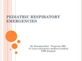
    ED MANAGEMENT OFACUTE ASTHMA Mental status Heart rate Oxygen saturation Degree of wheezing Inspiratory:expiratory ratio Accessory muscle use PEFR
  • 17.
    ED MANAGEMENT OFACUTE ASTHMA SABA by NEB or MDI-S Up to 3 SABA doses in 1 st hour Oral corticosteroids if incomplete response or if already taking at home
  • 18.
    ED MANAGEMENT OFACUTE ASTHMA D/C SABA q 4 hrs for 3-4 days Continue corticosteriod 3-10 days,if begun in ED Continue controller medications Initiate ICS if persistent asthma
  • 19.
    ED MANAGEMENT OFACUTE ASTHMA SABA and IB by NEB in 1 st hour Oral corticosteroids If emesis, consider parenteral corticosteriods
  • 20.
    ED MANAGEMENT OFACUTE ASTHMA Continue SABA q 30-45 mins or continuously Observe for 60-90 mins after SABA end If sustained response: D/C If minimal or no improvement : admit
  • 21.
    ED MANAGEMENT OFACUTE ASTHMA Continue SABA & IB by NEB —consider heliox Consider SQ or IM epinephrine or terbutaline IV corticosteroids Magnesium sulfate IV SABA infusion
  • 22.
    ED MANAGEMENT OFACUTE ASTHMA Admit to PICU : Continued severe respiratory distress Altered mental status Need for IV SABA Need for mechanical ventilation Admit others to hospital ward
  • 23.
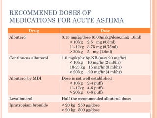
    RECOMMENED DOSES OFMEDICATIONS FOR ACUTE ASTHMA Drug Dose Albuterol 0.15 mg/kg/dose (0.03ml/kg/dose,max 1.0ml) < 10 kg 2.5 mg (0.5ml) 11-19kg 3.75 mg (0.75ml) > 20 kg 5 mg (1.0ml) Continuous albuterol 1.0 mg/kg/hr by NB (max 20 mg/hr) < 10 kg 10 mg/hr (2 ml/hr) 10-20 kg 15 mg/hr (3 ml/hr) > 20 kg 20 mg/hr (4 ml/hr) Albuterol by MDI Dose is not well established < 10 kg 2-4 puffs 11-19kg 4-6 puffs > 20 kg 6-8 puffs Levalbuterol Half the recommended albuterol doses Ipratropium bromide < 20 kg 250 µg/dose > 20 kg 500 µg/dose
  • 24.
    RECOMMENED DOSES OFMEDICATIONS FOR ACUTE ASTHMA(CONT.) Drug Dose L-epinephrine (1:1000) or terbutaline (1.0 mg/mg) 0.01ml/kg/dose SC or IM (max 0.4 ml) May be repeated every 10-15 min IV terbutaline 10 µg/kg bolus over 10 min,then 0.1-0.3 µg/kg/min infusion Every 30 min,may increase infusion by 0.3 µg/kg/min to a max of 5 µg/kg/min Prednisolone 2 mg/kg (max 60mg), in ED 1 mg/kg/dose bid, home therapy Dexamethasone 0.6mg/kg PO, 2 dose 24 hrs apart IV methylprednisolone 1-2 mg/kg (max 125 mg) IV magnesium sulfate 50-75 mg/kg over 20 min(max 2.5g)
  • 25.
    MILD SABA ifnot improve then q 20 mins in 1 st hours Mouthpiecess preferable for older child Decrease nasal filtering of drug Spacer must take 5-8 breaths to complete empty spacer If 3 dose  reevaluate
  • 26.
    SHORT ACTING BETA2-AGONISTSIN ACUTE ASTHMA IB= ipratropium bromide MDI-s=metered dose inhaler with spacer NHLBI=national,Heart,Lung and blood institute Mild Moderate Severe Delivery method Intermittent NEB or MDI-S Continuous by NEB for 1 hr, then reassess Consider SC or IM Continuous by NEB Comments Most patients will need 1-2 treatments Allow for MDI-S teaching No IB needed Not superior to MDI-S Easier to adhere to NHLBI guidelines for the first hr of therapy Concurrent IB therapy is more easily delivered Better outcomes in severe asthma
  • 27.
  • 28.
    BRONCHIOLITIS Seasonal disease: November &April Nearly all children : affected virus cause bronchiolitis at least once during first 2 years of life More common < 1 year
  • 29.
    Increased mucus secretion,cell death, and sloughing,  a peribronchiolar lymphocytic infiltrate and submucosal edema Combination of debris&edema produces  critical narrowing & obstruction of small airways .
  • 30.
    BRONCHIOLITIS M/C CAUSE: RESPIRATORY SYNCYTIAL VIRUS (RSV) Age < 3 mo Preterm GA < 34 wks Toxic or ill general appearance RR > 70/min SpO2 <95% CXR : atelectasis Breast feeding < 1 mo Maternal smoking RSV subtype A Male Air pollution environment Severity associate with
  • 31.
    SUGGEST BRONCHIOLITIS ASSESSMENTTOOL Mild Moderate Severe Feeding Normal Less Poor SaO2 in room air ≥ 95 % 92 -94 % < 92 % Respiratory rate(/min) < 60 60 - 70 > 70 Retraction None/ minimal Intercostal Substernal Accessory muscle use None None Neck or abdominal Wheeze None/minimal Moderate expiratory Severe inspiratory/expiratory, audible without stethoscope Air exchange Good, equal breath sound Localized decrease breath sound Multiple area of decrease breath sound
  • 32.
    ED MANAGEMENT OFBRONCHIOLITIS Improved Not Improved Improved Not Improved
  • 33.
    PROPHYLAXIS ED :No role preventive medication in ED Education Advice Palivizumab (Synagis) Monoclonal antibodies againt RSV Specific immune globulin Not effect acute process Effective in reducing hospitalization in high risk group (monthly IM during high prevalence month ) Recommended for most age < 24 month with Chronic lung disease Congenital heart disease Prematurity
  • 34.
    http://www.ginasthma.com/ Rosen 7th edition Tintinally 7 th edition
  • 35.
  • 36.
    PULSUS PARADOXUS ในการหายใจปกติพบว่า เมื่อมีการหายใจเข้า systolic blood pressure สามารถลดได้ประมาณ 10 mm.Hg  pulsus paradoxus เกิดขึ้นเมื่อมีความแตกต่างของ systolic blood pressure มากกว่า 12 mm.Hg ระหว่างการหายใจเข้า
  • 37.
    PULSUS PARADOXUS pulsusparadoxus สามารถเกิดขึ้นในผู้ป่วย COPD severe asthma pericardial tamponade aortic insufficiency poor LV compliance สามารถเกิด pulsus paradoxus โดยไม่มี pericardial fluid เพื่อต้องการวัด paradoxical pulse ผู้ป่วยควรนอน อยู่ในท่าสบาย ศีรษะ อยู่ประมาณ 30 ถึง 45 ( ซึ่งอาจทำได้ยากในผู้ป่วย COPD, severe asthma หรือ cardiac tamponade)
  • 38.
    เริ่มต้นการวัด โดย การ inflate cuff ขึ้นไปจนไม่ได้ยินเสียงอะไร จากนั้น deflate cuff จนกระทั่งได้ยินเสียงชีพจรเสียงแรก โดยที่เสียงนี้ จะ มีความสอดคล้องกับการหายใจออก ( คือได้ยินเมื่อหายใจออก และ หายไปเมื่อหายใจเข้า ซึ่งหมายความว่าเป็น systolic blood pressure ในช่วงหายใจออก จากนั้นให้ deflate cuff จนกระทั่งได้ยินเสียงชีพจรตลอด ( ทั้งตอนหายใจเข้าและหายใจออก ซึ่งหมายความว่าเป็น systolic blood pressure ในช่วงหายใจเข้า ) ด้วยวิธีการเช่นนี้ ทำให้เราสามารถตรวจพบ pulsus paradoxus ได้ถ้า ความแตกต่างมีมากพอ ถ้าความแตกต่างของ systolic blood pressure ระหว่างหายใจเข้าและหายใจออกมากกว่า 12 mm.Hg แสดงว่า paradoxical pulse สูง
  • 39.
    ผู้ป่วยส่วนใหญ่ที่มี tamponade มักมี paradoxical pulse มากกว่า 20-30 mm.Hg ระหว่างการหายใจ ซึ่งอาจมีข้อยกเว้นในผู้ป่วยที่มี pulse pressure แคบมากๆ เช่นในผู้ป่วยที่มี advanced tamponade  ที่อาจวัด paradoxical pulse ได้เป็นเท็จลวง คือประมาณ 5-15 mm.Hg เนื่องจาก systolic blood pressure อาจจะต่ำกว่า diastolic blood pressure ด้วยเหตุผลนี้ อัตราส่วนระหว่าง paradoxical pulse ต่อ pulse pressure จึงเป็นดัชนี ที่เชื่อถือได้มากกว่า paradoxical pulse โดยถือว่าเมื่อมากกว่า 50% ผิดปกติ Pulsus paradoxus มีความสัมพันธ์กับ cardiac output ที่ลดลงเนื่องจาก tamponade ใน pericardial effusion, pulsus paradoxus ที่มากกว่า 25 mm.Hg ทั้ง sensitive และ specific ต่อ severe มากกว่า mild tamponade
  • 40.
    ในผู้ป่วยเด็ก  pulsusparadoxus ถูกศึกษาเพื่อทำนายความรุนแรงในโรคปอด เช่น ในโรคหอหหืด พบว่า ถ้าผู้ป่วยมี pulsus paradoxus มากกว่า 15 mm.Hg จะมีอาการของโรคที่รุนแรงกว่า ถึงแม้ว่าจะมีโรคหลายชนิด ที่อาจระบุได้จากการตรวจ pulsus paradoxus แต่มักจะเป็นการยากโดยการวินิจฉัย pulsus paradoxus โดยใช้แต่ sphygmomanometer เพียงอย่างเดียว ในการศึกษาของ Jay และ คณะพบว่า แพทย์เวชศาสตร์ฉุกเฉินและแพทย์เวชศาสตร์วิกฤต ไม่สามารถวัด pulsus paradoxus ได้อย่างแม่นยำ โดยการใช้ sphygmomanometer  ดังนั้นจึงควรมีการพัฒนาเครื่องมืออื่นๆ ที่สามารถช่วยวินิจฉัย pulsus paradoxus
  • 41.
    HTTP://EMEDICINE.MEDSCAPE.COM/ARTICLE/800428-TREATMENT PEDIATRIC BRONCHIOLITIS: DIFFERENTIAL DIAGNOSES & WORKUP AUTHOR: MARK LOUDEN, MD, FACEP, ASSISTANT MEDICAL DIRECTOR, EMERGENCY DEPARTMENT, DUKE RALEIGH HOSPITAL CONTRIBUTOR INFORMATION AND DISCLOSURES UPDATED: DEC 27, 2010
  • 42.
  • 43.
  • 44.
    http://emedicine.medscape.com/article/800428-treatment Pediatric Bronchiolitis: Differential Diagnoses & Workup Author: Mark Louden, MD, FACEP, Assistant Medical Director, Emergency Department, Duke Raleigh Hospital Contributor Information and Disclosures Updated: Dec 27, 2010
  • 45.
    Croup / โรคหลอดลมอักเสบ ฮอร์โมนอะดรีนาลีน Racemic รักษาโรคซาง [14] [ 15] Racemic อะดรีนาลีนเป็นส่วนผสมของ dextrorotatory (D) 1:1 และ levorotatory (L) isomers ของอะดรีนาลีน [16] l แบบเป็นส่วนประกอบที่ใช้งานอยู่ [16] Racemic อะดรีนาลีนทำงานโดยการส่งเสริมของผู้รับในเส้นทางการบินกับผลลัพธ์ mucosal α - adrenergic vasoconstriction และลดอาการบวมน้ำ และสิ่งเร้าที่ β - adrenergic subglottic ผู้รับผลแห่งการผ่อนคลายกล้ามเนื้อเรียบหลอดลม [15]
  • 46.
  • 47.

Editor's Notes

  • #11 Pulsus paradoxus ในการหายใจปกติ พบว่า เมื่อมีการหายใจเข้า systolic blood pressure สามารถลดได้ประมาณ 10 mm.Hg  pulsus paradoxus เกิดขึ้นเมื่อมีความแตกต่างของ systolic blood pressure มากกว่า 12 mm.Hg ระหว่างการหายใจเข้า pulsus paradoxus สามารถเกิดขึ้นในผู้ป่วย COPD, severe asthma, pericardial tamponade, aortic insufficiency นอกจากนี้ poor LV compliance สามารถเกิด pulsus paradoxus โดยไม่มี pericardial fluid เพื่อต้องการวัด paradoxical pulse ผู้ป่วยควรนอน อยู่ในท่าสบาย ศีรษะ อยู่ประมาณ 30 ถึง 45 ( ซึ่งอาจทำได้ยากในผู้ป่วย COPD, severe asthma หรือ cardiac tamponade) เริ่มต้นการวัด โดย การ inflate cuff ขึ้นไปจนไม่ได้ยินเสียงอะไร จากนั้น deflate cuff จนกระทั่งได้ยินเสียงชีพจรเสียงแรก โดยที่เสียงนี้ จะ มีความสอดคล้องกับการหายใจออก ( คือได้ยินเมื่อหายใจออก และ หายไปเมื่อหายใจเข้า ซึ่งหมายความว่าเป็น systolic blood pressure ในช่วงหายใจออก จากนั้นให้ deflate cuff จนกระทั่งได้ยินเสียงชีพจรตลอด ( ทั้งตอนหายใจเข้าและหายใจออก ซึ่งหมายความว่าเป็น systolic blood pressure ในช่วงหายใจเข้า ) ด้วยวิธีการเช่นนี้ ทำให้เราสามารถตรวจพบ pulsus paradoxus ได้ถ้า ความแตกต่างมีมากพอ ถ้าความแตกต่างของ systolic blood pressure ระหว่างหายใจเข้าและหายใจออกมากกว่า 12 mm.Hg แสดงว่า paradoxical pulse สูง ผู้ป่วยส่วนใหญ่ที่มี tamponade มักมี paradoxical pulse มากกว่า 20-30 mm.Hg ระหว่างการหายใจ ซึ่งอาจมีข้อยกเว้นในผู้ป่วยที่มี pulse pressure แคบมากๆ เช่นในผู้ป่วยที่มี advanced tamponade  ที่อาจวัด paradoxical pulse ได้เป็นเท็จลวง คือประมาณ 5-15 mm.Hg เนื่องจาก systolic blood pressure อาจจะต่ำกว่า diastolic blood pressure ด้วยเหตุผลนี้ อัตราส่วนระหว่าง paradoxical pulse ต่อ pulse pressure จึงเป็นดัชนี ที่เชื่อถือได้มากกว่า paradoxical pulse โดยถือว่าเมื่อมากกว่า 50% ผิดปกติ Pulsus paradoxus มีความสัมพันธ์กับ cardiac output ที่ลดลงเนื่องจาก tamponade ใน pericardial effusion, pulsus paradoxus ที่มากกว่า 25 mm.Hg ทั้ง sensitive และ specific ต่อ severe มากกว่า mild tamponade ในผู้ป่วยเด็ก  pulsus paradoxus ถูกศึกษาเพื่อทำนายความรุนแรงในโรคปอด เช่น ในโรคหอหหืด พบว่า ถ้าผู้ป่วยมี pulsus paradoxus มากกว่า 15 mm.Hg จะมีอาการของโรคที่รุนแรงกว่า ถึงแม้ว่าจะมีโรคหลายชนิด ที่อาจระบุได้จากการตรวจ pulsus paradoxus แต่มักจะเป็นการยากโดยการวินิจฉัย pulsus paradoxus โดยใช้แต่ sphygmomanometer เพียงอย่างเดียว ในการศึกษาของ Jay และ คณะพบว่า แพทย์เวชศาสตร์ฉุกเฉินและแพทย์เวชศาสตร์วิกฤต ไม่สามารถวัด pulsus paradoxus ได้อย่างแม่นยำ โดยการใช้ sphygmomanometer  ดังนั้นจึงควรมีการพัฒนาเครื่องมืออื่นๆ ที่สามารถช่วยวินิจฉัย pulsus paradoxus
  • #30 Infection of bronchiolar respiratory and ciliated epithelial cells produces increased mucus secretion, cell death, and sloughing, followed by a peribronchiolar lymphocytic infiltrate and submucosal edema. The combination of debris and edema produces critical narrowing and obstruction of small airways.