Paul Ahern - Time of Flight Secondary Ion Mass Spectroscopy [ToF-SIMS] theory & practice
•
2 likes•2,002 views

Time of Flight (ToF) Secondary Ion Mass Spectroscopy (SIMS) is an extremely sensitive surface analysis technique where the mass to charge ratio of an ion or molecular fragment is determined by its velocity in the time domain. The commercially available ToF SIMS instruments available today have their roots in close to a century’s worth of academic research, and their ability to gather elemental and molecular information with excellent depth resolution and high sensitivity is incomparable.

More Related Content
What's hot
What's hot (20)
Similar to Paul Ahern - Time of Flight Secondary Ion Mass Spectroscopy [ToF-SIMS] theory & practice
Similar to Paul Ahern - Time of Flight Secondary Ion Mass Spectroscopy [ToF-SIMS] theory & practice (20)
Recently uploaded
Recently uploaded (20)
Paul Ahern - Time of Flight Secondary Ion Mass Spectroscopy [ToF-SIMS] theory & practice
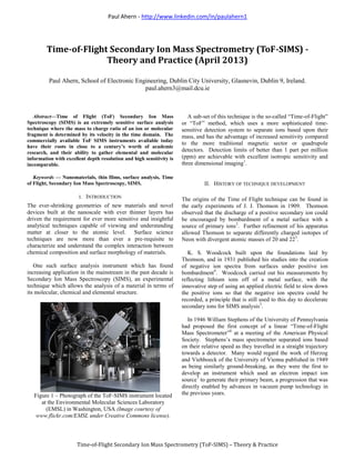
- 1. Paul Ahern - http://www.linkedin.com/in/paulahern1 Time-of-Flight Secondary Ion Mass Spectrometry (ToF-SIMS) – Theory & Practice Abstract—Time of Flight (ToF) Secondary Ion Mass Spectroscopy (SIMS) is an extremely sensitive surface analysis technique where the mass to charge ratio of an ion or molecular fragment is determined by its velocity in the time domain. The commercially available ToF SIMS instruments available today have their roots in close to a century’s worth of academic research, and their ability to gather elemental and molecular information with excellent depth resolution and high sensitivity is incomparable. Keywords — Nanomaterials, thin films, surface analysis, Time of Flight, Secondary Ion Mass Spectroscopy, SIMS. I. INTRODUCTION The ever-shrinking geometries of new materials and novel devices built at the nanoscale with ever thinner layers has driven the requirement for ever more sensitive and insightful analytical techniques capable of viewing and understanding matter at closer to the atomic level. Surface science techniques are now more than ever a pre-requisite to characterize and understand the complex interaction between chemical composition and surface morphology of materials. One such surface analysis instrument which has found increasing application in the mainstream in the past decade is Secondary Ion Mass Spectroscopy (SIMS), an experimental technique which allows the analysis of a material in terms of its molecular, chemical and elemental structure. Figure 1 – Photograph of the ToF-SIMS instrument located at the Environmental Molecular Sciences Laboratory (EMSL) in Washington, USA (Image courtesy of www.flickr.com/EMSL under Creative Commons license). A sub-set of this technique is the so-called “Time-of-Flight” or “ToF” method, which uses a more sophisticated time- sensitive detection system to separate ions based upon their mass, and has the advantage of increased sensitivity compared to the more traditional magnetic sector or quadrupole detectors. Detection limits of better than 1 part per million (ppm) are achievable with excellent isotropic sensitivity and three dimensional imaging1 . II. HISTORY OF TECHNIQUE DEVELOPMENT The origins of the Time of Flight technique can be found in the early experiments of J. J. Thomson in 1909. Thomson observed that the discharge of a positive secondary ion could be encouraged by bombardment of a metal surface with a source of primary ions2 . Further refinement of his apparatus allowed Thomson to separate differently charged isotopes of Neon with divergent atomic masses of 20 and 223 . K. S. Woodcock built upon the foundations laid by Thomson, and in 1931 published his studies into the creation of negative ion spectra from surfaces under positive ion bombardment4 . Woodcock carried out his measurements by reflecting lithium ions off of a metal surface, with the innovative step of using an applied electric field to slow down the positive ions so that the negative ion spectra could be recorded, a principle that is still used to this day to decelerate secondary ions for SIMS analysis5 . In 1946 William Stephens of the University of Pennsylvania had proposed the first concept of a linear “Time-of-Flight Mass Spectrometer”6 at a meeting of the American Physical Society. Stephens’s mass spectrometer separated ions based on their relative speed as they travelled in a straight trajectory towards a detector. Many would regard the work of Herzog and Viehboeck of the University of Vienna published in 1949 as being similarly ground-breaking, as they were the first to develop an instrument which used an electron impact ion source7 to generate their primary beam, a progression that was directly enabled by advances in vacuum pump technology in the previous years. Time-of-Flight Secondary Ion Mass Spectrometry (ToF-SIMS) - Theory and Practice (April 2013) Paul Ahern, School of Electronic Engineering, Dublin City University, Glasnevin, Dublin 9, Ireland. paul.ahern3@mail.dcu.ie
- 2. Paul Ahern - ie.linkedin.com/in/paulahern1 1 Time-of-Flight Secondary Ion Mass Spectrometry (ToF-SIMS) – Theory & Practice Figure 2 – Schematic from William E. Stephens original 1952 patent for a “Time of Flight” spectrometer8 . Meanwhile, Wiley and McLaren9 built upon Stephens’s study and via their Bendix Corporation released the first Time- of-Flight mass spectrometer with a similar “EI” or “electron ionisation” source in 1955, capable of sensitivities of less than 100 amu for the first time. Another notable development was the work of Richard Honig of RCA Laboratories in New Jersey, who through the search for new hot cathode materials for vacuum tubes led him to assemble first a two-stage mass spectrometer, and then in 1958 a full secondary ion mass spectrometry instrument10 . Further advances came in the 1960’s under the stewardship of NASA who funded Liebel and Herzog of Geophysics Corporation of America (GCA) Corp to create an analytical instrument11 to examine moon rock samples brought back to earth from the Apollo space flights. In 1967 Liebel, now of Applied Research Laboratories, created an instrument with an improved design12 which now used a duoplasmatron as the ion source and mass separation to improve the purity of the primary ion beam; the mass spectrometer design used a new arrangement with no entrance slit and an Einzel lens. In the early 1970’s Wittmaack13 demonstrated that the secondary ion yield could be increased to a high value using O2 + ions as the primary ion beam medium, and that there was a self-induction effect due to the mechanism of recoil implantation on the sample surface. The subsequent work by Magee14 working again with Honig at RCA Labs showed success with the first quadrupole mass analyser complemented by a high current density 40 Ar+ primary beam, and presented an instrument that was capable of speedy depth profiling where an ultra-high vacuum environment helped to maximize detection sensitivity. This was also the first time that the issue of the large amount of output data from SIMS as an analytical technique was addressed, as Magee’s system had computer automated control and acquisition modules. In the 1970’s Prof. Benninghoven15 and co-workers at the University of Münster developed the first “static” SIMS instrument, which was deemed to be much less harsh on the surface under analysis than its dynamic SIMS counterpart. The reason for the reduction in surface damage was the much lower dose density of the primary ion beam on the sample surface, with primary ion beam current densities of less than 1nA per cm2 being typical. This allowed slower rates of material removal but maintained the high sensitivity which ToF-SIMS would become noted for, as a sufficient amount of spectral information could be captured quickly before the sample surface was modified significantly16 , typically at levels of less than 1%. In common usage, though sometime the terms “static” and “Time of Flight” are used interchangeably to describe this subset of SIMS analysis techniques, they are actually very different. The main difference being that in Time of Flight SIMS, the atomic mass peaks are monitored and recorded more or less simultaneously, whereas in static SIMS only one peak can be measured at any one time. Benninghoven showed the static technique was very useful to analyse organic molecules which had been deposited onto conducting substrates. Subsequent improvements and refinements in the technique have been directly due to the advances that have been made in constructing high performance Time of Flight detectors, which has seen it become a key technique in the study of surfaces, especially those made up of organic materials. III. OVERVIEW OF TECHNIQUE To understand the operating principles of the ToF-SIMS technique it is useful if we summarise the basic steps of the process first, before we then break down the instrument into it's constituent parts and describe the phenomenon happening in each section in more detail. Figure 3 – Atomic scale representation of the ion sputtering process which takes place in SIMS when a primary ion beam impinges up a sample surface17 . Overview: In basic terms, the SIMS technique is one where a vacuum is created and within this vacuum region a beam of primary ions at an energy of hundreds to thousands of electron volts (eV) is accelerated onto a focussed spot on the surface of a sample to be analysed. Various elastic and inelastic processes occur at the surface (and subsurface) atomic layers,
- 3. Paul Ahern - ie.linkedin.com/in/paulahern1 2 Time-of-Flight Secondary Ion Mass Spectrometry (ToF-SIMS) – Theory & Practice giving rise to the liberation of ionised atoms (secondary ions) as part of a collision cascade, represented in figure 3. As the cascade emanates outwards from the path of the primary beam, its energy can affect atoms far away in the surface layer with sufficient kinetic energy to break their lattice bonds and be liberated from the surface as neutral ions, ionised atoms or molecular clusters18 . The secondary ions are extracted, accelerated, and fashioned into a beam and then traverse a flight path of a known length which is long enough that they have the opportunity to become time-focussed; lighter ions arriving at a mess spectrometer detector and being counted before their heaver contemporaries. Figure 4 – Simplified diagram showing the instrument set- up for secondary ion mass spectroscopy (SIMS)19 . SIMS has the ability to detect all elements in the periodic table, including Hydrogen. In SIMS, the analytical data is contained in the current signal of an ion with a certain mass: charge ratio, with little or no background signal to be removed from the spectrum. Therefore, signal to noise is good and the minimum detection limit in SIMS can be somewhere of the parts per million (ppm) to parts per billion (ppb) scale. A. Primary Ion Beam: The composition of the sample under investigation is of prime importance, as bombardment from the primary ion beam will give rise to the secondary ions which must then be resolved in time. There is no set primary ion beam medium which must be used for ToF-SIMS, and many instruments employ the use of more than one beam to give optimum material removal and sensitivity for the given sample material In terms of the atomic interaction of the primary ion beam and the sample surface, the key factor is the difference in electronegativity; that is, the ratio of the number of protons in the nucleus and the number and separation distance of valence electrons orbiting the atomic core. Consider the element Boron, which has two shells, with 2 electrons in the innermost {k} orbital and 3 in the outer {l} orbital. Compare Boron to Aluminium, which is directly below it on the periodic table and has 2 electrons in the {k} orbital, 8 in the{ l} orbital and 3 in the {m} orbital. The couloumbic attraction between the nucleus and the electrons is stronger in Boron than in Aluminium, as the atom is smaller and thus there is a smaller distance between the electrons and the nucleus, leading to a higher value of electronegativity (Boron has an electronegativity value of 2 "Pauling units", while Aluminium is 1.6). In reality, it can be seen that electronegativity values have a trend across the periodic table, increasing as you move to the top right hand corner, as illustrated in figure 5 below. Figure 5 – Pictorial representation of how electronegativity values vary across the periodic table. Electronegativity is the propensity for an atom to draw electrons from other atoms, and rises from left to right and from the bottom to the top of the periodic table. (Image © Wiley & Sons 2000, all rights reserved). The two most commonly used ion beam sources in ToF- SIMS, Oxygen and Caesium, are widespread because they generate a sufficiently high secondary ion yield for speedy depth profiling are respectively positively and negatively charged.; when using an Oxygen ion beam, the bombardment of ions on the sample surface will increase the number of positive ion fragments received at the detector, whilst for a Caesium source you will increase the number of negative ions collected. In specialised instruments which are designed specifically for image fine focussing techniques, a Gallium (Ga+ ) source may also be employed as it has a lower melting point and thus a higher achievable brightness which makes it worthwhile despite its relative lower sensitivity. Gallium ions are created in a liquid metal ion source (LMIS), as shown in figure 6 - the same method that is used to create a Caesium (Cs+ ) beam. Oxygen beams can be created by either using an electron impact source or a duoplasmatron. In the case of a LMIS, pulsing of the primary beam to achieve good mass resolution can be achieved by the inclusion of a set of deflection plates to quickly blank the beam on and off20 .
- 4. Paul Ahern - ie.linkedin.com/in/paulahern1 3 Time-of-Flight Secondary Ion Mass Spectrometry (ToF-SIMS) – Theory & Practice Figure 6 – Diagram of the parts of a liquid metal ion source (LMIS)21 with the parts as labelled (a) emerging metal ions (b) extractor plate (c) liquid metal film (d) capillary feed tube (e) liquid metal reservoir (f) crucible needle. In electron impact sources, oxygen or another noble gas flows into an ionisation region where a filament resides. Electrons from this filament are then accelerated by anodic grid while an extraction cathode (such as a Weinhalt aperture) accelerates the ions to the lens array where they are focussed and rastered if in imaging mode. In the case of a duoplasmatron, which is common in dynamic SIMS instruments also, plasma is formed in the extraction region and a pair of off-setted magnetic lenses is used to form the primary beam and tune it to a usable outline before it is extracted by the anode. Figure 7 – Block diagram of the primary ion column in a typical SIMS instrument22 . In instruments which have only one ion source, it is common to use an element in the middle of the electronegativity table which can successfully liberate both negative and positive ions at sufficient amounts. Recent advance have shown that the use of primary cluster ion sources such as those derived from Bismuth (Bi1+ , Bi3+ ) and polyatomic Carbon (C60+ ) have benefits when used to analyse biological samples, with Bi3+ showing the best surface sensitivity for lower atomic-mass molecular fragments23 . Other recent peptide analysis work with Argon (Arn+ ) primary ion beam sources24 has shown its usefulness in terms of minimising surface damage and allowing the user to maintain a constant sputter rate with an associated reduction in secondary ionization mechanisms. Once the ions have been formed, they must then be extracted and focussed into the primary beam before they are accelerated to the required energy25 . B. Beam / Sample Interaction: Once the primary ion beam has been formed, it is focussed and pulsed in short (typically between a 2ns and 20ns interval) onto the sample surface, giving rise to sputtering. One consequence of this pulsed ion beam regime is that a sufficient interval must be allowed so that the heaviest, and thus slowest-moving, secondary ions can vacate the detection region before the next bundle of secondary ion data can be accepted. Figure 8 – Diagram of some of the myriad and complex phenomena at play during the interaction between the primary ion beam and the sample in SIMS26 . The progression of the ion cascade started by a sole primary ion can be seen. Ideally the primary ion beam pulse is kept as short as possible, by using a device known as an electrodynamic pulse “buncher” in the primary ion column as shown in figure 9, to time focus the sputter pulse to a Gaussian profile of typically less than 1 nanosecond (1ns)27 . This primary beam pulse width value is termed tbeam, and one other consequence of this arrangement is that the AC current is only a smaller fraction of the DC current which further preserves the static conditions on the sample surface allowing for better chemical mapping.
- 5. Paul Ahern - ie.linkedin.com/in/paulahern1 4 Time-of-Flight Secondary Ion Mass Spectrometry (ToF-SIMS) – Theory & Practice Figure 9 – Simplified schematic diagram of the construction of a linear buncher for Time of Flight analysis. Ions with a small first ionisation potential will be readily ionised by the primary beam, while those with a greater potential will form positive ions much less readily; ions with high electron affinity will preferentially give rise to negative ions more readily. The timespan for the creation of these secondary ions is typically in the picosecond (ps) regime. Figure 10 – representation of total ion yield during sputtering, {S}, and a function of the incident ion energy {E}28 . Ion images are produced in a slightly different way, whereby the primary ion beam is rastered over the area of interest and the number of ions as a function of the {x,y} co- ordinates is presented. Secondary electrons generated as a by- product of his process can also be collected in a resident Everhardt-Thornley photomultiplier and used to generate a standard secondary electron image. The total sputter yield of secondary ions for a particular element depends on the incident ion energy as shown in figure 10 and also the angle of incidence when the primary beam strikes the sample surface as shown in figure 12; the vast bulk of the species evolved from the sputtered surface will actually be neutrals but it is only the very small positively or negatively charge portion of the secondary particle flux that is detected. A complete understanding of the formation of secondary ion species in ToF-SIMS has not yet been accepted, although many competing thesis have been proposed in the literature and have had limited success in predicting experimental results for a narrow subset of defined materials. i. Local Thermal Equilibrium (LTE) model: This (now defunct) historical model, sometimes also categorised as the “surface excitation model” proposed that underneath the bombardment area, surface plasma was created wherein the sputtered atoms became ionised. Under equilibrium conditions, the ionisation potential could be calculated by the use of the Saha-Eggert ionisation equation29 in the bombardment region. The only important factor was deemed to be the plasma temperature30 and this could be estimated by taking into account the amounts of each element present. The results from this model are strictly semi-quantitative as the exponential term in the S-E equation is compatible with quantum mechanical terms31 . As V. E. Krohn32 of the US Dept. of Energy laboratory in Argonne, Illinois summarised succinctly “Unfortunately, surface ionization is an equilibrium process, whereas secondary-ion emission is not.” ii. Electron Tunnelling model: This model of secondary ion formation (sometimes called the Schroeer model33 ) is based on quantum mechanical principles, and describes how the electron which sits in the conduction band (CB) has the ability to tunnel into the valence band (VB) of the ejected atom34 . The ionisation potential of the sputtered element governs the probability statistics that govern this phenomenon, as well as the adiabatic surface ionisation function and the velocity of the sputtered atom. However, in line with this model, the work function is autonomous of how the work function change, , is being induced - as long as the external ions in the primary beam used to induce does not change the chemical state of the target atoms35 , as shown in figure 11 below. Figure 11 – Simplified energy diagram of a charged particle separating from a metal surface36 . At the distance ZC, the atomic level intersects the Fermi level of the metal and charge exchange can occur by tunnelling.
- 6. Paul Ahern - ie.linkedin.com/in/paulahern1 5 Time-of-Flight Secondary Ion Mass Spectrometry (ToF-SIMS) – Theory & Practice iii. Broken Bond model: In this model, proposed initially by Šroubek37 the electronic temperature, Te, within the region of the collision cascade was calculated for different materials from a starting point of electron transport theory, and these values were compared to calculated values formulated in conjunction with empirical SIMS data. The resultant model tackles the process from the standpoint of the creation of ionic compounds in an idealised electronic lattice under bombardment from a primary ion beam of oxygen, and a prerequisite is that there be present an oxide layer on the surface of the sample to be analysed. In this model, the binding electrons stay with the oxygen atom and there is only emission of positively ionised species. Figure 12 – Secondary ion sputter yield plotted versus the angle of incidence, , of the primary ion beam (as measured referenced to the normal plane)38 . In the theoretical case whereby all the available secondary ions could be ionised by the incoming beam, and detected by the spectrometer, then the signal could be related to the specimen composition by the equation39 below: iS = iPS where iS is the sputtered ion current, is the atomic fraction of the element of interest, iP is the primary ion beam current, S the total effective sputter yield, and the specific sensitivity of the detector (which includes the previously explained effect of the incident angle of detection). Each spectrometer configuration will also have an inherent angular acceptance range and a reduction in mass resolution can be observed when angular divergence becomes too large. C. Secondary Ion Acceleration: Typically less than 1% of the sputtered ions from the primary beam are ionised, with the resultant cloud of ejected atomic and molecular secondary ions accelerated by a potential into the Time-of-Flight region. Since the lighter ions travel faster, they thus arrive at the ion detection module first and can be counted. The Time-of- Flight relationship can be understood simply as the travel time of a secondary ion being proportional to the square root of its mass, and by this mechanism all secondary ions can be isolated and detected discretely once they impinge upon the detector. The drawing out and collimating of the secondary ion signal emitted from the sample surface is by means of a combination of transfer and immersion lenses which control the image amplification and, by way of apertures, serve to limit the angular acceptance angle of the mass spectrometer detector. Secondary particles are accelerated by an applied constant voltage, Vacc such that they now have a fixed kinetic energy {E} which overrides their initial kinetic energy at source. In this case their accelerated velocity in the drift region dpends solely upon their mass, by the equation40 qVacc = E = ½ mv2 The overall secondary ion transmission can thus be greater than 40%. To further increase the detector effectiveness for dense ions (m>1,000 Da) a post –acceleration area (10-20 keV) can be sited between the end of the drift region and the entrance cone of the detector. D. Charge Neutralisation: Since many of the common samples analysed by ToF-SIMS are organic molecules or polymers and are non-conductive, a separate source of electrons is needed to provide a tuneable charge compensation function; this is achieved by producing low energy electrons and introducing them around the analysis area on the sample surface. A recurring issue in achieving good spectral quality with SIMS is adequate monitoring and control of the sample’s surface potential41 to ensure that as uniform an ion emission profile as possible is generated, as even a very slight increase in the electric field on the sample surface can significantly shift the energy level accepted by the energy filter into an undesirable region, whereupon the ion yield being generated is negligible and a impractical spectra with large intensity losses will be recorded. Typically a LaB6 or Tungsten filament electron source is used, and in some cases the removal of surface charge can be further facilitated by placing an earthed metal grid (such as a TEM grid) over or close to the area of interest on the sample surface. This method is useful as the pulsed low energy electrons automatically steady themselves further over time, as a positive surface potential serves to minimise the losses of
- 7. Paul Ahern - ie.linkedin.com/in/paulahern1 6 Time-of-Flight Secondary Ion Mass Spectrometry (ToF-SIMS) – Theory & Practice secondary ions which would serve to decrease the overall potential of the surface42 . Another complication with instruments that operate in the time domain such as ToF-SIMS is that charge neutralisation must typically done in a pulsed, cyclical manner in between the repeated sequences of secondary ion generation and acceleration. E. Mass Analysis Detection: Early SIMS instruments relied upon the quadrupole mass spectrometer for detection, consisting of four elements at equal distances whereby alternating pairs of DC and RF voltages were applied to act as a filter allowing only ions with a specific charge to mass ratio to traverse into the detector. However, the disadvantages of poor transmission (typically <1%, which decreases further with increasing mass number) and a” lossy” data collection method as it can only operate in scanning mode, meant that better detection methods were needed for ToF-SIMS to advance further. i. Reflectron Analyser Today the most common in commercial ToF-SIMS instrumentation, mass-reflectron analysers were first proposed by Russian physicists Mamyrin & Karataev43 in 1972. A modern reflectron detector offers a good balance of much enhanced mass resolution in a smaller equipment footprint44 . Figure 13 – Schematic representation of the nascent Time of Flight instrument (complete with reflectron) used by Benninghoven and co-workers at the University of Münster45 . The parts are labelled as shown – (a) Electron ion source (in this case, Ar+ ). (b) Ga+ liquid metal ion source (LMIS). (c) Sample holder (temperature controllable). (d) Secondary ion acceleration lens. (e) Reflectron of the gridless type design. (f) Mass spectrometer detector. The method of operation is that the secondary ions are accelerated towards an ion “mirror” which functions as an electrostatic reflector that then turns the ions and reflects them back towards the direction of the detector in a “folding” arrangement, preserving the time-focussed nature of the ions and enhancing the mass resolution by an order or magnitude. The travel time is governed by the equation - √ √ This setup offers mass resolution of the order of 10,000 amu when a deceleration & re-acceleration grid is used, but by moving to a gridless design superior mass resolution of up to 50,000 amu is reliably achievable46 . As measuring the usable secondary ion yield can be problematic, transmission is instead estimated using a Thomson distribution47 based on the incoming kinetic energy of the primary beam; however ,this approximation neglects any influence from detector efficiency. The detector itself is normally a combination of a Faraday cup and microchannel plate48 which contains an array of electron multipliers, similar in design to the historic channeltron. The signal amplification is by means of an electron cascade from the inner lining of leaded glass when it is struck by an incoming secondary ion with secondary excitation being provided by a phosphor screen. Figure 14 - Diagram of secondary ion detection in a ToF- SIMS instrument with a combination of a Faraday Cup and an Electron Multiplier49 ; the bias voltage is equally divided across the cathodes & anodes. To boost the detection of slow-moving, heavy ions a pre- acceleration region is often incorporated immediately before the detection plate. Between these two complimentary detectors the full dynamic range of the mass window can be sufficiently covered – the Faraday cup being useful at high count rates of >5x10⁴ c/s and the electron multiplier at lower intensities of <5x10⁶ c/s.
- 8. Paul Ahern - ie.linkedin.com/in/paulahern1 7 Time-of-Flight Secondary Ion Mass Spectrometry (ToF-SIMS) – Theory & Practice ii. TRIFT Analyser: In a nutshell, a TRIFT (or TRIple Focussing Time-of-flight) analyser uses three separate hemispherical electro static analysers (ESA’s) to give an effective 270 drift path between the secondary ion acceleration region and the detector. Each of these ESA’s continually refocuses the secondary ions so they can be resolved more accurately in the time domain at the detector, allowing the maximum angular and energy acceptance values. TRIFT detectors excel at gathering high secondary ion signal from all areas of the sample without appreciable “shadowing”, and the resultant ion images have higher depth of focus than reflectron captured images. High mass resolution can be obtained with a low signal background, especially on conductive samples with little topography such as the Silicon wafers used for integrated circuit manufacturing (This mass resolution can be of the order of ~ {m/m} >104 at 28 Daltons50 ). Figure 15 – Schematic of a typical set-up for static SIMS with a TRIFT mass spectrometer51 . The 270 defection path in the drift region can be clearly seen. (Image © Physical Electronic, MN, USA. All rights reserved) F. Data Reduction & Analysis: All commercially available SIMS instruments possess an in-built library of secondary ion data for comparison to the sample being analysed. Sometimes some ambiguity can be encountered when chemical compounds of a similar mass overlap; one simple way of sidestepping this limitation in a production environment is to supply known “good” samples, so that the spectrum of known material can be subtracted and only the difference signals of interest remain to be further analysed. If this is not possible, then another method of disambiguation is through the use of a complimentary analytical technique such as Auger Electron Spectroscopy (AES) or X-ray Photoelectron Spectroscopy (XPS) may be needed. There are numerous ways that the output data can be presented from ToF-SIMS analysis. The most basic and easy to understand is a standard {x-y} plot of positive or negative ion intensity (counts at the detector) vs. a linear mass scale of unified atomic mass units, (u), with the mass being recorded either as atomic or molecular mass: charge ration (m/q) 52 also called one “Dalton” – defined as “one twelfth of the mass of an unbound neutral atom of carbon-12 in its nuclear and electronic ground state” 53 , which is experimentally known to be 1.660538921 (73 ) × 10−27 kg. Sometimes it can be difficult to definitively allocate peaks to one specific material due to the influence of isotopic effects due to a large number of secondary isotopes for a particular element; the sensitivity of the ToF-SIMS technique also means that surface hydrocarbon contamination is readily seen and the data must be refined in a sensible way before meaningful analysis can proceed with the use of applicable standards to help understand the complex spectra. G. Vacuum System: Although not strictly a part of the analytical technique, it is worth mentioning the importance of the ultra high vacuum (UHV) environment and how it is generated. Many of the early advances in sensitivity improvement in mass spectroscopy can be related to improvements in the vacuum environment, he most notable of which came in the 1960's due to the invention of the turbo- molecular pump by Becker54 . These pumps allowed the realisation of an integrated vacuum system which allowed the sample surface to be maintained free of contamination, and maximised the inelastic mean free path that the primary and secondary ion beams could traverse without encountering losses due to gas phase scattering phenomena55 . The vacuum in the analysis chamber of a modern ToF- SIMS instrument can reach as low as 10-10 Torr and this is achieved through a holistic vacuum system arranged in series and using roughing pumps, turbo molecular pumps, ion pumps and sublimation gettering pumps, all monitored by different vacuum gauges and computer controlled. High temperature “baking out” is also needed periodically to maintain adequate system vacuum levels.
- 9. Paul Ahern - ie.linkedin.com/in/paulahern1 8 Time-of-Flight Secondary Ion Mass Spectrometry (ToF-SIMS) – Theory & Practice IV. SELECTED NOTABLE APPLICATIONS As previously described, the complex nature of Time of Flight spectral data means that often the technique is used in a comparative rather than an absolute sense, with samples analysed to show the differences between them as a method of side-stepping the arduous task of trying to understand all the uncertainties in the analytical results. ToF-SIMS excels rather at being a very sensitive technique with high surface specificity and resolution and it is in this niche that it has found use in fields such as nanoelectronics, composites, catalyst formulation, biomedical, and general failure analysis. A select few of these applications are detailed in the following sections. Semiconductor Analysis: ToF-SIMS has found a natural home in the labs of many semiconductor manufacturers where they leverage the tools analytical capability very successfully, especially in the arena of thin film analysis and defect metrology. The ability of SIMS to perform analysis using depth profiling has meant that three dimensional analyses of defects and structures has become commonplace, if a little time consuming. Several ToF-SIMS manufacturers provide instruments with large, fast entry load locks with differential pumping and five axes motorised stages which can hold and navigate a 12 inch silicon wafers. Figure 16 – Parallel ToF imaging used for defect identification and analysis56 . Ion maps of C- , Si3- , InO- and others used to show the presence and composition of a defect on a thin film transistor (TFT) array ( Imaged using a 25 keV Ga+ primary ion source over 128µm2 area). Indeed ToF-SIMS allows the analyst to understand delicate properties, for instance how far an implant has progressed into the silicon lattice or what exact layer of the fabrication process that a particle has been introduced. When used in conjunction with defect isolation information from in- fab metrology tools, ToF-SIMS can be an excellent method to understand subtle defects which arise in the manufacturing process and accurately pinpoint where they come from as depth profiling allows you to etch back to the layer where the defect was originally detected in-line even if the wafer has progressed all the way to end of line electrical test & sort. Figure 17 – A typical high mass resolution positive ion ToF-SIMS spectra of a contaminated silicon wafer57 . A plethora of different contaminants are shown, especially hydrocarbons which ToF-SIMS is especially sensitive to. More general analysis of molecular contamination of incoming virgin silicon and of thin films deposited during the fabrication process can also be done, and increasingly the results are used as part of routine process monitoring in certain sensitive parts of the process; for instance, when selective epitaxial growth is being carried out, to ensure the starting substrate is free from contamination; or to monitor surface oxidation levels on electroplated copper backend layers which have a narrow time window in which they can be allowed to progress to the next process step. Molecular Analysis: ToF-SIMS makes it possible to amply detect ppb levels of low-volatility molecules such as hydrocarbons, which is one of the reasons why it is so useful for molecular and polymer analysis at the nano scale. Everything from the composition of self-assembled monolayers, plant herbicides, interplanetary meteorites and photocopier paper have been analysed by ToF-SIMS. Polymer chemists have relied upon ToF-SIMS techniques for many years to understand how they can adjust a polymer’s composition through process alterations and variations in formulation. ToF-SIMS ion imaging allows them to visualise the molecular structure of the surface polymer while depth profiling brings an understanding of chemical distribution with submicrometer resolution, for instance when blend and copolymer thin films undergo understated modification to their molecular structure during annealing processes.
- 10. Paul Ahern - ie.linkedin.com/in/paulahern1 9 Time-of-Flight Secondary Ion Mass Spectrometry (ToF-SIMS) – Theory & Practice Figure 18 – Ion images obtained by ToF-SIMS from the “Nakhla” Martian meteorite58 . Pharmacological & Biomedical Analysis: ToF-SIMS is a common analytical technique in the field of pharmaceuticals as it is well suited to the parallel analysis of tablets and formulations which have numerous organic and inorganic ingredients. In the biomedical field, advances in charge neutralisation and data interpretation have seen ToF-SIMS become more widely utilised, especially in the analysis of biocompatible surfaces and coatings. Typically cluster ion sources such as bismuth are used to generate the secondary ions while caesium or oxygen are employed as a sputtering medium for rapid depth profiling. Figure 19 below shows a typical application, where ToF- SIMS is used to understand the coating homogeneity and drug loading of a stent with the aid of reducing inflammatory stent thrombosis. By calibrating the {x} axis from sputter time to depth, the film thicknesses can be readily measured. Figure 19 – Negative ion ToF-SIMS depth profile of a stent coated with Paclitaxel on a Parylene substrate59 . Biological samples can also be tackled through the use of appropriate pre-treatments of the sample. The major roadblock with adoption of ToF-SIMS in this field is the requirement for an ultra-high vacuum environment, and the still improving sensitivity for high mass ions which are typical in bio-molecular analysis specimens. Figure 20 shows high resolution parallel ion imaging by ToF-SIMS of human cancer cells. Figure 20 – Positive ion images by ToF-SIMS60 of human breast cancer cells pre-treated with 2-amino-1-methyl-6- phenylimidazo[4,5-b]pyridine and a fluorescent carbocyanine dye. Aside from ion imaging for qualitative analysis, depth profiling has also be used for the study of single or groups of cells where the high lateral resolution of the technique has been very beneficial. Breitenstein61 and co-workers used ToF- SIMS to profile the levels and types of amino acids present in
- 11. Paul Ahern - ie.linkedin.com/in/paulahern1 10 Time-of-Flight Secondary Ion Mass Spectrometry (ToF-SIMS) – Theory & Practice cells from a rat kidney, while Fletcher62 showed in 3D a similar result but constrained within a single freeze-dried cell using a primary beam of clustered C60 . Figure 21 – Fletcher’s 3D ToF-SIMS depth profile using C60 clusters of a single freeze-dried lipid cell, showing the amino acid fragments attributable to different proteins. V. CONCLUSION & FUTURE DIRECTION ToF-SIMS as an analytical technique can very successfully be used for the characterisation of nano-materials and films. Using it’s combination of depth profiling, ion imaging and static mode analysis it can give huge amounts of information relating to chemical, molecular and elemental composition of the sample under investigation. This has arisen due to decades of development of all the constituent parts which together make up the instrument; from the vacuum system to the ion source, an understanding of the interaction between the primary beam and the generation of secondary ions, charge neutralisation, signal detection and data analysis. The recent adoption of new techniques, such as the use of exotic cluster sources with highly sensitive mass fragmentation patterns for negative ion analysis of ultra thin layers at low impact energies, should pay dividends and ensure that ToF-SIMS remains a workhorse for nano- characterisation for the foreseeable future. In the longer term, the continuing efforts to refine new techniques related to ToF SIMS, such as the EU FP7 supported 3D NanoChemiscope tool housed in Switzerland which blends a Time of Flight detector with an AFM tip scanning approach to manipulate and analyse matter atom by atom for the creation of in-situ spatial 3D depth analysis, should see the skillsets of ToF mass spectrometry scientists remain useful and in demand for some time to come. In the rapidly advancing biological space, the advent of tandem mass spectroscopy has been applied to ToF instruments where ions have multiple disassociative steps taking place over time in a hybrid instrument with both a quadrupole and a Time of Flight detector. This approach has proven to be very useful in analysing molecular peptide chain arrays rapidly63 , and may one day be able to easily sequence a strand of DNA to check for genetic diseases. New instrumental designs are on the horizon that will allow a purely DC primary ion beam64 that has many advantages for use with clustered polyatomic sources. Across all disciplines the challenge remains of forging a deeper understanding of the shortcomings presented by low ionisation efficiency, and the data deconvolution processes necessitated by sputtering matrix effects in the sample. VI. ACKNOWLEDGMENT The author would like to thank Dr. Rajani K. Vijayaraghavan of the School of Electronic Engineering in Dublin City University for her proposal of this review topic, as well as her patience in explaining many of the important theoretical principles and concepts that underlie the ToF-SIMS analytical technique as a tool for nanomaterial characterisation.
- 12. Paul Ahern - ie.linkedin.com/in/paulahern1 11 Time-of-Flight Secondary Ion Mass Spectrometry (ToF-SIMS) – Theory & Practice VII. REFERENCES 1 Hutter, H., Dynamic Secondary Ion Mass Spectrometry (SIMS), Surface & Thin Film Analysis (Second Edition) edited by Prof. G. Friedbacher and Dr. H. Bubert, 2011 Wiley- VCH Verlag GmbH. 2 Thomson, J. J., Positive Rays, Philos. Mag. 1909, 16, 657- 91. 3 Thomson, J. J., Rays of Positive Electricity, Proc. R. Soc. London, A 1913, 89, 1-20. 4 Woodcock, K.S. 1931. The Emission of Negative Ions under the Bombardment of Positive Ions. Physical Review, 38(9), pp.1696-1703. 5 Honig, R.E. 1985. The development of secondary ion mass spectrometry (SIMS): A retrospective. International Journal of Mass Spectrometry and Ion Processes, 66(1), pp.31-54. 6 Stephens, W. E. Phys. Rev. 1946, 69, 691. 7 Herzog, R. F. K., Viehboeck, F (1949). "Ion source for mass spectrography". Phys. Rev. 76 (6): 855–856. 8 Emmons, B. 2012. Mass Spectrometry. World Technologies. 9 Wiley, W.C. and McLaren, I.H. 1955. Time-of-Flight Mass Spectrometer with Improved Resolution. Review of Scientific Instruments, 26(12), pp.1150-1157. 10 Honig, R. E. (1958). "Sputtering of surfaces by positive ion beams of low energy". J. Appl. Phys. 29: 549–555. 11 Liebl, H.J. and Herzog, R.F.K. 1963. Sputtering Ion Source for Solids. Journal of Applied Physics, 34(9), pp.2893-2896. 12 Liebl, H. 1967. Ion Microprobe Mass Analyzer. Journal of Applied Physics, 38(13), pp.5277-5283. 13 Wittmaack, K. 1975. Pre-equilibrium variation of the secondary ion yield. International Journal of Mass Spectrometry and Ion Physics, 17(1), pp.39-50. 14 Magee, C.W., Harrington, W.L. and Honig, R.E. 1978. Secondary ion quadrupole mass spectrometer for depth profiling---design and performance evaluation. Review of Scientific Instruments, 49(4), pp.477-485. 15 Benninghoven, A. 1969. Analysis of Submonolayers on Silver by Negative Secondary Ion Emission. Physica Status Solidi (b), 34(2), pp.K169-K171. 16 Arlinghaus, H. F., Static Secondary Ion Mass Spectrometry (SSIMS), Surface & Thin Film Analysis (Second Edition) edited by Prof. G. Friedbacher and Dr. H. Bubert, 2011 Wiley- VCH Verlag GmbH. 17 Jenett, H. and Bubert, H. 2002. Surface and thin film analysis: principles, instrumentation, applications. Weinheim: Wiley-VCH. 18 Jenett, H. and Bubert, H. Ibid. 19 Chung, Y. 2001. Practical guide to surface science and spectroscopy. San Diego, CA; London: Academic Press. 20 Vickerman, J.C. and Gilmore, I.S.(.S. 2009. Surface analysis: the principal techniques. 2nd ed. ed. Chichester, U.K: Wiley. 21 Jenett, H. and Bubert, H. Ibid. 22 Morgan, A. E., ibid. 23 Dubey, M., Brison, J., Grainger, D.W. and Castner, D.G. 2011. Comparison of Bi1+, Bi3+ and C60+ primary ion sources for ToF-SIMS imaging of patterned protein samples. Surface and Interface Analysis, 43(1-2), pp.261-264. 24 Rabbani, S., Barber, A.M., Fletcher, J.S., Lockyer, N.P. and Vickerman, J.C. 2011. TOF-SIMS with Argon Gas Cluster Ion Beams: A Comparison with C60+. Analytical Chemistry, 83(10), pp.3793-3800. 25 Morgan, A. E., SIMS, Characterization of Semiconductor Materials, Principles, and Methods, Vol. 1, G. E. McGuire, ed., Noyes Publications, 3-6 Park Ridge, NJ (1989) 48. 26 O'Connor, D.J., Smart, R.S.C.(.S.C. and Sexton, B.A. 2003. Surface analysis methods in materials science. 2nd ed. ed. Berlin; New York: Springer. 27 Green, F.M., Gilmore, I.S. and Seah, M.P. 2006. TOF- SIMS: Accurate Mass Scale Calibration. Journal of the American Society for Mass Spectrometry, 17(4), pp.514-523. 28 Chung, Y., ibid. 29 Anderson, C. A. Hinthorne J. R. 1973. Anal. Chem. 45 pp. 1421. 30 Rüdenauer, F. and Steiger, W. 1976. Quantitative evaluation of SIMS-spectra using Saha-Eggert type equations. Vacuum, 26(12), pp.537-543. 31 Tamaki, S. 1985. Improvement in quantitative correction in SIMS using Saha-Eggert equation. Microchimica Acta 87, pp 1-13. 32 Krohn, V. 1981. Alternative Approach to the Surface- Excitation Model, 33 Schroeer, J.M., Rodin, T., and Bradley, R. 1973. Surf. Sci., 34, 511. 34 Jenett, H. and Bubert, H. Ibid. 35 Yu, M. 1984. Electron Tunneling and the Emission of Secondary Ions from Silicon Surfaces IN: Benninghoven, A., Okano, J., Shimizu, R. and Werner, H.W. (eds.) Secondary Ion Mass Spectrometry SIMS IV. Springer Berlin Heidelberg, pp.60-62. 36 Yu, M. L., Lang, N. 1983. D.Mechanisms of atomic ion emission during sputtering. Nucl.Instrum. Methods B14, pp. 403. 37 Sroubek, Z. Phys. Rev. B. 1983 25 pp. 604. 38 Chung, Y., ibid. 39 Chung, Y., ibid. 40 Brune, D. 1997. Surface characterization: a user's sourcebook. Chichester: Wiley-VCH. 41 Briggs, D. and Seah, M.P. 1983; 1992. Practical surface analysis. 2nd ed. ed. Chichester: John Wiley and Sons. 42 O'Connor, D.J., Smart, R.S.C.(.S.C. and Sexton, B.A. 2003. Ibid. 43 V.I. Karataev, B.A. Mamyrin and D.V. Shmikk. Sov. Phys.- Technol. Phys. 16 (1972), p. 1177. 44 Vickerman, J.C. and Briggs, D. 2001. ToF-SIMS: Surface Analysis by Mass Spectrometry. IM. 45 Jenett, H. and Bubert, H. 2002. Ibid. 46 Vickerman, J.C., Brown, A. and Reed, N.M. 1989. Secondary ion mass spectrometry: principles and applications. Clarendon Press. 47 Briggs, D. and Seah, M.P. 1983; 1992. Ibid. 48 Vickerman, J.C. and Gilmore, I.S.(.S. 2009. Ibid.
- 13. Paul Ahern - ie.linkedin.com/in/paulahern1 12 Time-of-Flight Secondary Ion Mass Spectrometry (ToF-SIMS) – Theory & Practice 49 Werner, H. W., 1978. Introduction to SIMS, in Electron and Ion Spectroscopy of Solids, L. Fiermans, et al., eds., Plenum Press, pp. 324. 50 Brune, D. 1997. Ibid. 51 Bertrand, P. and Lu-Tao, W. 1996. Time-of-Flight Secondary Ion Mass Spectrometry (ToF-SIMS) IN: Benoit, D., Bresse, J., Vantdack, L., Werner, H. and Wernisch, J. (eds.) Microbeam and Nanobeam Analysis. Springer Vienna, pp.167-182. 52 Jenett, H. and Bubert, H. Ibid. 53 International Bureau of Weights and Measures (2006), The International System of Units (SI) (8th ed.), p. 126, ISBN 92- 822-2213-6. 54 Robert M. Besançon, ed. (1990). "Vacuum Techniques". The Encyclopedia of Physics (3rd ed.). Van Nostrand Reinhold, New York. pp. 1278–1284. ISBN 0-442-00522-9. 55 Watson, J.T. and Sparkman, O.D. 2008. Introduction to mass spectrometry: instrumentation, applications and strategies for data interpretation. GB: John Wiley & Sons Ltd. 56 Grehl, T. 2003. Improvementin TOF-SIMS Instrumentation for Analytical Application and Fundamental Research, Universität Münster. 57 Jenett, H. and Bubert, H. Ibid. 58 Stephan, T. 2001. TOF-SIMS in cosmochemistry. Planetary and Space Science, 49(9), pp.859-906. 59 Bege, N., Steinmüller, S.O., Kalinowski, M., Reul, R., Klaus, S., Petersen, H., Curdy, C., Janek, J. and Kissel, T. 2012. Drug eluting stents based on Poly(ethylene carbonate): Optimization of the stent coating process. European Journal of Pharmaceutics and Biopharmaceutics, 80(3), pp.562-570. 60 Quong, J.N., Knize, M.G., Kulp, K.S. & Wu, K.J. 2004, Molecule-specific imaging analysis of carcinogens in breast cancer cells using time-of-flight secondary ion mass spectrometry, Applied Surface Science, vol. 231-232, pp. 424- 427. 61 Breitenstein, D., Rommel, C.E., Möllers, R., Wegener, J. & Hagenhoff, B. 2007, The chemical composition of animal cells and their intracellular compartments reconstructed from 3D mass spectrometry, Angewandte Chemie - International Edition, vol. 46, no. 28, pp. 5332-5335. 62 Fletcher, J.S., Lockyer, N.P., Vaidyanathan, S. and Vickerman, J.C. 2007. TOF-SIMS 3D biomolecular imaging of Xenopus laevis oocytes using buckminsterfullerene (C60) primary ions. Analytical Chemistry, 79(6), pp.2199-2206. 63 FAU, P.M. and N, W. 2011. Lipid imaging with time-of- flight secondary ion mass spectrometry (ToF-SIMS). Biochim Biophys Acta. 1811(11) pp. 976-90. 64 FAU, F.J., FAU, L.N. and JC, V. Developments in molecular SIMS depth profiling and 3D imaging of biological systems using polyatomic primary ions. 2011. Mass Spectrom Rev. 30(1):pp. 142-74.