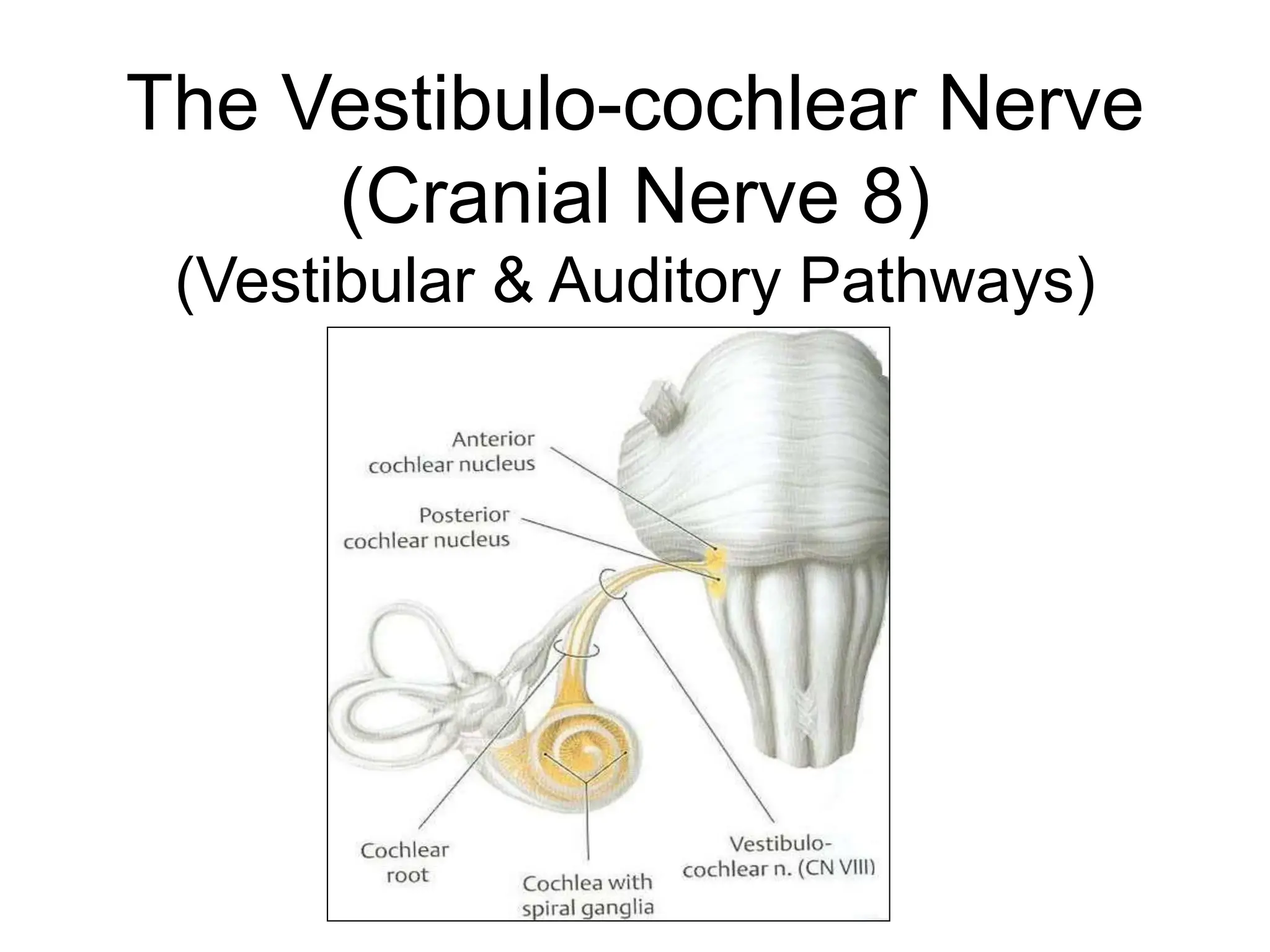
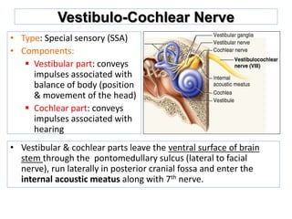
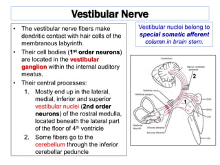
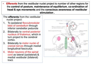
The Vestibulo-cochlear Nerve
(Cranial Nerve 8)
(Vestibular & Auditory Pathways)
Functional Components of Peripheral Nerves
Spinal Nerves
• General somatic afferents
(GSA)
• General somatic efferents
(GSE)
• General visceral afferents
(GVA)
• General viscera efferents
(GVE)
All spinal nerves carry all the
four components
Cranial Nerves
• GSA
• GSE
• GVA
• GVE
• Special somatic afferents (SSA)
• Special viscera afferents (SVA)
• Special visceral efferents (SVE)
NOT all cranial nerves carry all these
components
Vestibulo-Cochlear Nerve
• Type: Special sensory (SSA)
• Components:
 Vestibular part: conveys
impulses associated with
balance of body (position
& movement of the head)
 Cochlear part: conveys
impulses associated with
hearing
• Vestibular & cochlear parts leave the ventral surface of brain
stem through the pontomedullary sulcus (lateral to facial
nerve), run laterally in posterior cranial fossa and enter the
internal acoustic meatus along with 7th nerve.
Vestibular Nerve
• The vestibular nerve fibers make
dendritic contact with hair cells of the
membranous labyrinth.
• Their cell bodies (1st order neurons)
are located in the vestibular
ganglion within the internal auditory
meatus.
• Their central processes:
1. Mostly end up in the lateral,
medial, inferior and superior
vestibular nuclei (2nd order
neurons) of the rostral medulla,
located beneath the lateral part
of the floor of 4th ventricle
2. Some fibers go to the
cerebellum through the inferior
cerebellar peduncle
Vestibular nuclei belong to
special somatic afferent
column in brain stem.
1
2
The efferents from the vestibular
nuclei project:
1. To ipsilateral flocculonodular
lobe of cerebellum through
inferior cerebellar peduncle
2. Bilaterally to ventral posterior
nucleus of thalamus, which in
turn project to the cerebral
cortex.
3. Bilaterally to motor nuclei of
cranial nerves through medial
longitudinal fasciculus
4. Motor neurons of the spinal
cord as lateral (ipsilateral) &
medial vestibular (bilateral)
tract.
2
4
3
1
• Efferents from the vestibular nuclei project to number of other regions for
the control of posture, maintenance of equilibrium, co-ordination of
head & eye movements and the conscious awareness of vestibular
stimulation .
Medial Longitudinal Fasciculus
• Extends through out the brain
stem and formed of both
descending & ascending fibers
• Projects bilaterally
• Has two components:
The ascending component
establishes connections with
the nuclei of the Occulomotor,
Trochlear & Abducent nerves
(motor nuclei for extraoccular
muscles) for coordination of
head & eye movements.
The descending component
extends into the spinal cord as
the medial vestibulospinal tract
Vestibulospinal Tracts
• Vestibulospinal fibers influence
the activity of spinal motor
neurons concerned with the
control of body posture and
balance
• Two tracts: lateral & medial
• Lateral arises from lateral
vestibular (Deiter’s) nucleus,
descends ipsilaterally
• Medial is the descending part of
the medial longitudinal fasciculus,
projects bilaterally
Vestibular Cortex
• Located in the
lower part of
postcentral gyrus
(head area).
• Responsible for
conscious
awareness of
vestibular
sensation.
Auditory Pathway
• It is a multisynaptic pathway
• There are several locations between medulla and the
thalamus where axons may synapse and not all the
fibers behave in the same manner.
• Representation of cochlea is bilateral at all levels above
cochlear nuclei.
Cochlear (Auditory) Nerve
• The cochlear nerve fibers
make dendritic contact with
hair cells of the organ of
Corti within the cochlear
duct of the inner ear.
• Their cell bodies (1st order
neurons) are located within
the cochlea in the spiral
ganglion.
• Their central processes
terminate in the dorsal and
ventral cochlear nuclei (2nd
order neurons), which lie
close to the inferior
cerebellar peduncle (ICP)
Cochlear nuclei belong to
special somatic afferent
column in brain stem.
ICP
ICP
• From the cochlear nuclei,
2nd order neurons ascend
into the pons, where:
 Some fibers cross the
midline in trapezoid body
(1) and terminate in the
nucleus of trapezoid body
or in the contralateral
superior olivary nucleus (2)
 Some fibers run ipsilaterally
and terminate in the
superior olivary nucleus
• From the superior olivary
nuclei, ascending fibers
comprise the lateral
lemniscus (3), which runs
through tegmentum of
pons and terminate in the
inferior colliculus (4) of the
mdibrain.
2
2
1
3
4
3
4
• Some axons within lateral
lemniscus terminate in
small nucleus of the
lateral lemniscus (5)
• The inferior colliculi
project to medial
geniculate nuclei of
thalamus (6)
• The axons originating in
the medial geniculate
nucleus (auditory
radiation) pass through
sublenticular part of the
internal capsule to the
primary auditory cortex
(Brodmann’s areas 41,
42) located in the dorsal
surface of the superior
temporal gyrus (Heschl’s
gyri) (7)
5 5
3
6
6
7 7
3
2
2
1
4
4
Auditory radiation
• The region
surrounding the
primary auditory
cortex is known as
the auditory
association cortex or
Wernick’s area
(Brodmann’s areas 22)
• Wernick’s area is
related to
recognition and
processing of
language by the
brain
• Superior olivary nucleus sends olivocochlear fibers to
end in organ of Corti through the vestibulocochlear
nerve. These fibers are inhibitory in function and
serve to modulate transmission to the cochlear
nerve
• Superior olivary nucleus & the nucleus of the lateral
lemniscus establish reflex connections with motor
neurons of trigeminal and facial motor nuclei
mediating contraction of tensor tympani and
stapedius muscles in response to loud noise
• Inferior colliculi establish reflex connections with
motor neurons in the cervical spinal segments
(tectospinal tract) for the movement of head and
neck in response to auditory stimulation
Clinical Notes
• Lesion of vestibulocochlear nerve produces deafness
(disturbnce of cochlear nerve functions), tinnitis, vertigo,
dizziness, nausea, nystagmus, loss of balance and ataxia
(disturbnce of vestibular nerve functions)
Acoustic neuroma: a benign tumour of 8th nerve leads to
compression of the nerve leading to attacks of dizziness, and
profound deafness and ataxia
• The representation of cochlea is essentially bilateral at all
levels rostral to the cochlear nuclei
• Lesions anywhere along the pathway usually have no
obvious effect on hearing.
• Deafness is essentially only caused by damage to the middle
ear, cochlea, or auditory nerve.
11Cranial nerve 8 (Vestibulo-cochlear).ppt

11Cranial nerve 8 (Vestibulo-cochlear).ppt

  • 1.
    The Vestibulo-cochlear Nerve (CranialNerve 8) (Vestibular & Auditory Pathways)
  • 2.
    Functional Components ofPeripheral Nerves Spinal Nerves • General somatic afferents (GSA) • General somatic efferents (GSE) • General visceral afferents (GVA) • General viscera efferents (GVE) All spinal nerves carry all the four components Cranial Nerves • GSA • GSE • GVA • GVE • Special somatic afferents (SSA) • Special viscera afferents (SVA) • Special visceral efferents (SVE) NOT all cranial nerves carry all these components
  • 3.
    Vestibulo-Cochlear Nerve • Type:Special sensory (SSA) • Components:  Vestibular part: conveys impulses associated with balance of body (position & movement of the head)  Cochlear part: conveys impulses associated with hearing • Vestibular & cochlear parts leave the ventral surface of brain stem through the pontomedullary sulcus (lateral to facial nerve), run laterally in posterior cranial fossa and enter the internal acoustic meatus along with 7th nerve.
  • 4.
    Vestibular Nerve • Thevestibular nerve fibers make dendritic contact with hair cells of the membranous labyrinth. • Their cell bodies (1st order neurons) are located in the vestibular ganglion within the internal auditory meatus. • Their central processes: 1. Mostly end up in the lateral, medial, inferior and superior vestibular nuclei (2nd order neurons) of the rostral medulla, located beneath the lateral part of the floor of 4th ventricle 2. Some fibers go to the cerebellum through the inferior cerebellar peduncle Vestibular nuclei belong to special somatic afferent column in brain stem. 1 2
  • 5.
    The efferents fromthe vestibular nuclei project: 1. To ipsilateral flocculonodular lobe of cerebellum through inferior cerebellar peduncle 2. Bilaterally to ventral posterior nucleus of thalamus, which in turn project to the cerebral cortex. 3. Bilaterally to motor nuclei of cranial nerves through medial longitudinal fasciculus 4. Motor neurons of the spinal cord as lateral (ipsilateral) & medial vestibular (bilateral) tract. 2 4 3 1 • Efferents from the vestibular nuclei project to number of other regions for the control of posture, maintenance of equilibrium, co-ordination of head & eye movements and the conscious awareness of vestibular stimulation .
  • 6.
    Medial Longitudinal Fasciculus •Extends through out the brain stem and formed of both descending & ascending fibers • Projects bilaterally • Has two components: The ascending component establishes connections with the nuclei of the Occulomotor, Trochlear & Abducent nerves (motor nuclei for extraoccular muscles) for coordination of head & eye movements. The descending component extends into the spinal cord as the medial vestibulospinal tract
  • 7.
    Vestibulospinal Tracts • Vestibulospinalfibers influence the activity of spinal motor neurons concerned with the control of body posture and balance • Two tracts: lateral & medial • Lateral arises from lateral vestibular (Deiter’s) nucleus, descends ipsilaterally • Medial is the descending part of the medial longitudinal fasciculus, projects bilaterally
  • 8.
    Vestibular Cortex • Locatedin the lower part of postcentral gyrus (head area). • Responsible for conscious awareness of vestibular sensation.
  • 9.
    Auditory Pathway • Itis a multisynaptic pathway • There are several locations between medulla and the thalamus where axons may synapse and not all the fibers behave in the same manner. • Representation of cochlea is bilateral at all levels above cochlear nuclei.
  • 10.
    Cochlear (Auditory) Nerve •The cochlear nerve fibers make dendritic contact with hair cells of the organ of Corti within the cochlear duct of the inner ear. • Their cell bodies (1st order neurons) are located within the cochlea in the spiral ganglion. • Their central processes terminate in the dorsal and ventral cochlear nuclei (2nd order neurons), which lie close to the inferior cerebellar peduncle (ICP) Cochlear nuclei belong to special somatic afferent column in brain stem. ICP ICP
  • 11.
    • From thecochlear nuclei, 2nd order neurons ascend into the pons, where:  Some fibers cross the midline in trapezoid body (1) and terminate in the nucleus of trapezoid body or in the contralateral superior olivary nucleus (2)  Some fibers run ipsilaterally and terminate in the superior olivary nucleus • From the superior olivary nuclei, ascending fibers comprise the lateral lemniscus (3), which runs through tegmentum of pons and terminate in the inferior colliculus (4) of the mdibrain. 2 2 1 3 4 3 4
  • 12.
    • Some axonswithin lateral lemniscus terminate in small nucleus of the lateral lemniscus (5) • The inferior colliculi project to medial geniculate nuclei of thalamus (6) • The axons originating in the medial geniculate nucleus (auditory radiation) pass through sublenticular part of the internal capsule to the primary auditory cortex (Brodmann’s areas 41, 42) located in the dorsal surface of the superior temporal gyrus (Heschl’s gyri) (7) 5 5 3 6 6 7 7 3 2 2 1 4 4 Auditory radiation
  • 13.
    • The region surroundingthe primary auditory cortex is known as the auditory association cortex or Wernick’s area (Brodmann’s areas 22) • Wernick’s area is related to recognition and processing of language by the brain
  • 14.
    • Superior olivarynucleus sends olivocochlear fibers to end in organ of Corti through the vestibulocochlear nerve. These fibers are inhibitory in function and serve to modulate transmission to the cochlear nerve • Superior olivary nucleus & the nucleus of the lateral lemniscus establish reflex connections with motor neurons of trigeminal and facial motor nuclei mediating contraction of tensor tympani and stapedius muscles in response to loud noise • Inferior colliculi establish reflex connections with motor neurons in the cervical spinal segments (tectospinal tract) for the movement of head and neck in response to auditory stimulation
  • 15.
    Clinical Notes • Lesionof vestibulocochlear nerve produces deafness (disturbnce of cochlear nerve functions), tinnitis, vertigo, dizziness, nausea, nystagmus, loss of balance and ataxia (disturbnce of vestibular nerve functions) Acoustic neuroma: a benign tumour of 8th nerve leads to compression of the nerve leading to attacks of dizziness, and profound deafness and ataxia • The representation of cochlea is essentially bilateral at all levels rostral to the cochlear nuclei • Lesions anywhere along the pathway usually have no obvious effect on hearing. • Deafness is essentially only caused by damage to the middle ear, cochlea, or auditory nerve.