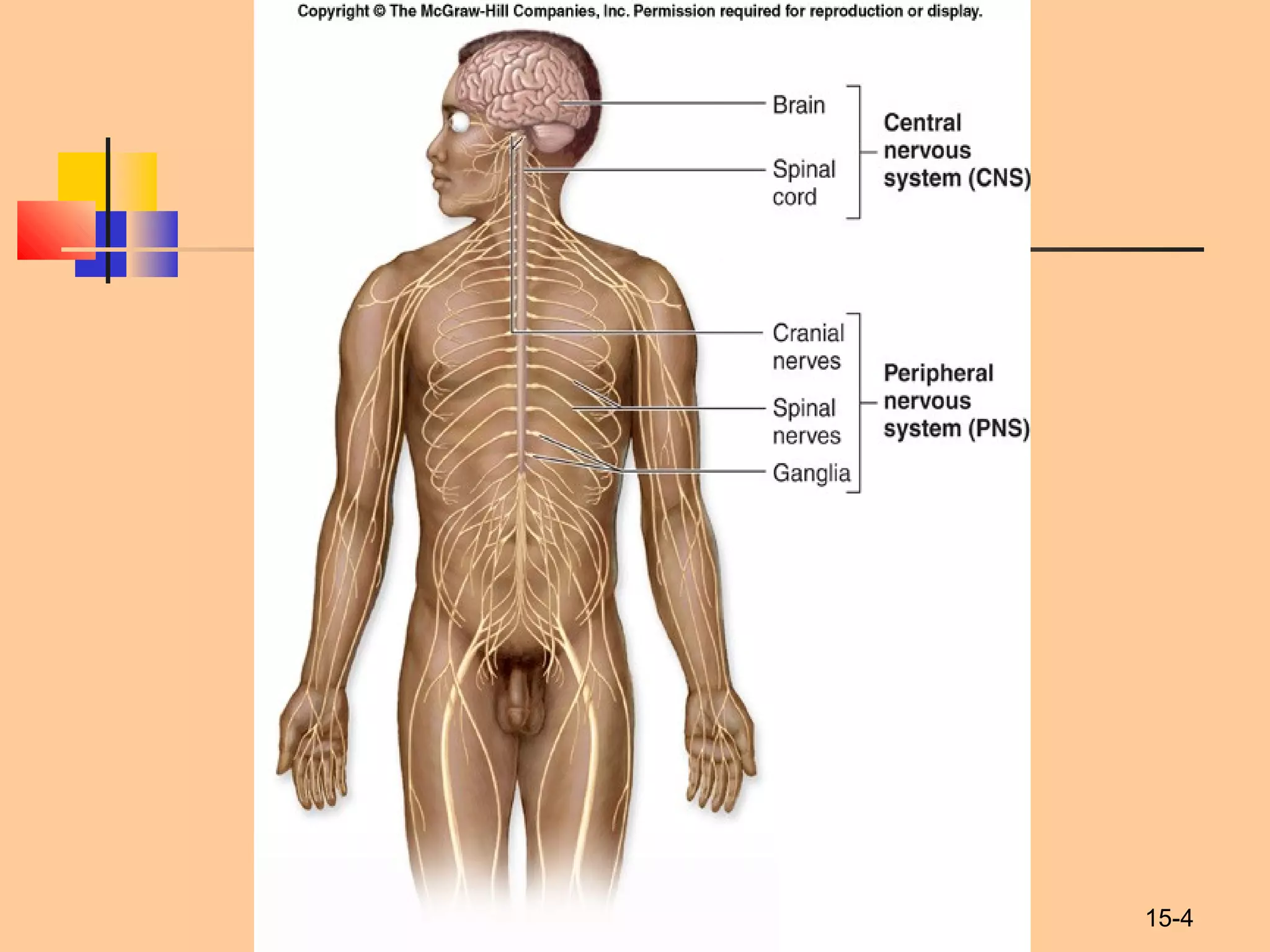
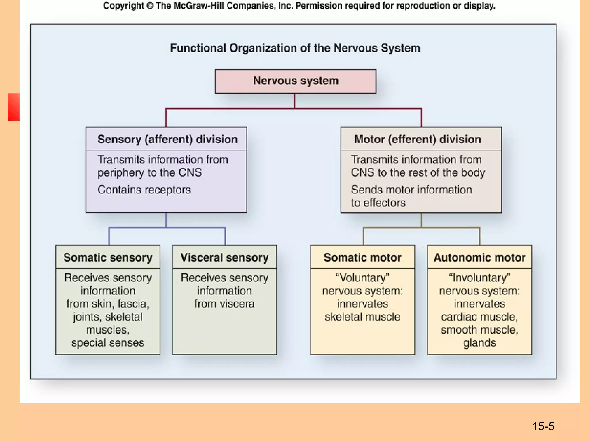
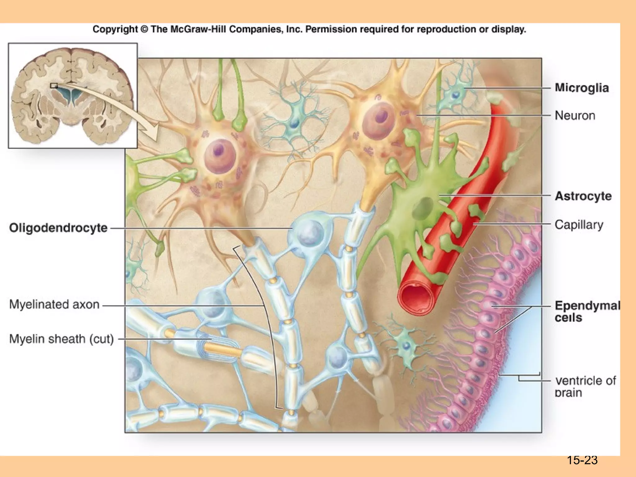
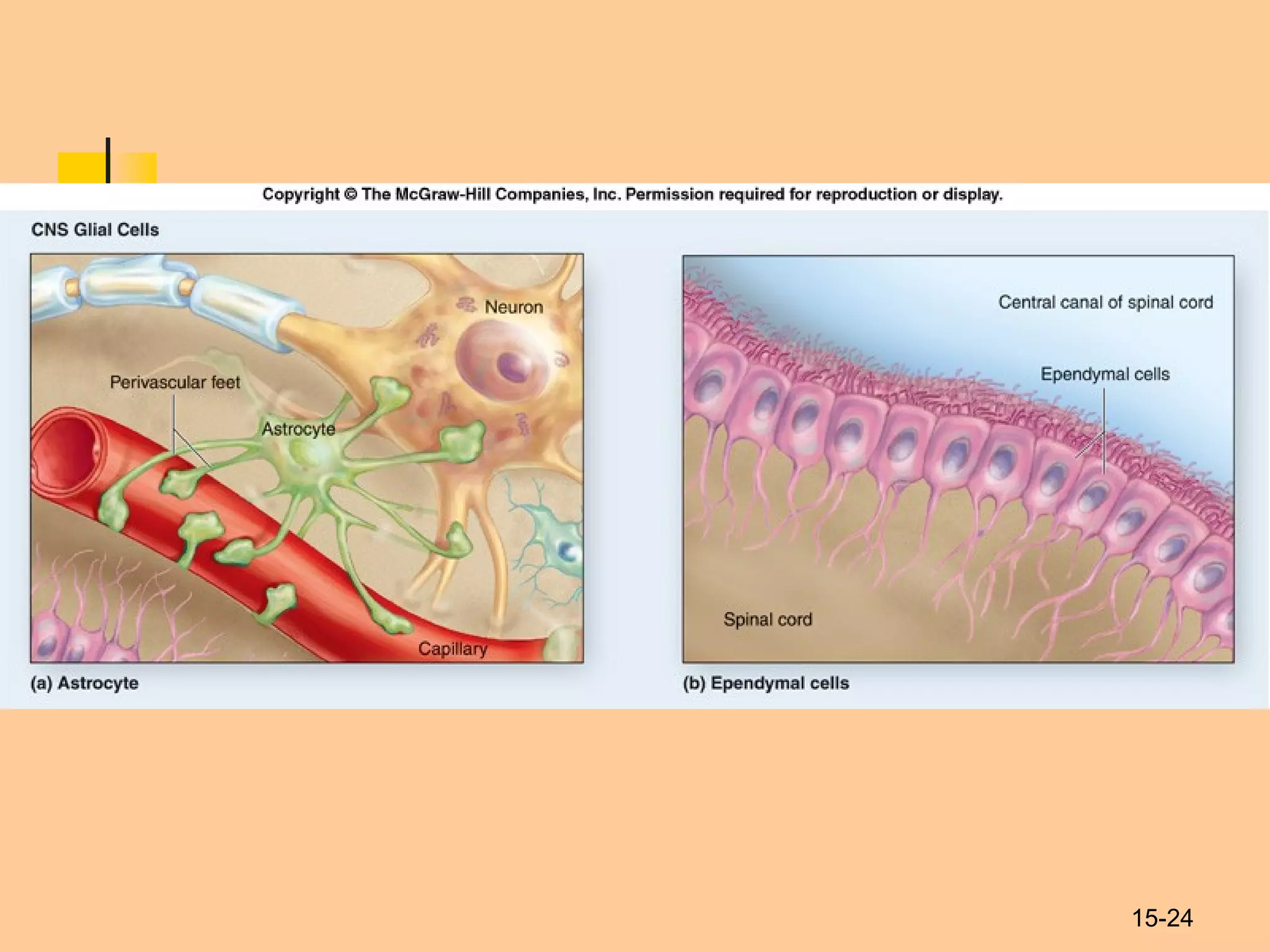
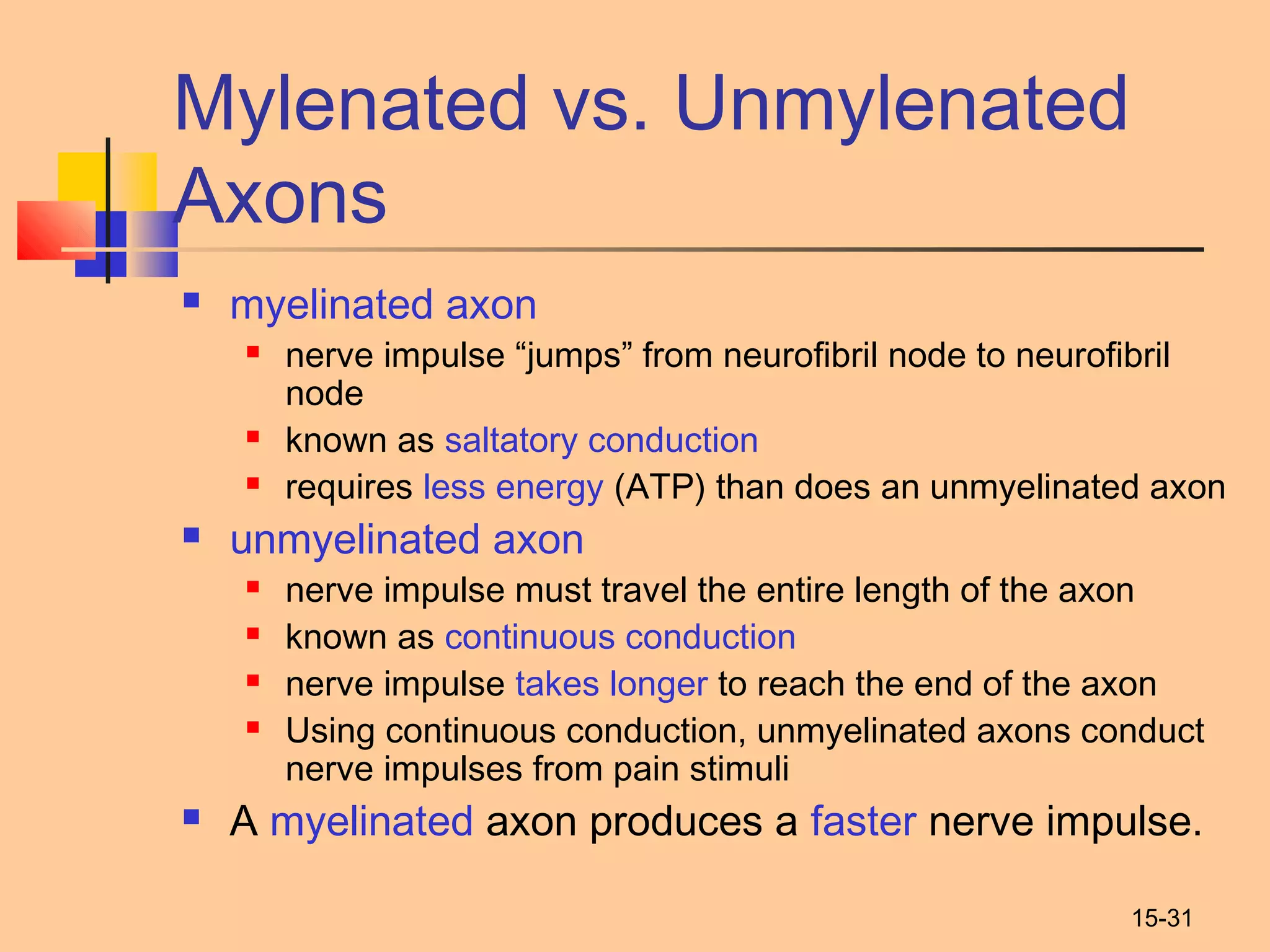
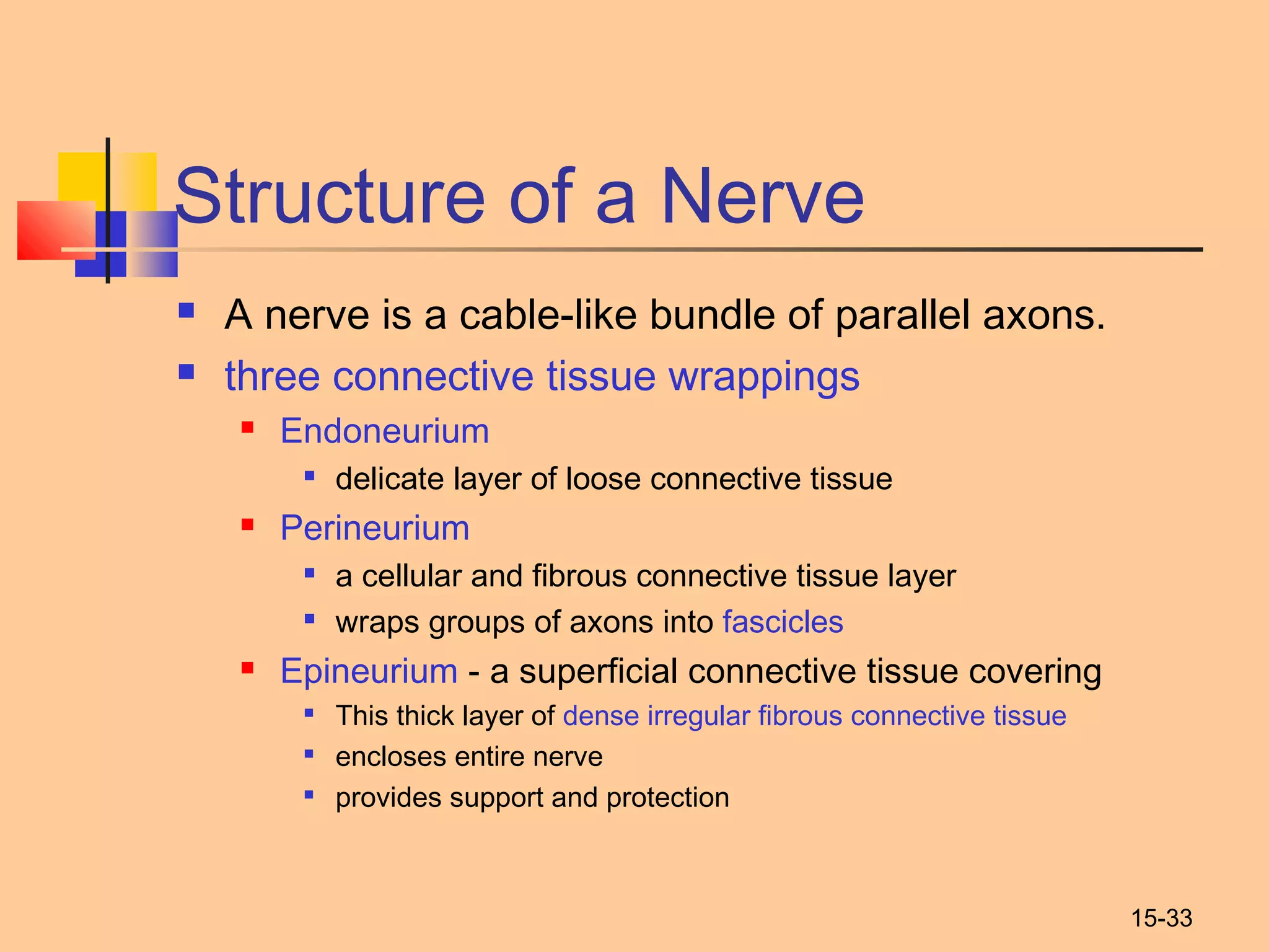
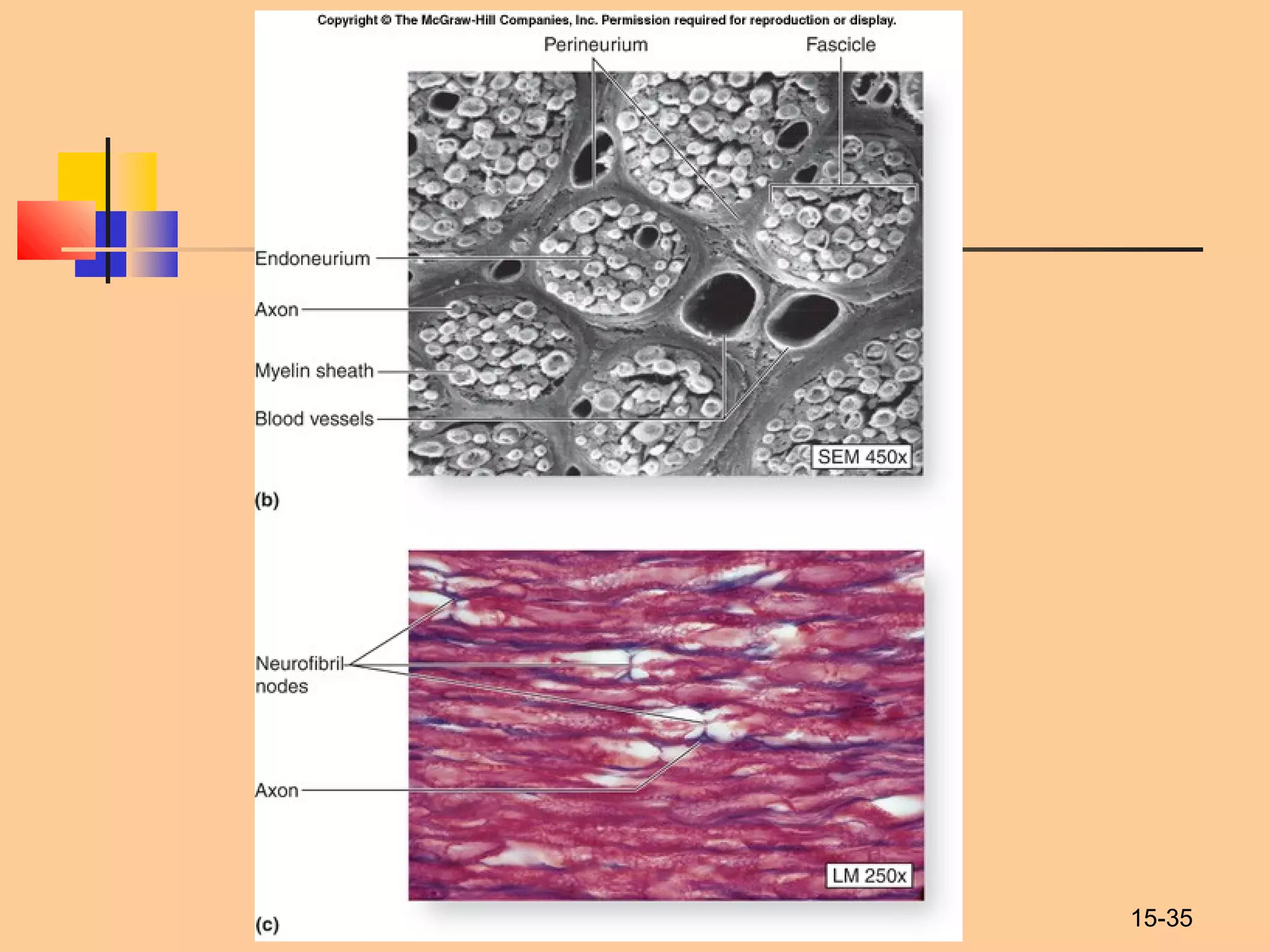
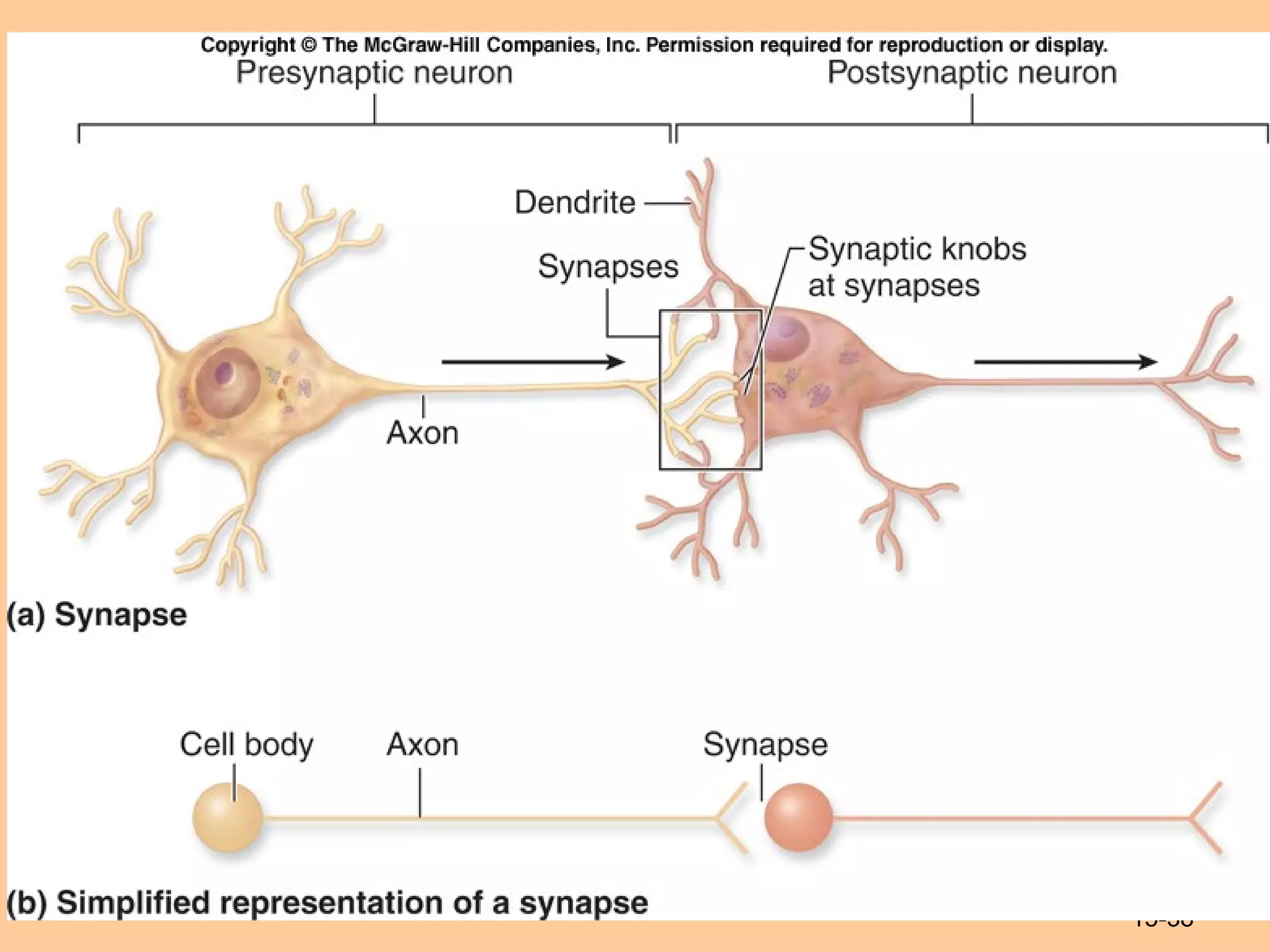
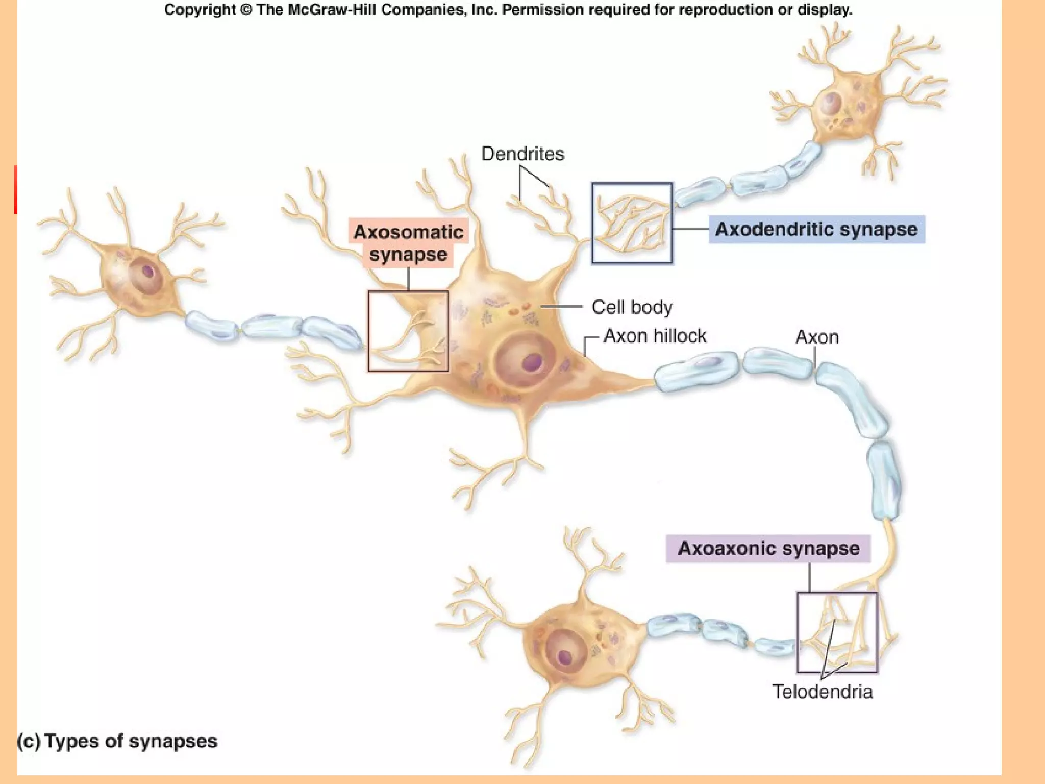
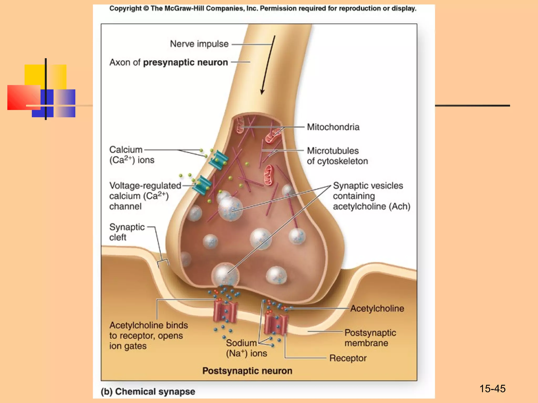
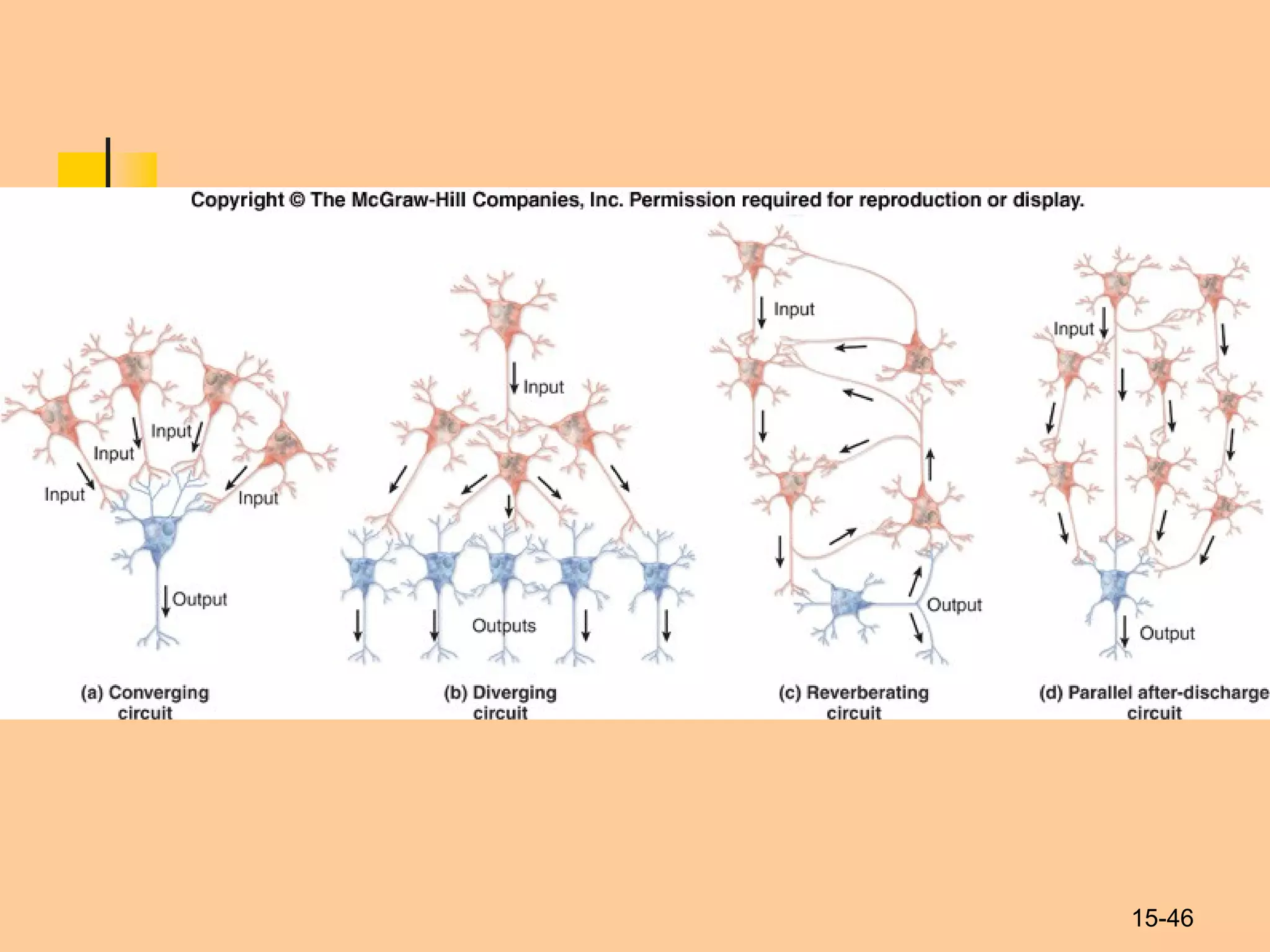
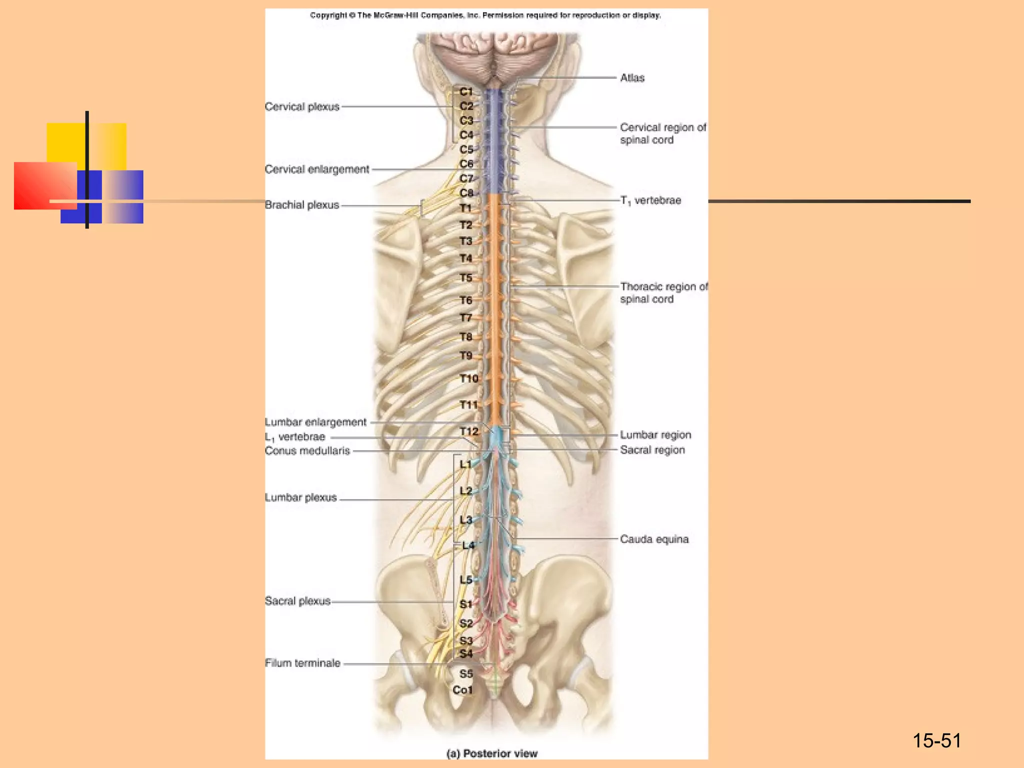
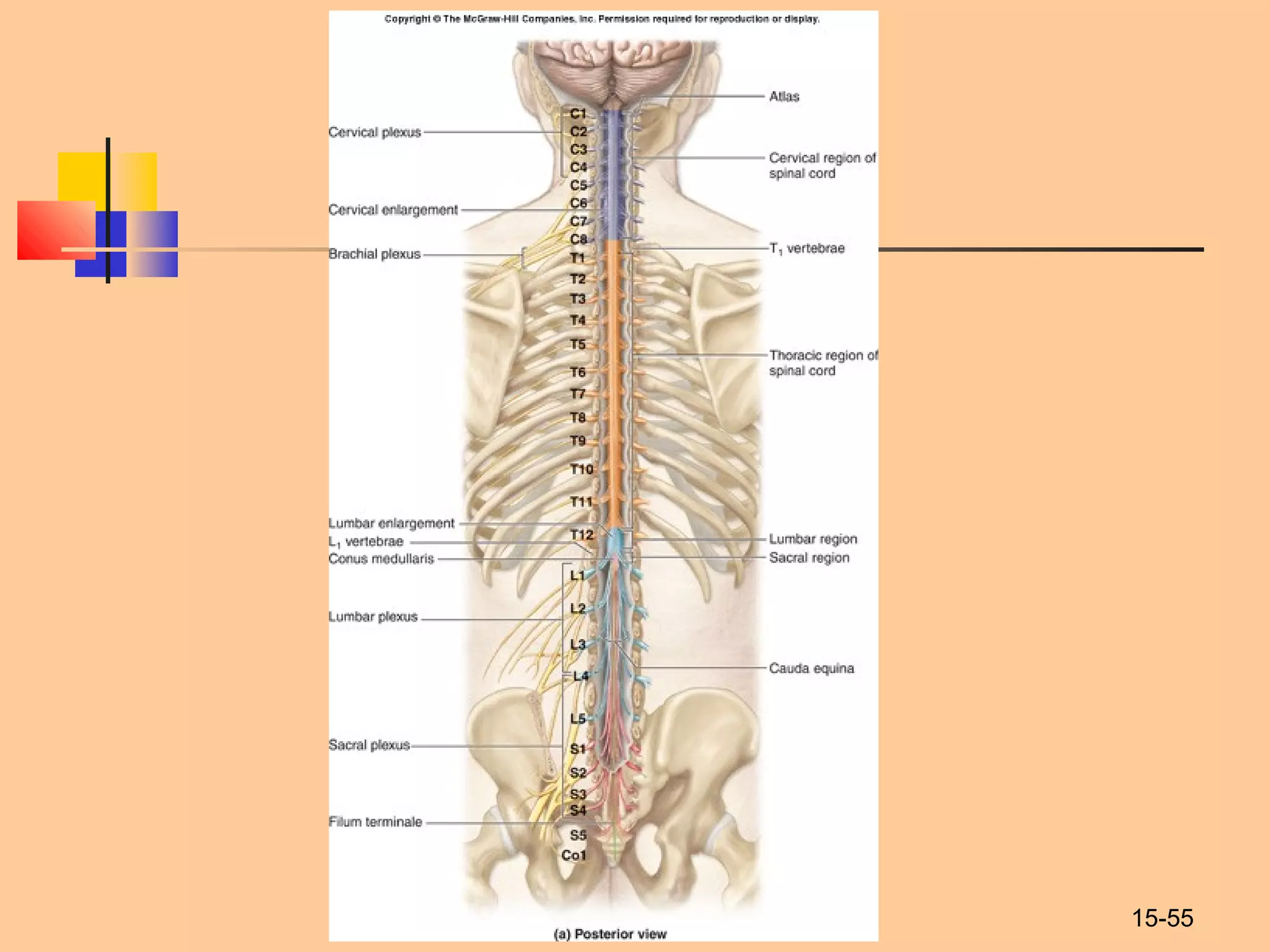
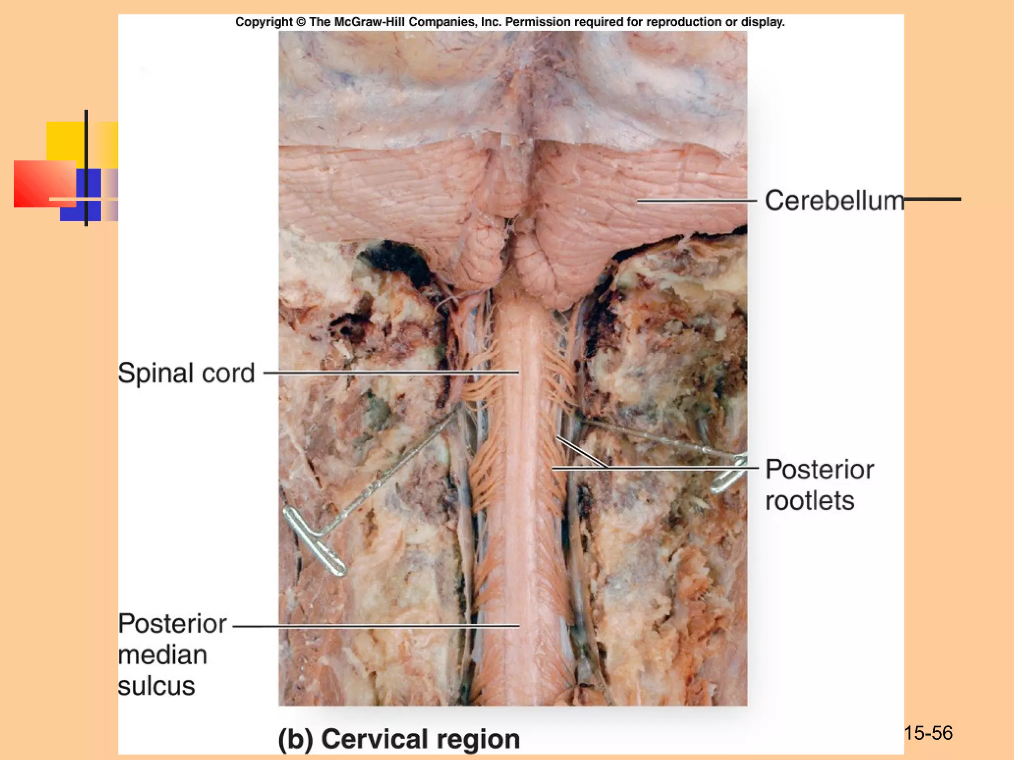
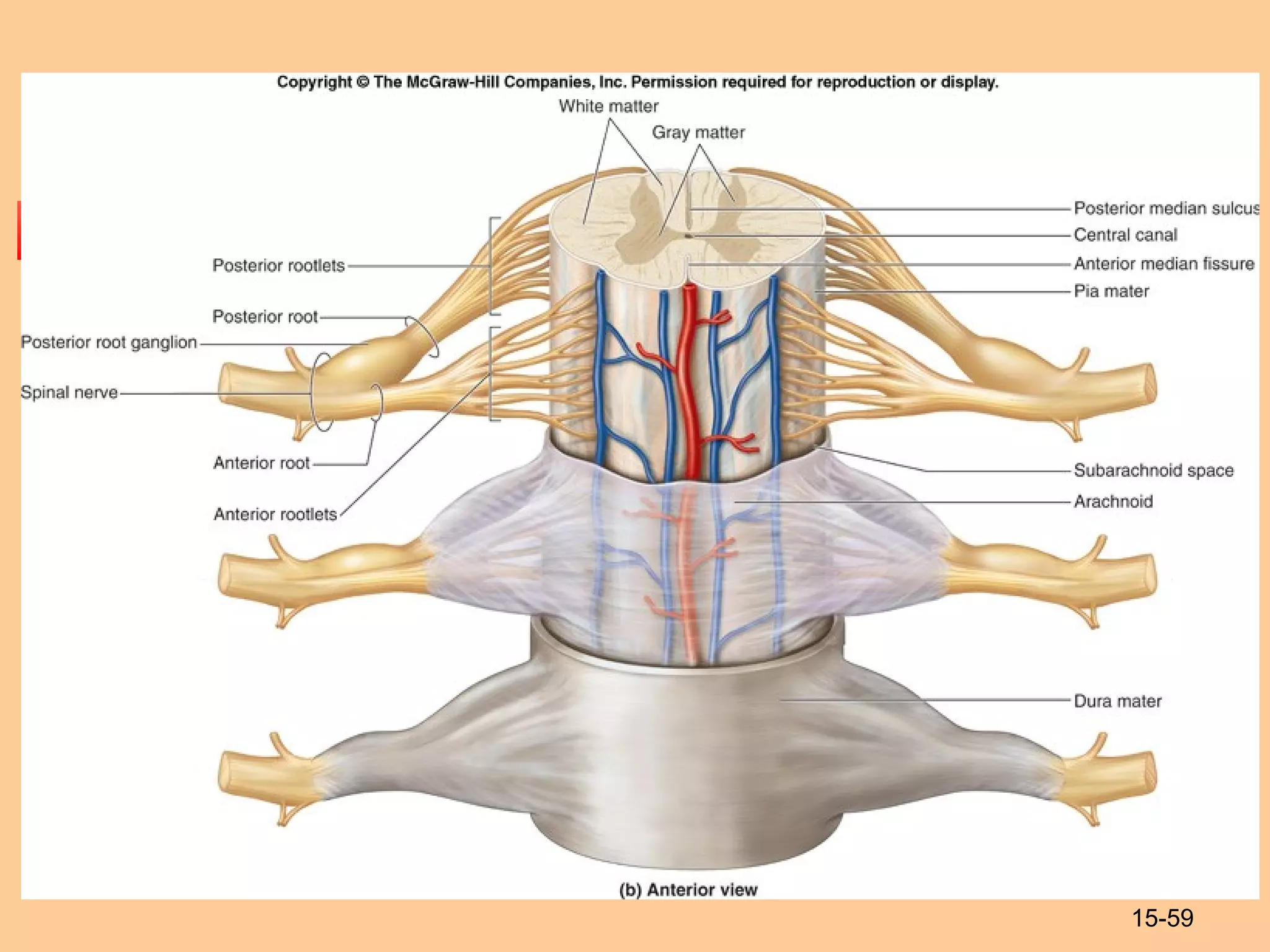
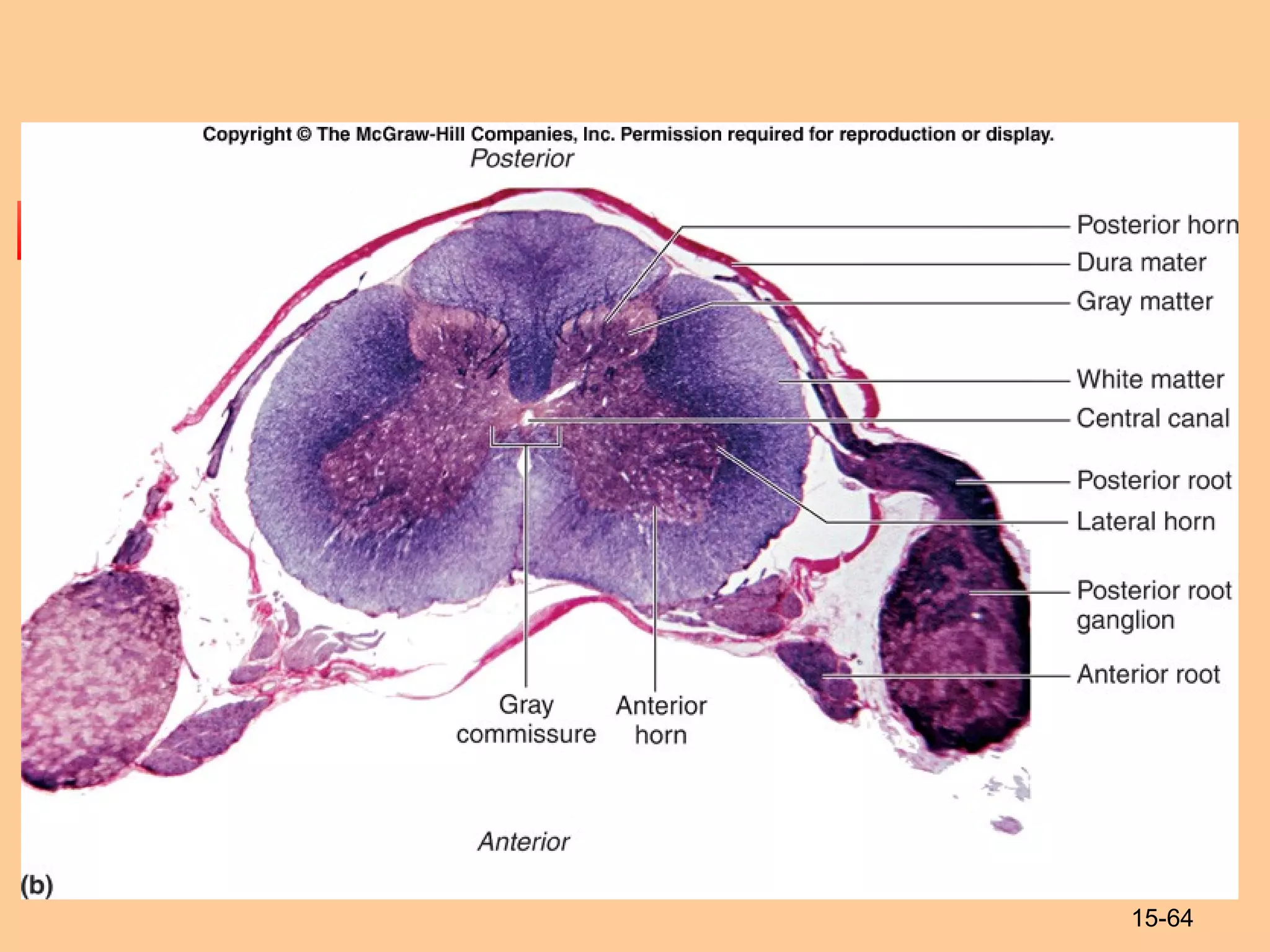
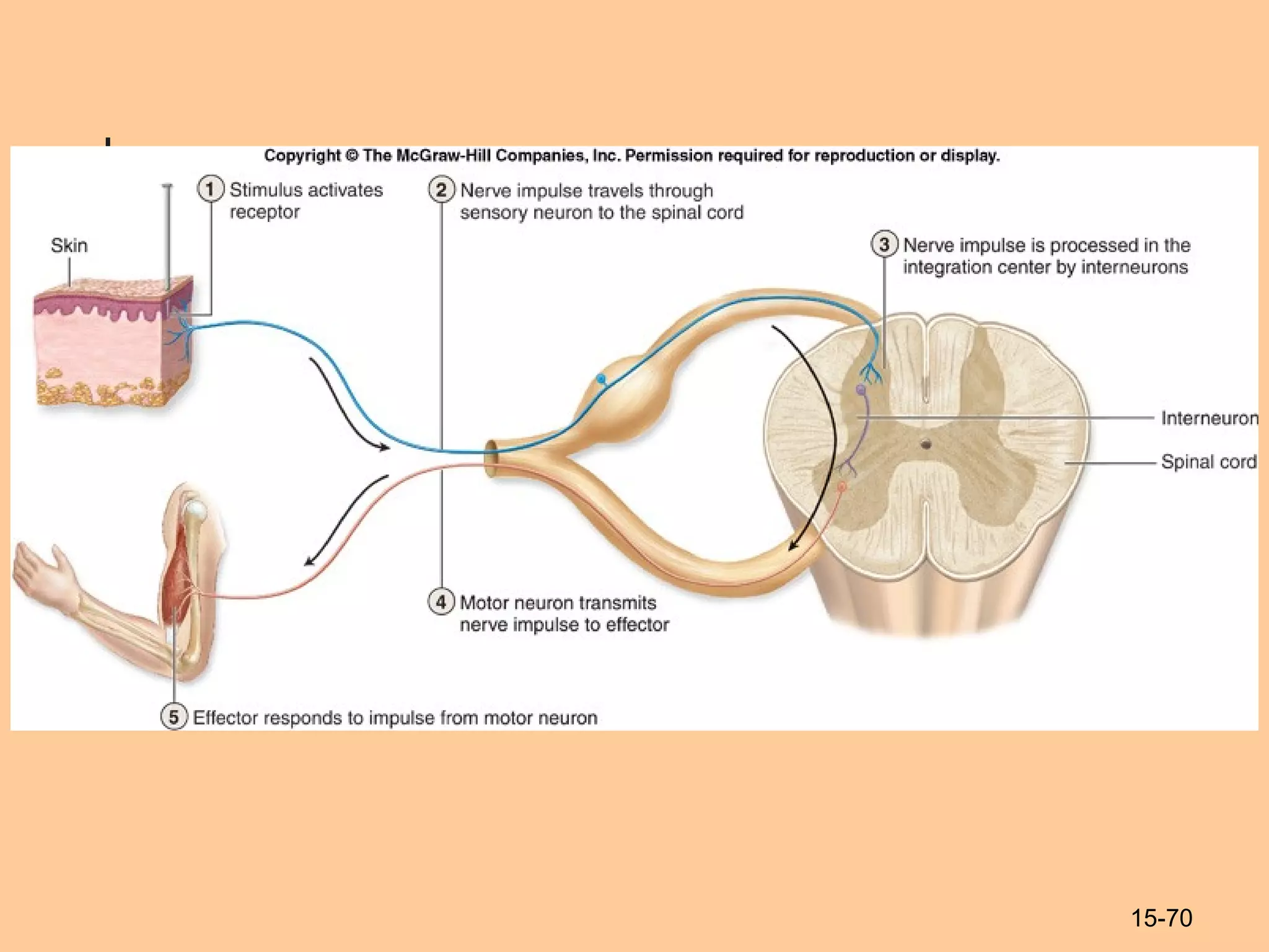
1
Ivano-Frankivsk National Medical
University
The Department of Human anatomy
The Nervous
System
Prepared by PhDPrepared by PhD
Tetyana Knyazevych - ChornaTetyana Knyazevych - Chorna
15-2
The Nervous System
 The body’s primary communication and
control system.
 Can be divided according to:
 Structural categories
 Functional categories.
15-3
Nervous System: Structural
Organization
Structural subdivisions of the nervous system:
 Central nervous system (CNS)
 brain and spinal cord
 Peripheral nervous system (PNS)
 cranial nerves (nerves that extend from the brain)
 spinal nerves (nerves that extend from the spinal cord)
 ganglia (clusters of neuron cell bodies (somas) located
outside the CNS)
15-4
15-5
15-6
 The central nervous system forms from the
embryonic neural tube that developes from
ectoderm.
 The neural plate gives rise to neural groove,
that later form the neural tube.
Development
15-7
Spinal Cord Development
 The cranial (superior) part of the neural
tube expands and develops into the
brain.
 The caudal (inferior) part of the neural
tube forms the spinal cord.
15-8
15-9
15-10
15-11
15-12
15-13
Nerve Cells
 Nervous Tissue
 Two distinct cell types

Neurons
 excitable cells
 initiate and transmit nerve impulses

Glial cells
 nonexcitable cells
 support and protect the neurons
15-14
Characteristics of Neurons
 Neurons have a high metabolic rate.
 Neurons have extreme longevity.
 Neurons typically are non-mitotic.
15-15
Neuron Structure
 Neurons come in all shapes and sizes
 All neurons share certain basic structural features.
 typical neuron:
 Cell body (soma, perikaryon)
 Dendrites
 Axon
15-16
15-17
15-18
Classifications of Neurons
 Neurons vary widely in morphology and
location.
 classified based on

structure

function.
 Structural classification: number of processes
extending from the cell body.
 unipolar neuron has a single process
 bipolar neurons have two processes
 multipolar neurons have three or more processes
15-19
15-20
Functional Classification
 Sensory afferent neurons: receptor to CNS
 Motor efferent neurons: CNS to effector
 Interneurons (association neurons): facilitate
communication between sensory and motor neurons.
15-21
15-22
Glial Cells
 Also called neuroglia
 Occur within both the CNS and the PNS.
 are smaller than neurons
 are capable of mitosis.
 do not transmit nerve impulses.
 Glial cells
 physically protect neurons
 help nourish neurons
 provide a supporting framework for all the nervous tissue.
 Glial cells far outnumber neurons.
 Glial cells account for about half the volume of the
nervous system.
15-23
15-24
15-25
Glial Cells of the PNS
 Satellite cells:
 Flattened cells
 Cover somas in ganglia
 Separate soma from surrounding tissue
fluid

Regulate exchange.
 Neurolemmocytes (Schwann cells)
 Myelination in the PNS
15-26
Myelination
 Process by which part of an axon is
wrapped with a myelin sheath
 Forms a protective fatty coating
 Has a glossy-white appearance.
 The myelin sheath:
 supports the axon
 protects the axon
 insulates an axon
15-27
Myelination
 No change in voltage can occur across the
membrane in the insulated portion of an axon.
 Voltage change occurs at the nodes
 Neurolemmocytes: form myelin sheaths in PNS
 Oligodendrocytes: form myelin sheaths in the CNS
15-28
15-29
15-30
15-31
Mylenated vs. Unmylenated
Axons
 myelinated axon
 nerve impulse “jumps” from neurofibril node to neurofibril
node
 known as saltatory conduction
 requires less energy (ATP) than does an unmyelinated axon
 unmyelinated axon
 nerve impulse must travel the entire length of the axon
 known as continuous conduction
 nerve impulse takes longer to reach the end of the axon
 Using continuous conduction, unmyelinated axons conduct
nerve impulses from pain stimuli
 A myelinated axon produces a faster nerve impulse.
15-32
15-33
Structure of a Nerve
 A nerve is a cable-like bundle of parallel axons.
 three connective tissue wrappings
 Endoneurium

delicate layer of loose connective tissue
 Perineurium

a cellular and fibrous connective tissue layer

wraps groups of axons into fascicles
 Epineurium - a superficial connective tissue covering

This thick layer of dense irregular fibrous connective tissue

encloses entire nerve

provides support and protection
15-34
15-35
15-36
Nerves
 Nerves are organs of the PNS.
 Sensory (afferent) nerves convey sensory information
to the CNS.
 Motor (efferent) nerves convey motor impulses from
the CNS to the muscles and glands.
 Mixed nerves: both sensory and motor
 Axons terminate as they contact other neurons,
muscle cells, or gland cells.
 An axon transmits a nerve impulse at a specialized
junction with another neuron called synapse.
15-37
Synapses
 Presynaptic neurons
 transmit nerve impulses toward a synapse.
 Postsynaptic neurons
 conduct nerve impulses away from the synapse.
 Axons may establish synaptic contacts with
any portion of the surface of another neuron
 except those regions that are myelinated.
15-38
15-39
Types of synapses: based on contacts
 axodendritic
 axosomatic
 axoaxonic
15-40
15-41
Main types of synapses
 Electrical synapses
 Gap junctions
 Chemical synapses
 Use neurotransmitters
15-42
Electrical Synapses
 Electrical synapses are not very common in
mammals.
 In humans, these synapses occur primarily between
smooth muscle cells where quick, uniform innervation
is essential.
 Electrical synapses are also located in cardiac
muscle.
15-43
15-44
Chemical Synapses
 Most numerous type of synapse
 Facilitates interactions
 between neurons
 between neurons and effectors.
 These are cell junctions
 Presynaptic membrane:
 releases a signaling molecule called a neurotransmitter,
such as acetylcholine (ACh).
 Other types of neurons use other neurotransmitters.
 Postsynaptic membrane:
 Contains receptors for neurotransmitters
15-45
15-46
15-47
The Spinal Cord
 Link between the brain and the body.
 Exhibits some functional independence from
the brain.
 The spinal cord and spinal nerves serve two
functions:
 pathway for sensory and motor impulses
 responsible for reflexes
15-48
Structure of the Spinal Cord
 Typical adult spinal cord
 ranges between 42 and 45 centimeters (cm) (16 to
18 inches) in length.
 In cross section
 roughly cylindrical
 slightly flattened both posteriorly and anteriorly.
 External surface has two longitudinal
depressions:
 the posterior (dorsal) median sulcus
 the anterior (ventral) median fissure
15-49
15-50
Regions of the Spinal Cord
 The cervical region
 continuous with the medulla oblongata
 contains neurons whose axons form the cervical spinal
nerves (8)
 The thoracic region
 attached to this region are the thoracic spinal nerves (12)
 The lumbar region
 contains the neurons for the lumbar spinal nerves (5)
 The sacral region
 contains the neurons for the sacral spinal nerves (5)
 The coccygeal region
 one pair of coccygeal spinal nerves arises from this region
15-51
15-52
Structure of the Spinal Cord
 The spinal cord is shorter than the vertebral canal
that houses it.
 Conus medullaris:
 tapered inferior end of the spinal cord
 marks the official “end” of the spinal cord proper.
 Cauda equina
 Inferior to conus medularis
 nerve roots (groups of axons) that project inferiorly from the
spinal cord.
 Filum terminale
 Within the cauda equina
 thin strand of pia mater
 helps anchor the conus medullaris to the coccyx.
15-53
15-54
Structure of the Spinal Cord
 The spinal cord is associated with 31 pairs of spinal
nerves
 Connect the CNS to:
 receptors
 effectors (muscle and glands)
 Each side of the spinal cord contains:
 8 cervical nerves (called C1–C8)
 12 thoracic nerves (T1–T12)
 5 lumbar nerves (L1–L5)
 5 sacral nerves (S1–S5)
 1 coccygeal nerve (Co1)
15-55
15-56
15-57
Arrangement and Functions of
the Spinal Meninges
 Are continuous with the cranial meninges.
 Structures that encircle the spinal cord, listed from
superficial to deep are:
 vertebra
 epidural space
 dura mater
 subdural space
 arachnoid
 subarachnoid space
 pia mater
15-58
15-59
15-60
Location and Distribution of
Gray Matter
 In the spinal cord, it is centrally located.
 Its shape resembles a letter H or a butterfly.
 The gray matter may be subdivided into the following
components:
 anterior horns
 lateral horns
 posterior horns
 the gray commissure
15-61
Location and Distribution of
White Matter
 The white matter of the spinal cord is
external to the gray matter.
 Three regions.
 Composed of tracts

Ascending

Descending
 A posterior funiculus:

lies between the posterior gray horns and the
posterior median sulcus.
15-62
Location and Distribution of
White Matter
 Lateral funiculus.
 Anterior funiculus
 between the anterior gray horns and the
anterior median fissure.
 The anterior funiculi are interconnected
by the white commissure.
15-63
15-64
15-65
Reflexes
 A reflex is a response:
 Rapid, automatic
 involuntary reactions of effectors to a stimulus.
 Properties.
 a stimulus

required to initiate a response to sensory input
 a rapid response

requires that few neurons be involved

synaptic delay be minimal
 an automatic response occurs the same way every time
 An involuntary response requires no intent or pre-awareness of
the reflex activity.
 Reflexes usually can not be suppressed.
 Awareness of the stimulus occurs after the reflex action
 in time to correct or avoid a potentially dangerous situation.
15-66
Components of a Reflex Arc
 The neural “wiring” of a single reflex.
 Always begins at a receptor in the PNS
 Sensory afferent
 Communicates with the CNS.
 May involve interneurons
 Ends at a peripheral effector (muscle or
gland)
 Motor efferent
15-67
Ipsilateral and Contralateral
Reflex Arcs
 Ipsilateral:
 both the receptor and effector organs of the reflex
are on the same side of the spinal cord.
 Contralateral
 the sensory impulses from a receptor organ cross
over through the spinal cord to activate effector
organs in the opposite side
15-68
Monosynaptic Reflexes
 The simplest of all reflexes.
 No interneurons.
 The patellar (knee-jerk) reflex is a
monosynaptic reflex
 physicians use to assess the functioning of the
spinal cord.
 tap the patellar ligament with a reflex hammer
 muscle spindles in the quadriceps muscles are
stretched.
 Produces a noticeable kick of the leg.
15-69
Polysynaptic Reflexes
 Have more complex neural pathways
 exhibit a number of synapses
 involve interneurons within the reflex arc.
 Has more components
 more prolonged delay between stimulus and response.
15-70
15-71
15-72
Stretch Reflexes
 Monosynaptic reflex that monitors and regulates
skeletal muscle length.
 When a stimulus results in the stretching of a muscle,
that muscle reflexively contracts.
 The patellar (knee-jerk) reflex is an example of a
stretch reflex.
 The stimulus (the tap on the patellar tendon) initiates
contraction of the quadriceps femoris muscle and
extension of the knee joint.
15-73
15-74
Golgi Tendon Reflex
 Prevents skeletal muscles from tensing excessively.
 Golgi tendon organs are nerve endings located within
tendons near a muscle–tendon junction.
 activation of the Golgi tendon organ signal interneurons in
the spinal cord, which in turn inhibit the actions of the motor
neurons
 The associated muscle is allowed to relax, thus
protecting the muscle and tendon from excessive
tension damage.
15-75
15-76
Reflex Testing in a Clinical
Setting
 Reflexes can be used to test specific muscle groups
and specific spinal nerves or segments of the spinal
cord.
 Consistently abnormal reflex response may indicate
damage to the nervous system or muscles.
 A reflex response may be normal, hypoactive, or
hyperactive.
15-77
Thank you for attention!

Nervous system

  • 1.
    1 Ivano-Frankivsk National Medical University TheDepartment of Human anatomy The Nervous System Prepared by PhDPrepared by PhD Tetyana Knyazevych - ChornaTetyana Knyazevych - Chorna
  • 2.
    15-2 The Nervous System The body’s primary communication and control system.  Can be divided according to:  Structural categories  Functional categories.
  • 3.
    15-3 Nervous System: Structural Organization Structuralsubdivisions of the nervous system:  Central nervous system (CNS)  brain and spinal cord  Peripheral nervous system (PNS)  cranial nerves (nerves that extend from the brain)  spinal nerves (nerves that extend from the spinal cord)  ganglia (clusters of neuron cell bodies (somas) located outside the CNS)
  • 4.
  • 5.
  • 6.
    15-6  The centralnervous system forms from the embryonic neural tube that developes from ectoderm.  The neural plate gives rise to neural groove, that later form the neural tube. Development
  • 7.
    15-7 Spinal Cord Development The cranial (superior) part of the neural tube expands and develops into the brain.  The caudal (inferior) part of the neural tube forms the spinal cord.
  • 8.
  • 9.
  • 10.
  • 11.
  • 12.
  • 13.
    15-13 Nerve Cells  NervousTissue  Two distinct cell types  Neurons  excitable cells  initiate and transmit nerve impulses  Glial cells  nonexcitable cells  support and protect the neurons
  • 14.
    15-14 Characteristics of Neurons Neurons have a high metabolic rate.  Neurons have extreme longevity.  Neurons typically are non-mitotic.
  • 15.
    15-15 Neuron Structure  Neuronscome in all shapes and sizes  All neurons share certain basic structural features.  typical neuron:  Cell body (soma, perikaryon)  Dendrites  Axon
  • 16.
  • 17.
  • 18.
    15-18 Classifications of Neurons Neurons vary widely in morphology and location.  classified based on  structure  function.  Structural classification: number of processes extending from the cell body.  unipolar neuron has a single process  bipolar neurons have two processes  multipolar neurons have three or more processes
  • 19.
  • 20.
    15-20 Functional Classification  Sensoryafferent neurons: receptor to CNS  Motor efferent neurons: CNS to effector  Interneurons (association neurons): facilitate communication between sensory and motor neurons.
  • 21.
  • 22.
    15-22 Glial Cells  Alsocalled neuroglia  Occur within both the CNS and the PNS.  are smaller than neurons  are capable of mitosis.  do not transmit nerve impulses.  Glial cells  physically protect neurons  help nourish neurons  provide a supporting framework for all the nervous tissue.  Glial cells far outnumber neurons.  Glial cells account for about half the volume of the nervous system.
  • 23.
  • 24.
  • 25.
    15-25 Glial Cells ofthe PNS  Satellite cells:  Flattened cells  Cover somas in ganglia  Separate soma from surrounding tissue fluid  Regulate exchange.  Neurolemmocytes (Schwann cells)  Myelination in the PNS
  • 26.
    15-26 Myelination  Process bywhich part of an axon is wrapped with a myelin sheath  Forms a protective fatty coating  Has a glossy-white appearance.  The myelin sheath:  supports the axon  protects the axon  insulates an axon
  • 27.
    15-27 Myelination  No changein voltage can occur across the membrane in the insulated portion of an axon.  Voltage change occurs at the nodes  Neurolemmocytes: form myelin sheaths in PNS  Oligodendrocytes: form myelin sheaths in the CNS
  • 28.
  • 29.
  • 30.
  • 31.
    15-31 Mylenated vs. Unmylenated Axons myelinated axon  nerve impulse “jumps” from neurofibril node to neurofibril node  known as saltatory conduction  requires less energy (ATP) than does an unmyelinated axon  unmyelinated axon  nerve impulse must travel the entire length of the axon  known as continuous conduction  nerve impulse takes longer to reach the end of the axon  Using continuous conduction, unmyelinated axons conduct nerve impulses from pain stimuli  A myelinated axon produces a faster nerve impulse.
  • 32.
  • 33.
    15-33 Structure of aNerve  A nerve is a cable-like bundle of parallel axons.  three connective tissue wrappings  Endoneurium  delicate layer of loose connective tissue  Perineurium  a cellular and fibrous connective tissue layer  wraps groups of axons into fascicles  Epineurium - a superficial connective tissue covering  This thick layer of dense irregular fibrous connective tissue  encloses entire nerve  provides support and protection
  • 34.
  • 35.
  • 36.
    15-36 Nerves  Nerves areorgans of the PNS.  Sensory (afferent) nerves convey sensory information to the CNS.  Motor (efferent) nerves convey motor impulses from the CNS to the muscles and glands.  Mixed nerves: both sensory and motor  Axons terminate as they contact other neurons, muscle cells, or gland cells.  An axon transmits a nerve impulse at a specialized junction with another neuron called synapse.
  • 37.
    15-37 Synapses  Presynaptic neurons transmit nerve impulses toward a synapse.  Postsynaptic neurons  conduct nerve impulses away from the synapse.  Axons may establish synaptic contacts with any portion of the surface of another neuron  except those regions that are myelinated.
  • 38.
  • 39.
    15-39 Types of synapses:based on contacts  axodendritic  axosomatic  axoaxonic
  • 40.
  • 41.
    15-41 Main types ofsynapses  Electrical synapses  Gap junctions  Chemical synapses  Use neurotransmitters
  • 42.
    15-42 Electrical Synapses  Electricalsynapses are not very common in mammals.  In humans, these synapses occur primarily between smooth muscle cells where quick, uniform innervation is essential.  Electrical synapses are also located in cardiac muscle.
  • 43.
  • 44.
    15-44 Chemical Synapses  Mostnumerous type of synapse  Facilitates interactions  between neurons  between neurons and effectors.  These are cell junctions  Presynaptic membrane:  releases a signaling molecule called a neurotransmitter, such as acetylcholine (ACh).  Other types of neurons use other neurotransmitters.  Postsynaptic membrane:  Contains receptors for neurotransmitters
  • 45.
  • 46.
  • 47.
    15-47 The Spinal Cord Link between the brain and the body.  Exhibits some functional independence from the brain.  The spinal cord and spinal nerves serve two functions:  pathway for sensory and motor impulses  responsible for reflexes
  • 48.
    15-48 Structure of theSpinal Cord  Typical adult spinal cord  ranges between 42 and 45 centimeters (cm) (16 to 18 inches) in length.  In cross section  roughly cylindrical  slightly flattened both posteriorly and anteriorly.  External surface has two longitudinal depressions:  the posterior (dorsal) median sulcus  the anterior (ventral) median fissure
  • 49.
  • 50.
    15-50 Regions of theSpinal Cord  The cervical region  continuous with the medulla oblongata  contains neurons whose axons form the cervical spinal nerves (8)  The thoracic region  attached to this region are the thoracic spinal nerves (12)  The lumbar region  contains the neurons for the lumbar spinal nerves (5)  The sacral region  contains the neurons for the sacral spinal nerves (5)  The coccygeal region  one pair of coccygeal spinal nerves arises from this region
  • 51.
  • 52.
    15-52 Structure of theSpinal Cord  The spinal cord is shorter than the vertebral canal that houses it.  Conus medullaris:  tapered inferior end of the spinal cord  marks the official “end” of the spinal cord proper.  Cauda equina  Inferior to conus medularis  nerve roots (groups of axons) that project inferiorly from the spinal cord.  Filum terminale  Within the cauda equina  thin strand of pia mater  helps anchor the conus medullaris to the coccyx.
  • 53.
  • 54.
    15-54 Structure of theSpinal Cord  The spinal cord is associated with 31 pairs of spinal nerves  Connect the CNS to:  receptors  effectors (muscle and glands)  Each side of the spinal cord contains:  8 cervical nerves (called C1–C8)  12 thoracic nerves (T1–T12)  5 lumbar nerves (L1–L5)  5 sacral nerves (S1–S5)  1 coccygeal nerve (Co1)
  • 55.
  • 56.
  • 57.
    15-57 Arrangement and Functionsof the Spinal Meninges  Are continuous with the cranial meninges.  Structures that encircle the spinal cord, listed from superficial to deep are:  vertebra  epidural space  dura mater  subdural space  arachnoid  subarachnoid space  pia mater
  • 58.
  • 59.
  • 60.
    15-60 Location and Distributionof Gray Matter  In the spinal cord, it is centrally located.  Its shape resembles a letter H or a butterfly.  The gray matter may be subdivided into the following components:  anterior horns  lateral horns  posterior horns  the gray commissure
  • 61.
    15-61 Location and Distributionof White Matter  The white matter of the spinal cord is external to the gray matter.  Three regions.  Composed of tracts  Ascending  Descending  A posterior funiculus:  lies between the posterior gray horns and the posterior median sulcus.
  • 62.
    15-62 Location and Distributionof White Matter  Lateral funiculus.  Anterior funiculus  between the anterior gray horns and the anterior median fissure.  The anterior funiculi are interconnected by the white commissure.
  • 63.
  • 64.
  • 65.
    15-65 Reflexes  A reflexis a response:  Rapid, automatic  involuntary reactions of effectors to a stimulus.  Properties.  a stimulus  required to initiate a response to sensory input  a rapid response  requires that few neurons be involved  synaptic delay be minimal  an automatic response occurs the same way every time  An involuntary response requires no intent or pre-awareness of the reflex activity.  Reflexes usually can not be suppressed.  Awareness of the stimulus occurs after the reflex action  in time to correct or avoid a potentially dangerous situation.
  • 66.
    15-66 Components of aReflex Arc  The neural “wiring” of a single reflex.  Always begins at a receptor in the PNS  Sensory afferent  Communicates with the CNS.  May involve interneurons  Ends at a peripheral effector (muscle or gland)  Motor efferent
  • 67.
    15-67 Ipsilateral and Contralateral ReflexArcs  Ipsilateral:  both the receptor and effector organs of the reflex are on the same side of the spinal cord.  Contralateral  the sensory impulses from a receptor organ cross over through the spinal cord to activate effector organs in the opposite side
  • 68.
    15-68 Monosynaptic Reflexes  Thesimplest of all reflexes.  No interneurons.  The patellar (knee-jerk) reflex is a monosynaptic reflex  physicians use to assess the functioning of the spinal cord.  tap the patellar ligament with a reflex hammer  muscle spindles in the quadriceps muscles are stretched.  Produces a noticeable kick of the leg.
  • 69.
    15-69 Polysynaptic Reflexes  Havemore complex neural pathways  exhibit a number of synapses  involve interneurons within the reflex arc.  Has more components  more prolonged delay between stimulus and response.
  • 70.
  • 71.
  • 72.
    15-72 Stretch Reflexes  Monosynapticreflex that monitors and regulates skeletal muscle length.  When a stimulus results in the stretching of a muscle, that muscle reflexively contracts.  The patellar (knee-jerk) reflex is an example of a stretch reflex.  The stimulus (the tap on the patellar tendon) initiates contraction of the quadriceps femoris muscle and extension of the knee joint.
  • 73.
  • 74.
    15-74 Golgi Tendon Reflex Prevents skeletal muscles from tensing excessively.  Golgi tendon organs are nerve endings located within tendons near a muscle–tendon junction.  activation of the Golgi tendon organ signal interneurons in the spinal cord, which in turn inhibit the actions of the motor neurons  The associated muscle is allowed to relax, thus protecting the muscle and tendon from excessive tension damage.
  • 75.
  • 76.
    15-76 Reflex Testing ina Clinical Setting  Reflexes can be used to test specific muscle groups and specific spinal nerves or segments of the spinal cord.  Consistently abnormal reflex response may indicate damage to the nervous system or muscles.  A reflex response may be normal, hypoactive, or hyperactive.
  • 77.