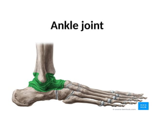
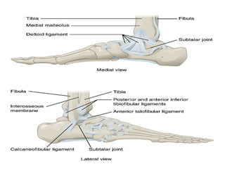
Ankle joint
• The ankle joint is a synovial joint located in the
lower limb.
• It is formed by the bones of the leg (tibia and
fibula) and the foot (talus).
• Functionally, it is a hinge type joint, permitting
dorsiflexion and plantarflexion of the foot.
• Articulating Surfaces
• The ankle joint is formed by three bones; the
tibia and fibula of the leg, and the talus of the
foot:
• The tibia and fibula are bound together by
strong tibiofibular ligaments.
• Together, they form a bracket shaped socket,
covered in hyaline cartilage.
• This socket is known as a mortise.
• The body of the talus fits snugly into the
mortise formed by the bones of the leg.
• The articulating part of the talus is wedge
shaped – it is broad anteriorly, and narrow
posteriorly:
• Dorsiflexion – the anterior part of the talus is
held in the mortise, and the joint is more stable.
• Plantarflexion – the posterior part of the talus is
held in the mortise, and the joint is less stable.
• Ligaments
• There are two main sets of ligaments, which originate from
each malleolus.
• Medial Ligament
• The medial ligament (or deltoid ligament) is attached to the
medial malleolus (a bony prominence projecting from the
medial aspect of the distal tibia).
• It consists of four ligaments, which fan out from the malleolus,
attaching to the talus, calcaneus and navicular bones.
• The primary action of the medial ligament is to resist over-
eversion of the foot.
Lateral Ligament
The lateral ligament originates from the lateral malleolus (a
bony prominence projecting from the lateral aspect of the
distal fibula).
It resists over-inversion of the foot, and is comprised of three
distinct and separate ligaments:
Anterior talofibular – spans between the lateral malleolus
and lateral aspect of the talus.
Posterior talofibular – spans between the lateral malleolus
and the posterior aspect of the talus.
Calcaneofibular – spans between the lateral malleolus and
the calcaneus.
• Movements and Muscles Involved
• The ankle joint is a hinge type joint, with
movement permitted in one plane.
• Thus, plantarflexion and dorsiflexion are the
main movements that occur at the ankle joint.
• Eversion and inversion are produced at the other
joints of the foot, such as the subtalar joint.
• Plantarflexion – produced by the muscles in
the posterior compartment of the leg
(gastrocnemius, soleus, plantaris and posterior
tibialis).
• Dorsiflexion – produced by the muscles in the
anterior compartment of the leg (tibialis
anterior, extensor hallucis longus and extensor
digitorum longus).
• Neurovascular Supply
• The arterial supply to the ankle joint is derived
from the malleolar branches of the anterior
tibial, posterior tibial and fibular arteries.
• Innervation is provided by tibial, superficial
fibular and deep fibular nerves.
• Clinical Relevance - Ankle Sprain
• An ankle sprain refers to partial or complete tears in the
ligaments of the ankle joint. It usually occurs via
excessive inversion to a plantarflexed and weight-
bearing foot.
• The lateral ligament is more likely to be damaged for
two main reasons:
• The lateral ligament is weaker than the medial ligament.
• The lateral ligament resists inversion.
• The anterior talofibular ligament is the lateral ligament
most at risk of irreversible damage
• Pott’s Fracture-Dislocation
• A Pott’s fracture is a term used to describe a bimalleolar
(medial and lateral malleoli) or trimalleolar (medial and
lateral malleoli, and distal tibia) fracture.
• This type of injury is produced by forced eversion of the
foot. It occurs in a series of stages:
• Forced eversion pulls on the medial ligaments, producing
an avulsion fracture of the medial malleolus.
• The talus moves laterally, breaking off the lateral malleolus.
• The tibia is then forced anteriorly, shearing off the distal
and posterior part against the talus
Ankle joint which are interesting to study

Ankle joint which are interesting to study

  • 1.
  • 2.
    • The anklejoint is a synovial joint located in the lower limb. • It is formed by the bones of the leg (tibia and fibula) and the foot (talus). • Functionally, it is a hinge type joint, permitting dorsiflexion and plantarflexion of the foot.
  • 3.
    • Articulating Surfaces •The ankle joint is formed by three bones; the tibia and fibula of the leg, and the talus of the foot: • The tibia and fibula are bound together by strong tibiofibular ligaments. • Together, they form a bracket shaped socket, covered in hyaline cartilage. • This socket is known as a mortise.
  • 5.
    • The bodyof the talus fits snugly into the mortise formed by the bones of the leg. • The articulating part of the talus is wedge shaped – it is broad anteriorly, and narrow posteriorly: • Dorsiflexion – the anterior part of the talus is held in the mortise, and the joint is more stable. • Plantarflexion – the posterior part of the talus is held in the mortise, and the joint is less stable.
  • 6.
    • Ligaments • Thereare two main sets of ligaments, which originate from each malleolus. • Medial Ligament • The medial ligament (or deltoid ligament) is attached to the medial malleolus (a bony prominence projecting from the medial aspect of the distal tibia). • It consists of four ligaments, which fan out from the malleolus, attaching to the talus, calcaneus and navicular bones. • The primary action of the medial ligament is to resist over- eversion of the foot.
  • 8.
    Lateral Ligament The lateralligament originates from the lateral malleolus (a bony prominence projecting from the lateral aspect of the distal fibula). It resists over-inversion of the foot, and is comprised of three distinct and separate ligaments: Anterior talofibular – spans between the lateral malleolus and lateral aspect of the talus. Posterior talofibular – spans between the lateral malleolus and the posterior aspect of the talus. Calcaneofibular – spans between the lateral malleolus and the calcaneus.
  • 9.
    • Movements andMuscles Involved • The ankle joint is a hinge type joint, with movement permitted in one plane. • Thus, plantarflexion and dorsiflexion are the main movements that occur at the ankle joint. • Eversion and inversion are produced at the other joints of the foot, such as the subtalar joint.
  • 10.
    • Plantarflexion –produced by the muscles in the posterior compartment of the leg (gastrocnemius, soleus, plantaris and posterior tibialis). • Dorsiflexion – produced by the muscles in the anterior compartment of the leg (tibialis anterior, extensor hallucis longus and extensor digitorum longus).
  • 11.
    • Neurovascular Supply •The arterial supply to the ankle joint is derived from the malleolar branches of the anterior tibial, posterior tibial and fibular arteries. • Innervation is provided by tibial, superficial fibular and deep fibular nerves.
  • 12.
    • Clinical Relevance- Ankle Sprain • An ankle sprain refers to partial or complete tears in the ligaments of the ankle joint. It usually occurs via excessive inversion to a plantarflexed and weight- bearing foot. • The lateral ligament is more likely to be damaged for two main reasons: • The lateral ligament is weaker than the medial ligament. • The lateral ligament resists inversion. • The anterior talofibular ligament is the lateral ligament most at risk of irreversible damage
  • 13.
    • Pott’s Fracture-Dislocation •A Pott’s fracture is a term used to describe a bimalleolar (medial and lateral malleoli) or trimalleolar (medial and lateral malleoli, and distal tibia) fracture. • This type of injury is produced by forced eversion of the foot. It occurs in a series of stages: • Forced eversion pulls on the medial ligaments, producing an avulsion fracture of the medial malleolus. • The talus moves laterally, breaking off the lateral malleolus. • The tibia is then forced anteriorly, shearing off the distal and posterior part against the talus