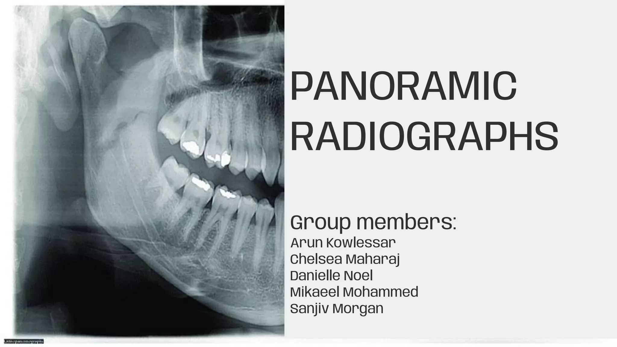
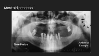
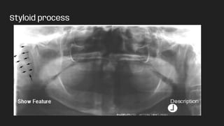
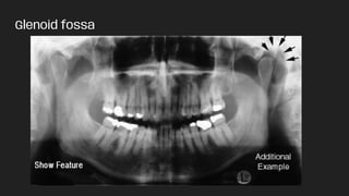
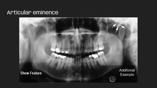
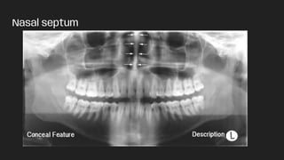
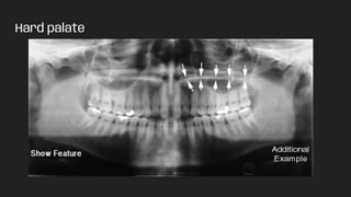
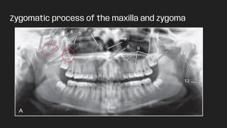
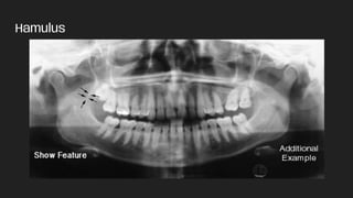
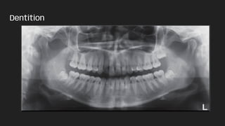
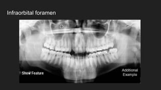
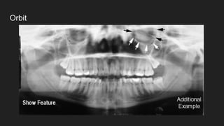
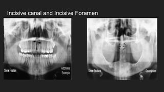
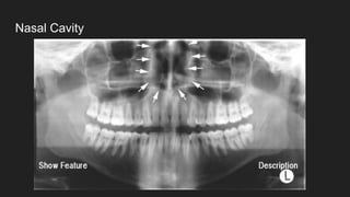
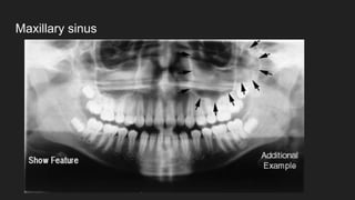
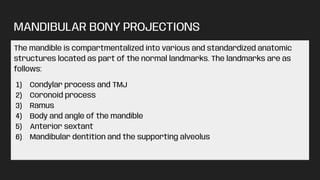
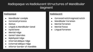
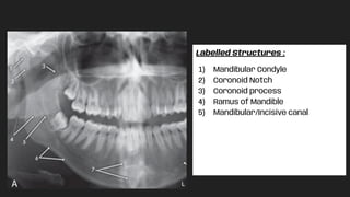
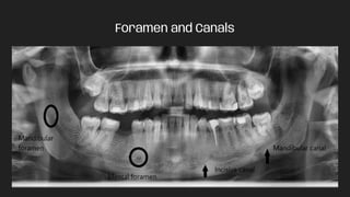
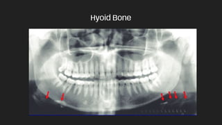
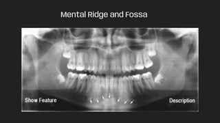
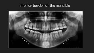
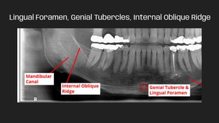
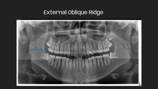
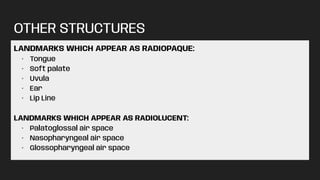
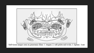
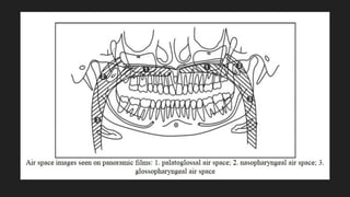
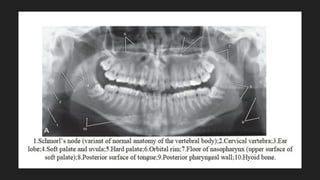
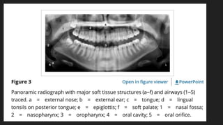
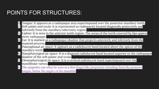
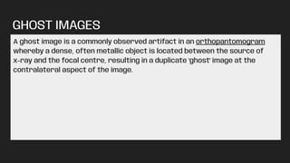
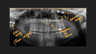
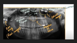
This document discusses various extra-oral radiographic techniques including:
1. Mandibular oblique lateral, true lateral, submento-vertex, occipitomental, postero-anterior, and reverse Towne projections.
2. The occipitomental view shows the facial skeleton, maxillary sinuses, and avoids superimposition of dense skull bones.
3. Linear tomography creates a tomographic cut where structures above and below are blurred while the focal plane is sharp. Multidirectional tomography is needed for a thin tomographic layer.
This document discusses panoramic radiography, including its history, advantages, procedure details, and principles of image formation. Panoramic radiography uses a rotating x-ray beam and receptor to create a single image of the facial structures, including teeth and supporting bones. It provides broad anatomic coverage with a low radiation dose compared to full-mouth intraoral x-rays. Proper patient positioning is needed to place the dental arches within the "focal trough" where structures will be reasonably defined.
The document discusses panoramic radiography, including:
- The focal trough is a curved zone where structures appear clearly on panoramic films.
- Panoramic machines rotate an x-ray tube and film cassette around the patient's head to produce a single image of the jaws.
- Positioning errors can produce artifacts that obscure anatomy, such as incorrect lip/tongue placement or improper Frankfort plane alignment. Precise patient positioning is important for diagnostic quality.
This document discusses common artifacts and positioning errors seen on panoramic radiographs. It describes ghost images, which are duplicate images caused when an object is penetrated twice by x-rays. It also discusses errors like open lips obscuring teeth, improper positioning of the chin resulting in overlapping structures, and movement during exposure causing blurring or duplication. Positioning the patient correctly in relation to the focal trough and keeping the spine straight are important to avoid errors.
Thorough knowledge of the indications of various extra oral techniques allows accurate and timely diagnosis of various maxillofacial pathologies. Further, we can arrive at a diagnosis with minimum number of x-rays there by reducing patient exposure to radiation.
Radiographic evaluation of midface fracturejyoti sharma
This document discusses the radiographic evaluation of midface fractures. It describes the LeFort classifications of midface fractures including LeFort I, II, and III fractures. LeFort I involves a horizontal maxillary fracture. LeFort II is a pyramidal fracture through the maxilla and nasal bones. LeFort III is a craniofacial disjunction that separates the midface from the cranium. Clinical features and radiographic findings are provided for each type of fracture. Radiographic evaluation includes panoramic imaging, CT scans, and MRI which are useful for detecting fractures and complications. Physical examination involves inspecting the head, eyes, ears, nose, throat, and neck for signs of midface trauma.
Panoramic radiography produces a single tomographic image of the facial structures including the maxillary and mandibular arches. It uses the principle of reciprocal movement of an X-ray source and image receptor around a central point or plane called the image layer. Multiple centers of rotation are used to maintain the dental arches within the focal trough. Panoramic radiography provides a broad anatomical view with less radiation than full-mouth intraoral films but has less resolution and potential for superimposition artifacts. It is useful for trauma evaluation, orthodontic treatment planning, and detection of lesions or developmental anomalies too large for intraoral films.
Technique and projection errors, exposure errors, and processing errors are the main categories of errors that can occur in dental radiographs. Technique errors include issues with patient preparation, film placement, and projection geometry. Exposure errors result in under or over exposure of the film. Processing errors stem from chemical or handling issues during film development and fixing. Addressing the root causes of errors in technique, exposure, and processing can help produce diagnostic quality radiographs and avoid unnecessary radiation exposure or wasted time and materials.
- Extraoral radiographs are used to examine large areas of the skull and jaws when intraoral films cannot be used. This document discusses various extraoral radiographic techniques including panoramic, skull, mandible, maxillary sinus, and temporomandibular joint views.
- Panoramic radiographs produce a single tomographic image of the facial structures and are commonly used. Skull views like lateral cephalograms evaluate facial growth while other views examine the skull vault or sinuses.
- Mandible views include lateral obliques of the body and ramus as well as posteroanterior projections. Maxillary sinus views use modifications of the Water's view.
- Temporomandibular
The document discusses various types of extraoral radiographs including lateral jaw projections, posteroanterior views, cephalometric radiographs, Water's views, reverse Towne projections, and submentovertex projections. It describes the purposes, techniques, patient positioning, and anatomical structures visualized for each type of extraoral radiograph. Extraoral radiographs are used to examine large areas of the jaws, skull, sinuses, and temporomandibular joints as well as to detect fractures, lesions, and developmental abnormalities.
This document discusses dental radiography and x-rays. It defines key terms like radiograph and radiology. It describes the components of a dental x-ray unit and different types of radiographs like bitewings and panoramic x-rays. It discusses techniques like paralleling, bisecting angle technique and SLOB technique. It also covers topics like indications for radiographs, radiation safety, and advantages and disadvantages of digital radiography compared to conventional film.
This document provides an overview of panoramic radiography. It discusses the history and development of panoramic radiography, the principles behind it including image layer and rotation center, equipment used, procedures for taking panoramic x-rays, common errors, clinical indications, advantages, and limitations. Panoramic radiography allows visualization of all teeth and supporting structures on a single film with a relatively low radiation dose.
The document describes several anatomical landmarks of the maxilla and mandible that are visible on dental radiographs. Key maxillary landmarks include the median palatine suture, nasal fossa, nasal septum, anterior nasal spine, incisive foramen, maxillary sinus, malar bone, maxillary tuberosity, hamular process, and nasolacrimal duct. Mandibular landmarks include the lingual foramen, genial tubercles, mental ridge, mental foramen, mental fossa, external and internal oblique lines, mylohyoid line, mandibular foramen, inferior dental canal, and submandibular gland fossa. These landmarks appear as radiopaque or
This document provides an overview of orthopantomography (OPG) and lateral cephalometric radiography. It defines OPG as a technique that produces a single tomographic image of the facial structures including the dental arches and supporting bones. The document lists several advantages of OPG including its broad coverage, low radiation dose, and short examination time. It describes the principles of panoramic image formation using reciprocal movement of the x-ray source and image receptor. Key diagnostic regions and structures visible on OPG images are identified. Common errors, artifacts, and positioning considerations are discussed. Lateral cephalometry is introduced as useful for orthodontic diagnosis and treatment planning by allowing evaluation of skeletal and dental abnormalities. References on
This document discusses infections that can occur in the five masticatory spaces. It describes the boundaries, contents, causes, clinical features, and treatment for infections of the pterygomandibular space, submasseteric space, and the three temporal spaces (superficial, infratemporal, and deep). Infections in these spaces can spread between adjacent spaces and present with symptoms like trismus, swelling, and pain. Treatment involves incision and drainage through intraoral, extraoral, or combined approaches depending on the specific infected space.
Differential diagnosis of swellings of head & neckSaleh Bakry
This document provides a differential diagnosis for various types of head and neck swellings that may be encountered during clinical examination. It discusses the steps that should be taken to properly identify and characterize lesions, including health history, examination of the lesion, radiographic examination, and biopsy if needed. Numerous potential diagnoses are provided for different locations of swellings including midline, lateral neck, pericoronal, periapical, interradicular, multilocular, and solitary lesions. Specific cysts, tumors, and other conditions are outlined depending on factors such as patient age, lesion location and characteristics.
Peripheral and central giant cell granulomaRijuwana77
This document discusses two types of non-epithelial tumours of the oral cavity: peripheral giant cell granuloma and central giant cell granuloma. Peripheral giant cell granuloma originates from the periodontal membrane or alveolar bone and presents as a soft tissue nodule composed of multinucleated giant cells. Central giant cell granuloma is a rare, benign, intraosseous lesion most commonly found in the mandible of young people that causes expansion of the bone and resorption of tooth roots. Both lesions contain proliferation of multinucleated giant cells and other cells and may require surgical excision, with central giant cell granuloma having a higher rate of recurrence.
Differential diagnosis of cysts of jawsSk Aziz Ikbal
This document discusses the differential diagnosis of various cysts of the jaws. It describes the clinical presentation and radiographic appearance of odontogenic keratocyst, eruption cyst, gingival cyst of adult, lateral periodontal cyst, radicular cyst, nasopalatine duct cyst, residual cyst, calcifying epithelial odontogenic cyst, dentigerous cyst, and globulomaxillary cyst. Key information provided includes location, symptoms, appearance on imaging, and conditions included in the differential diagnosis for each cyst type.
This document provides information on extraoral radiographic techniques. It discusses various extraoral views including lateral oblique, cephalometric, submentovertex, and zygomatic arch views. For each view, it describes the positioning of the patient's head, placement of the radiographic cassette and film, and path of the x-ray beam. It also discusses the components and function of screen-film systems used in extraoral radiography, including intensifying screens, screen speeds, and the advantages of Ektavision film over T-Mat film. Common cephalometric landmarks and their use in orthodontic assessment are also summarized.
Panoramic radiography produces a single tomographic image of the facial structures including the maxilla, mandible, and supporting bones. It works by employing tomography with a slit beam x-ray that moves in an arc around the patient. This technique allows for broad anatomical coverage with less radiation than intraoral films, but with less detail and potential for superimposition. The image can be interpreted by examining landmarks of the maxilla, mandible, air spaces, and soft tissues visible on the radiograph.
This document provides information on the radiographic appearance of structures in dental radiographs. It describes which structures appear radiopaque or radiolucent. Key radiopaque structures include enamel, dentin, cementum, lamina dura, alveolar crest, cancellous bone, genial tubercles, and mental ridge. Radiolucent structures include the pulp, periodontal ligament space, nutrient canals, lingual foramen, symphysis, mental fossa, and mandibular canal. Supporting structures like the lamina dura, alveolar crest, periodontal space, and cancellous bone are also detailed. Common mandibular landmarks are defined, along with how they appear
This document provides an overview of orthopantomography (OPG), also known as panoramic imaging. It discusses the principles of panoramic image formation using reciprocal movement of the x-ray source and image receptor around a central rotation point. Key components of panoramic machines and films are described. Proper patient positioning is emphasized to obtain images of the mandible, midfacial region, dentition and soft tissues within the image layer. Advantages include broad coverage with low radiation dose, while disadvantages include potential for distortion and lack of detail. Indications for OPG use include dental trauma, implants, orthodontics and lesion evaluation.
This document discusses the diagnosis, imaging, and treatment of zygomatic complex fractures. Signs and symptoms include periorbital ecchymosis, edema, pain, numbness, step deformity, trismus, subconjunctival hemorrhage, unequal pupillary levels, and diplopia. Imaging like coronal and axial CT scans with 3D reconstruction are beneficial. Treatment depends on the degree of fracture displacement and may include closed reduction without surgery for minimal displacement, open reduction with fixation using approaches like Gillies temporal or intraoral for arch or complex fractures, or open reduction with plate fixation at 1-4 points for complex fractures along with orbital floor reconstruction.
This document discusses panoramic x-rays and their use in dental practice. It describes how panoramic x-rays provide a wider view than traditional dental x-rays and allow visualization of gross anatomy and pathology. The document outlines advantages like field size and lower radiation dose compared to full mouth x-rays. Disadvantages discussed include potential image quality issues from distortion and superimposition. Proper patient positioning within the focal trough is emphasized as important for diagnostic image quality.
Panoramic radiography provides a wide view of the dental arches and associated structures using a rotating x-ray beam. It was developed starting in the 1920s to image the entire jaw at once. Modern panoramic machines use tomography to produce a single focused plane, known as the focal trough. This allows for detailed imaging of teeth and jaw structures while minimizing radiation exposure compared to full mouth x-rays. Panoramic images can reveal both normal anatomy as well as abnormalities, though some structures may appear as doubled "ghost images" due to the scanning technique.
This document discusses various radiographic projections used in extra oral radiology of the skull and temporomandibular joint. It describes several standard projections including occipitomental, posteroanterior, and lateral skull views. It explains the positioning techniques and main clinical indications for each view, such as detecting fractures or investigating sinuses. Radiographs have limitations for the temporomandibular joint but can show bone and joint relationships. Special imaging is needed to examine soft tissues of the TMJ.
This document discusses imaging modalities for the temporomandibular joint (TMJ). It begins by introducing the anatomy and components of the TMJ. For osseous structures, imaging options include panoramic radiography, plain film radiography, computed tomography (CT), and cone beam CT. Panoramic radiography is useful for detecting gross bony changes but does not show detail or joint positions. CT and cone beam CT provide three-dimensional bone images but not of soft tissues. For soft tissues like the articular disc, magnetic resonance imaging (MRI) is the best option, as it clearly depicts disc position and abnormalities. The document reviews the techniques and indications for various imaging modalities of both osseous
Radiographic anatomical landmarks By Dr. Armaan SinghDr. Armaan Singh
The document discusses various anatomical landmarks that are visible on dental radiographs. It begins by describing the radiographic appearance of tooth structures like enamel, dentin, cementum and the pulp. It then discusses supporting structures like the periodontal ligament space, lamina dura, alveolar crest and trabecular bone. Finally, it outlines the radiographic features of anatomical landmarks in the maxilla like the intermaxillary suture, anterior nasal spine, maxillary sinus and zygomatic process. It also describes landmarks in the mandible such as the mental foramen, mandibular canal, mylohyoid ridge and coronoid process.
This document discusses the anatomy and considerations for prosthodontics related to the tongue. It begins with an introduction and overview of the development, anatomy, histology, applied anatomy, anomalies, and prosthodontic reconstruction of the tongue. The anatomy section describes the parts and surfaces of the tongue, including the papillae and muscles. It also discusses the vascular supply, lymphatic drainage and nerve innervation. The prosthodontic section notes considerations for impressions, tongue size and position, teeth setting, and the role of the tongue in denture retention. It describes the use of a mandibular tongue prosthesis for reconstructed patients.
The Indian Dental Academy is the Leader in continuing dental education , training dentists in all aspects of dentistry and
offering a wide range of dental certified courses in different formats.for more details please visit
www.indiandentalacademy.com
The document describes various normal anatomical landmarks visible on dental radiographs, including teeth, supporting bone structures, and sinuses. It explains the radiographic appearance and density of structures like enamel, dentin, bone, and air-filled spaces. Landmarks discussed include the nasal septum, maxillary sinus, mental foramen, mandibular canal, and zygomatic process among others.
This document summarizes the anatomy of the face, including:
- The skin of the face is thick, elastic and highly vascular. It allows for rapid spread of edema.
- The facial skeleton contains 14 bones including the mandible, maxilla, nasal and zygomatic bones.
- Facial muscles originate from the mesoderm and function to open and close the 3 main orifices of the face. They include muscles of the nose, mouth and around the orbit.
The document summarizes key aspects of the tongue, teeth, and lymph drainage in the head and neck region. It describes the muscles and papillae of the tongue. It outlines the structure, layers, types and clinical importance of teeth. It details the regional and deep cervical lymph nodes that drain the head and neck area, including the mastoid, parotid, buccal, occipital, submandibular, submental, anterior cervical, superficial cervical, retropharyngeal, and laryngeal lymph nodes.
This document provides an overview of radiographic interpretation for periapical and panoramic dental x-rays. It discusses the objectives of understanding normal dental anatomy and interpreting pathology under radiographs. Key views covered include periapical and panoramic x-rays. The document outlines the normal radiographic anatomy seen in periapical and panoramic views and provides guidance on interpreting radiographs, including steps to localize, observe, consider generally, interpret, and correlate findings. Common dental structures and conditions that can be evaluated on radiographs are described, such as caries, pulp calcification, bone loss, restorations, and lesions.
Surgical anatomy of periodontal structures,Ankita Jain
This document provides an overview of the surgical anatomy of the periodontal structures, including the mandible and maxilla. It describes the anatomy, age-related changes, muscles, blood supply and nerves associated with these structures. Key anatomical spaces are also discussed, which are important for understanding the spread of infections in the head and neck region.
INTRODUCTIONSalivary glands are compound tubuloacinar, exocrine gland and the ducts opens in the oral cavity.
Salivary glands secretes a fluid called saliva that coats the teeth and the mucosa.
Saliva is a complex fluid, produced by the salivary glands, the most important function of which is to maintain the well- being of mouth.
Individuals with a deficiency of salivary secretion experience difficulty in eating, speaking, and swallowing and become prone to mucosal infections and dental caries.
Cephalometrics is the analysis of cephalometric radiographs to evaluate craniofacial structures and relationships. It is important for pediatric dentists to diagnose and treat developing malocclusions. Cephalometric landmarks are points on radiographs that guide measurements and plane construction. Common landmarks include nasion, sella, point A, point B, and pogonion. Tracings are made on acetate paper over radiographs using a sharp pencil to mark landmarks and assess skeletal and dental measurements and proportions.
Cephalometrics is the analysis and measurements made on cephalometric radiographs, which are standardized x-rays of the head used in orthodontics. Cephalometrics involves identifying anatomical landmarks, tracing radiographs, and analyzing relationships between craniofacial structures using reference planes and angular and linear measurements. It is an important diagnostic tool for orthodontists to evaluate dental and skeletal abnormalities, plan treatment, and assess treatment outcomes.
Teeth are composed primarily of dentin with an enamel cap and cementum layer. Enamel appears radiopaque due to its high mineral content. Dentin has lower mineral content and appears less radiopaque than enamel. The enamel-dentin junction is a distinct interface. The periodontal ligament appears as a radiolucent space between the root and lamina dura. The lamina dura is a thin radiopaque layer surrounding the tooth socket. Anatomical landmarks of the maxilla and mandible include the nasal fossa, maxillary sinus, mental foramen, and mandibular canal.
This document provides an overview of imaging modalities used in ENT and summarizes the anatomy and common pathologies of the ear, nose, sinuses, and throat. It discusses the basic anatomy of the ear including the external, middle, and inner ear. Common ENT pathologies are described such as chronic otitis media, cholesteatoma, otosclerosis, deviated septum, sinus polyps, and angiofibroma. Imaging findings of these conditions on modalities like CT and MRI are shown. The anatomy and diseases of the nose, sinuses, throat are also reviewed.
The document provides information about canine radiographs from the Royal Veterinary College. It includes radiographs and descriptions of the skull, mandible, tympanic bullae, frontal sinuses, and larynx from different views and angles. Users can click on the anatomy and radiographs to view labels and descriptions of the structures visible in each image.
This document describes various instruments used in otorhinolaryngology. It discusses nasal speculums like the Thudicum and Killian speculums used to examine the nose. Indirect laryngoscopy mirrors of varying sizes are described for examining the larynx. Other instruments mentioned include the Higginson syringe for nasal irrigation, osteotomes and chisels for bone cutting, curettes for bone removal, and various aural speculums for examining the ear canal.
Cell Therapy Expansion and Challenges in Autoimmune DiseaseHealth Advances
There is increasing confidence that cell therapies will soon play a role in the treatment of autoimmune disorders, but the extent of this impact remains to be seen. Early readouts on autologous CAR-Ts in lupus are encouraging, but manufacturing and cost limitations are likely to restrict access to highly refractory patients. Allogeneic CAR-Ts have the potential to broaden access to earlier lines of treatment due to their inherent cost benefits, however they will need to demonstrate comparable or improved efficacy to established modalities.
In addition to infrastructure and capacity constraints, CAR-Ts face a very different risk-benefit dynamic in autoimmune compared to oncology, highlighting the need for tolerable therapies with low adverse event risk. CAR-NK and Treg-based therapies are also being developed in certain autoimmune disorders and may demonstrate favorable safety profiles. Several novel non-cell therapies such as bispecific antibodies, nanobodies, and RNAi drugs, may also offer future alternative competitive solutions with variable value propositions.
Widespread adoption of cell therapies will not only require strong efficacy and safety data, but also adapted pricing and access strategies. At oncology-based price points, CAR-Ts are unlikely to achieve broad market access in autoimmune disorders, with eligible patient populations that are potentially orders of magnitude greater than the number of currently addressable cancer patients. Developers have made strides towards reducing cell therapy COGS while improving manufacturing efficiency, but payors will inevitably restrict access until more sustainable pricing is achieved.
Despite these headwinds, industry leaders and investors remain confident that cell therapies are poised to address significant unmet need in patients suffering from autoimmune disorders. However, the extent of this impact on the treatment landscape remains to be seen, as the industry rapidly approaches an inflection point.
share - Lions, tigers, AI and health misinformation, oh my!.pptxTina Purnat
• Pitfalls and pivots needed to use AI effectively in public health
• Evidence-based strategies to address health misinformation effectively
• Building trust with communities online and offline
• Equipping health professionals to address questions, concerns and health misinformation
• Assessing risk and mitigating harm from adverse health narratives in communities, health workforce and health system
8 Surprising Reasons To Meditate 40 Minutes A Day That Can Change Your Life.pptxHolistified Wellness
We’re talking about Vedic Meditation, a form of meditation that has been around for at least 5,000 years. Back then, the people who lived in the Indus Valley, now known as India and Pakistan, practised meditation as a fundamental part of daily life. This knowledge that has given us yoga and Ayurveda, was known as Veda, hence the name Vedic. And though there are some written records, the practice has been passed down verbally from generation to generation.
5-hydroxytryptamine or 5-HT or Serotonin is a neurotransmitter that serves a range of roles in the human body. It is sometimes referred to as the happy chemical since it promotes overall well-being and happiness.
It is mostly found in the brain, intestines, and blood platelets.
5-HT is utilised to transport messages between nerve cells, is known to be involved in smooth muscle contraction, and adds to overall well-being and pleasure, among other benefits. 5-HT regulates the body's sleep-wake cycles and internal clock by acting as a precursor to melatonin.
It is hypothesised to regulate hunger, emotions, motor, cognitive, and autonomic processes.
Mercurius is named after the roman god mercurius, the god of trade and science. The planet mercurius is named after the same god. Mercurius is sometimes called hydrargyrum, means ‘watery silver’. Its shine and colour are very similar to silver, but mercury is a fluid at room temperatures. The name quick silver is a translation of hydrargyrum, where the word quick describes its tendency to scatter away in all directions.
The droplets have a tendency to conglomerate to one big mass, but on being shaken they fall apart into countless little droplets again. It is used to ignite explosives, like mercury fulminate, the explosive character is one of its general themes.
The skin is the largest organ and its health plays a vital role among the other sense organs. The skin concerns like acne breakout, psoriasis, or anything similar along the lines, finding a qualified and experienced dermatologist becomes paramount.
Clinic ^%[+27633867063*Abortion Pills For Sale In Tembisa Central19various
Clinic ^%[+27633867063*Abortion Pills For Sale In Tembisa Central Clinic ^%[+27633867063*Abortion Pills For Sale In Tembisa CentralClinic ^%[+27633867063*Abortion Pills For Sale In Tembisa CentralClinic ^%[+27633867063*Abortion Pills For Sale In Tembisa CentralClinic ^%[+27633867063*Abortion Pills For Sale In Tembisa Central
Travel Clinic Cardiff: Health Advice for International TravelersNX Healthcare
Travel Clinic Cardiff offers comprehensive travel health services, including vaccinations, travel advice, and preventive care for international travelers. Our expert team ensures you are well-prepared and protected for your journey, providing personalized consultations tailored to your destination. Conveniently located in Cardiff, we help you travel with confidence and peace of mind. Visit us: www.nxhealthcare.co.uk
10 Benefits an EPCR Software should Bring to EMS Organizations Traumasoft LLC
The benefits of an ePCR solution should extend to the whole EMS organization, not just certain groups of people or certain departments. It should provide more than just a form for entering and a database for storing information. It should also include a workflow of how information is communicated, used and stored across the entire organization.
Does Over-Masturbation Contribute to Chronic Prostatitis.pptxwalterHu5
In some case, your chronic prostatitis may be related to over-masturbation. Generally, natural medicine Diuretic and Anti-inflammatory Pill can help mee get a cure.
Travel vaccination in Manchester offers comprehensive immunization services for individuals planning international trips. Expert healthcare providers administer vaccines tailored to your destination, ensuring you stay protected against various diseases. Conveniently located clinics and flexible appointment options make it easy to get the necessary shots before your journey. Stay healthy and travel with confidence by getting vaccinated in Manchester. Visit us: www.nxhealthcare.co.uk
26. MANDIBULAR BONY PROJECTIONS
The mandible is compartmentalized into various and standardized anatomic
structures located as part of the normal landmarks. The landmarks are as
follows:
1) Condylar process and TMJ
2) Coronoid process
3) Ramus
4) Body and angle of the mandible
5) Anterior sextant
6) Mandibular dentition and the supporting alveolus
35. OTHER STRUCTURES
LANDMARKS WHICH APPEAR AS RADIOPAQUE:
· Tongue
· Soft palate
· Uvula
· Ear
· Lip Line
LANDMARKS WHICH APPEAR AS RADIOLUCENT:
· Palatoglossal air space
· Nasopharyngeal air space
· Glossopharyngeal air space
36.
37.
38.
39.
40. POINTS FOR STRUCTURES:
1. Tongue: It appears as a radiopaque area superimposed over the posterior maxillary teeth.
2. Soft palate and uvula: It is represented as radiopacity located diagonally posteriorly and
inferiorly from the maxillary tuberosity region.
3. Lipline: It is seen in the anterior teeth region. The areas of the teeth covered by lips appear
more radiopaque.
4. Ear: It is marked as a radiopaque shadow that projects anteriorly and inferiorly from the
mastoid process.
5. Palatoglossal air space: It appears as a radiolucent band located above the apices of the
maxillary teeth.
6. Nasopharyngeal air space: It is a diagonal radiolucent band located superior to the radiopaque
shadow of the soft palate and uvula.
7. Glossopharyngeal air space: It is a vertical radiolucent band superimposed over the
mandibular ramus.
8. The epiglottis can often be seen as a thin finger-like projection extending from the posterior
tongue, below the angles of the mandible
41. GHOST IMAGES
A ghost image is a commonly observed artifact in an orthopantomogram
whereby a dense, often metallic object is located between the source of
x-ray and the focal centre, resulting in a duplicate 'ghost' image at the
contralateral aspect of the image.