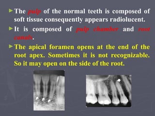
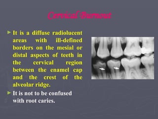
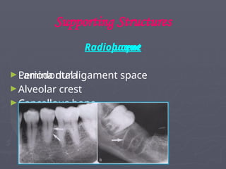
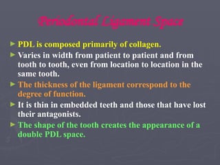
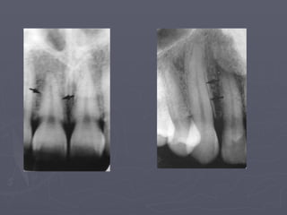
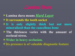
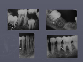
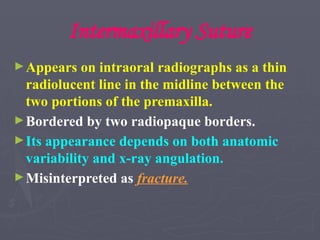
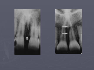
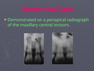
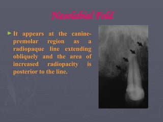
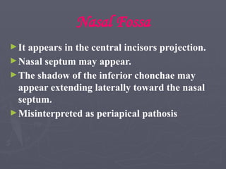
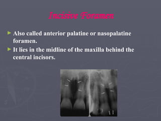
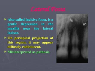
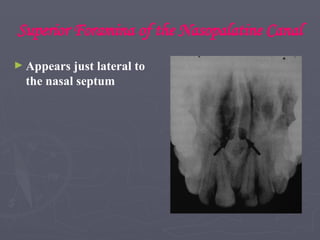
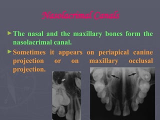
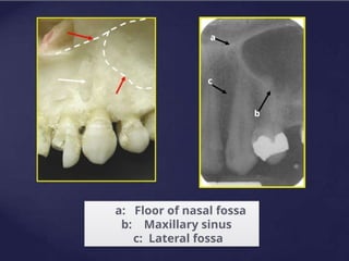
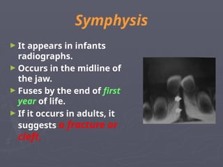
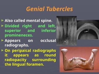
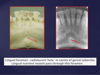
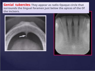
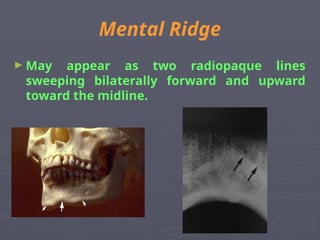
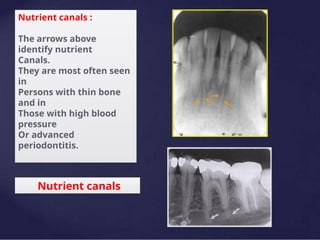
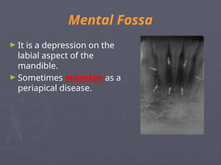
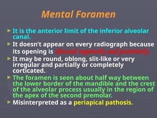
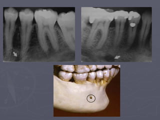
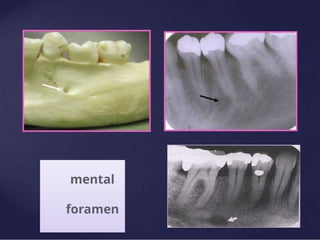
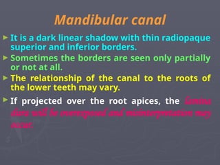
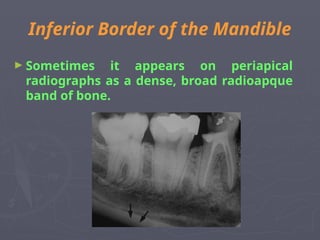
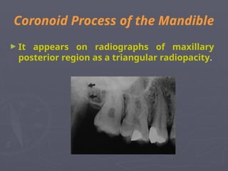
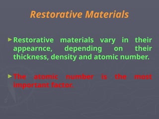
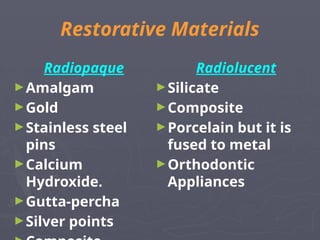
Normal Radiographic
Anatomy
Dr .Hidayah Elyas
► The radiographic recognition of disease requires a
sound knowledge of the radiographic appearance of
normal structures.
► Intelligent diagnosis mandates an appreciation of
the wide range of variation in the appearance of
normal anatomic structures.
Teeth
► Teeth are composed primarily of dentin, with an
enamel cap over the coronal portion and a thin layer
of cementum over the root surface.
► Enamel cap appears the most radiopaque.
► Dentin is less radiopaque and is roughly comparable
to the bone.
► The enamelo-dentinal junction appears as a distinct
interface that separates those two structures.
► Cementum is not usually apparent.
►The pulp of the normal teeth is composed of
soft tissue consequently appears radiolucent.
►It is composed of pulp chamber and root
canals.
►The apical foramen opens at the end of the
root apex. Sometimes it is not recognizable.
So it may open on the side of the root.
• In a mature tooth, the shape of the pulp
chamber and canals may change, with aging
occurs a gradual deposition of secondary
dentin which begins apically, proceeds
coronally and may lead to pulp obliteration.
• Trauma to the teeth also may stimulate
dentin production.
► Caries, restoration, attrition or erosion may
cause pulp obliteration.
Cervical Burnout
► It is a diffuse radiolucent
areas with ill-defined
borders on the mesial or
distal aspects of teeth in
the cervical region
between the enamel cap
and the crest of the
alveolar ridge.
► It is not to be confused
with root caries.
Supporting Structures
Radiolucent
►Periodontal ligament space
Radiopaque
►Lamina dura
►Alveolar crest
►Cancellous bone
Periodontal Ligament Space
► PDL is composed primarily of collagen.
► Varies in width from patient to patient and from
tooth to tooth, even from location to location in the
same tooth.
► The thickness of the ligament correspond to the
degree of function.
► It is thin in embedded teeth and those that have lost
their antagonists.
► The shape of the tooth creates the appearance of a
double PDL space.
Lamina Dura
►Lamina dura means Hard Layer
►It surrounds the tooth socket
►It is only slightly thick but not more
mineralized than the cancellous bone
►The thickness varies with the amount of
occlusal stress.
►Wider in heavy occlusion.
►Its presence is of valuable diagnostic feature
Alveolar Crest
► It is a thin radiopaque
line that extends between
the teeth.
► The level of the alveolar
crest is considered
normal when it is not
more than 1.5mm from
the cemento-enamel
junction.
► In anterior region it is
reduced to a point.
► Posteriorly, it is flat.
Cancellous bone
► Also called trabecular bone or spongiosa.
► Lies between the cortical plates in both jaws.
► It is composed of a thin radiopaque plates and rods
surrounding many small radiolucent pockets of
marrow.
► There is considerable variation between patients and
in the same patient.
► To evaluate a trabecular pattern, the practitioner
should examine the trabecular distribution, size and
density and compare them throughout both jaws.
Maxilla
Radiolucent Landmarks
1- Intermaxillary
suture
2- Nasal Fossa
3- Incisive foramen
4- Lateral Fossa
5- Superior foramina
of the nasopalatine
canals
Radiopaque Landmarks
1- Anterior nasal spine
2- Shadow of the nose
3- Zygomatic process
and zygomatic bone
4- Nasolabial fold
5- Pterygoid plates
Intermaxillary Suture
►Appears on intraoral radiographs as a thin
radiolucent line in the midline between the
two portions of the premaxilla.
►Bordered by two radiopaque borders.
►Its appearance depends on both anatomic
variability and x-ray angulation.
►Misinterpreted as fracture.
Anterior Nasal Spine
►Demonstrated on a periapical radiograph
of the maxillary central incisors.
Shadow of the Nose
► The soft tissue of the tip of
the nose may appear as a
radiopaque sharp border in
the projections of the
maxillary and lateral
incisors, superimposed over
the roots of these teeth.
Nasolabial Fold
► It appears at the canine-
premolar region as a
radiopaque line extending
obliquely and the area of
increased radiopacity is
posterior to the line.
Nasal Fossa
►It appears in the central incisors projection.
►Nasal septum may appear.
►The shadow of the inferior chonchae may
appear extending laterally toward the nasal
septum.
►Misinterpreted as periapical pathosis
Incisive Foramen
► Also called anterior palatine or nasopalatine
foramen.
► It lies in the midline of the maxilla behind the
central incisors.
Lateral Fossa
► Also called incisive fossa, is a
gentle depression in the
maxilla near the lateral
incisor.
► On periapical projection of
this region, it may appear
diffusely radiolucent.
► Misinterpreted as pathosis.
Superior Foramina of the Nasopalatine Canal
► Appears just lateral to
the nasal septum
Nasolacrimal Canals
►The nasal and the maxillary bones form the
nasolacrimal canal.
►Sometimes it appears on periapical canine
projection or on maxillary occlusal
projection.
a: Floor of nasal fossa
b: Maxillary sinus
c: Lateral fossa
a
b
c
Zygomatic Process and Zygomatic Bone
►Extension of the lateral maxillary surface in
the region of the apices of the first and second
molars.
►On periapical radiographs it appears a U-
shaped radiopaque line.
Maxillary Sinus
►The sinus is considered as a three sided
pyramid. The base (medial wall) adjacent to
the nasal cavity and the apex extending
laterally into the zygomatic process.
Maxillary sinus :
The maxillary sinus with
Its thin bony walls, its
thin mucosa, and its vast
Air space, produce an
Extremely dark image
Deep to the maxillary
Teeth.
Its outlines, particularly
its
Floor, are clearly
delineated
By delicate radio-opaque
lines
Pneumanization :expansion of sinus wall into
surrounding bone, usually in an area where
teeth have been lost prematurely.
It increases with age .
Pterygoid Plates
►The medial and lateral plates lie immediately
posterior to the tuberosity of the maxilla.
Mandible
Radiolucent Landmarks
►Symphysis
►Mental Fossa
►Mental Foramen
►Mandibular Canal
►Nutrient Canals
►Submandibular gland Fossa
Radiopaque Landmarks
►Genial Tubercles
►Mental Ridge
►Mylohyoid ridge
►External oblique ridge
►Inferior border of the mandible
►Coronoid process
Symphysis
► It appears in infants
radiographs.
► Occurs in the midline of
the jaw.
► Fuses by the end of first
year of life.
► If it occurs in adults, it
suggests a fracture or
cleft.
Genial Tubercles
► Also called mental spine.
► Divided right and left,
superior and inferior
promineneces.
► Appears on occlusal
radiographs.
► On peripaical radiographs
it appears as round
radiopacity surrounding
the lingual foramen.
Lingual foramen : radiolucent ‘hole ’ in centre of genial tubercles.
Lingual nutrient vessels pass through this foramen .
Genial tubercles: They appear as radio Opaque circle that
surrounds the lingual foramen just below the apices of the Of
the incisors.
Mental Ridge
► May appear as two radiopaque lines
sweeping bilaterally forward and upward
toward the midline.
Nutrient canals :
The arrows above
identify nutrient
Canals.
They are most often seen
in
Persons with thin bone
and in
Those with high blood
pressure
Or advanced
periodontitis.
Nutrient canals
Mental Fossa
► It is a depression on the
labial aspect of the
mandible.
► Sometimes mistaken as a
periapical disease.
Mental Foramen
► It is the anterior limit of the inferior alveolar
canal.
► It doesn’t appear on every radiograph because
its opening is directed superiorly and posteriorly.
► It may be round, oblong, slit-like or very
irregular and partially or completely
corticated.
► The foramen is seen about half way between
the lower border of the mandible and the crest
of the alveolar process usually in the region of
the apex of the second premolar.
► Misinterpreted as a periapical pathosis.
mental
foramen
Mandibular canal
► It is a dark linear shadow with thin radiopaque
superior and inferior borders.
► Sometimes the borders are seen only partially
or not at all.
► The relationship of the canal to the roots of
the lower teeth may vary.
► If projected over the root apices, the lamina
dura will be overexposed and misinterpretation may
occur.
Submandibular Gland Fossa
► It occurs on the lingual surface of the
mandibular body just below the myelohyoid
ridge.
► May be misinterpreted as a bony lesion.
Myelohyoid Ridge
►Irregular crest of bone on the lingual
surface of the mandibular body.
►Extending from the lower third molars
to the lower border of the mandible in
the region of the chin.
External Oblique Ridge
► It is the continuation of the
anterior border of the
mandibular ramus.
► It follows an antero-
inferior course lateral to
the alveolar process.
Inferior Border of the Mandible
► Sometimes it appears on periapical
radiographs as a dense, broad radioapque
band of bone.
Coronoid Process of the Mandible
► It appears on radiographs of maxillary
posterior region as a triangular radiopacity.
Restorative Materials
►Restorative materials vary in their
appearnce, depending on their
thickness, density and atomic number.
►The atomic number is the most
important factor.
Restorative Materials
Radiopaque
►Amalgam
►Gold
►Stainless steel
pins
►Calcium
Hydroxide.
►Gutta-percha
►Silver points
Radiolucent
►Silicate
►Composite
►Porcelain but it is
fused to metal
►Orthodontic
Appliances
7-Normal Radiographic Anatomy-updated..pptx

7-Normal Radiographic Anatomy-updated..pptx

  • 1.
  • 2.
    ► The radiographicrecognition of disease requires a sound knowledge of the radiographic appearance of normal structures. ► Intelligent diagnosis mandates an appreciation of the wide range of variation in the appearance of normal anatomic structures.
  • 3.
    Teeth ► Teeth arecomposed primarily of dentin, with an enamel cap over the coronal portion and a thin layer of cementum over the root surface. ► Enamel cap appears the most radiopaque. ► Dentin is less radiopaque and is roughly comparable to the bone. ► The enamelo-dentinal junction appears as a distinct interface that separates those two structures. ► Cementum is not usually apparent.
  • 5.
    ►The pulp ofthe normal teeth is composed of soft tissue consequently appears radiolucent. ►It is composed of pulp chamber and root canals. ►The apical foramen opens at the end of the root apex. Sometimes it is not recognizable. So it may open on the side of the root.
  • 6.
    • In amature tooth, the shape of the pulp chamber and canals may change, with aging occurs a gradual deposition of secondary dentin which begins apically, proceeds coronally and may lead to pulp obliteration. • Trauma to the teeth also may stimulate dentin production. ► Caries, restoration, attrition or erosion may cause pulp obliteration.
  • 7.
    Cervical Burnout ► Itis a diffuse radiolucent areas with ill-defined borders on the mesial or distal aspects of teeth in the cervical region between the enamel cap and the crest of the alveolar ridge. ► It is not to be confused with root caries.
  • 8.
    Supporting Structures Radiolucent ►Periodontal ligamentspace Radiopaque ►Lamina dura ►Alveolar crest ►Cancellous bone
  • 9.
    Periodontal Ligament Space ►PDL is composed primarily of collagen. ► Varies in width from patient to patient and from tooth to tooth, even from location to location in the same tooth. ► The thickness of the ligament correspond to the degree of function. ► It is thin in embedded teeth and those that have lost their antagonists. ► The shape of the tooth creates the appearance of a double PDL space.
  • 11.
    Lamina Dura ►Lamina durameans Hard Layer ►It surrounds the tooth socket ►It is only slightly thick but not more mineralized than the cancellous bone ►The thickness varies with the amount of occlusal stress. ►Wider in heavy occlusion. ►Its presence is of valuable diagnostic feature
  • 13.
    Alveolar Crest ► Itis a thin radiopaque line that extends between the teeth. ► The level of the alveolar crest is considered normal when it is not more than 1.5mm from the cemento-enamel junction. ► In anterior region it is reduced to a point. ► Posteriorly, it is flat.
  • 14.
    Cancellous bone ► Alsocalled trabecular bone or spongiosa. ► Lies between the cortical plates in both jaws. ► It is composed of a thin radiopaque plates and rods surrounding many small radiolucent pockets of marrow. ► There is considerable variation between patients and in the same patient. ► To evaluate a trabecular pattern, the practitioner should examine the trabecular distribution, size and density and compare them throughout both jaws.
  • 16.
    Maxilla Radiolucent Landmarks 1- Intermaxillary suture 2-Nasal Fossa 3- Incisive foramen 4- Lateral Fossa 5- Superior foramina of the nasopalatine canals Radiopaque Landmarks 1- Anterior nasal spine 2- Shadow of the nose 3- Zygomatic process and zygomatic bone 4- Nasolabial fold 5- Pterygoid plates
  • 17.
    Intermaxillary Suture ►Appears onintraoral radiographs as a thin radiolucent line in the midline between the two portions of the premaxilla. ►Bordered by two radiopaque borders. ►Its appearance depends on both anatomic variability and x-ray angulation. ►Misinterpreted as fracture.
  • 19.
    Anterior Nasal Spine ►Demonstratedon a periapical radiograph of the maxillary central incisors.
  • 20.
    Shadow of theNose ► The soft tissue of the tip of the nose may appear as a radiopaque sharp border in the projections of the maxillary and lateral incisors, superimposed over the roots of these teeth.
  • 21.
    Nasolabial Fold ► Itappears at the canine- premolar region as a radiopaque line extending obliquely and the area of increased radiopacity is posterior to the line.
  • 22.
    Nasal Fossa ►It appearsin the central incisors projection. ►Nasal septum may appear. ►The shadow of the inferior chonchae may appear extending laterally toward the nasal septum. ►Misinterpreted as periapical pathosis
  • 24.
    Incisive Foramen ► Alsocalled anterior palatine or nasopalatine foramen. ► It lies in the midline of the maxilla behind the central incisors.
  • 25.
    Lateral Fossa ► Alsocalled incisive fossa, is a gentle depression in the maxilla near the lateral incisor. ► On periapical projection of this region, it may appear diffusely radiolucent. ► Misinterpreted as pathosis.
  • 26.
    Superior Foramina ofthe Nasopalatine Canal ► Appears just lateral to the nasal septum
  • 27.
    Nasolacrimal Canals ►The nasaland the maxillary bones form the nasolacrimal canal. ►Sometimes it appears on periapical canine projection or on maxillary occlusal projection.
  • 28.
    a: Floor ofnasal fossa b: Maxillary sinus c: Lateral fossa a b c
  • 29.
    Zygomatic Process andZygomatic Bone ►Extension of the lateral maxillary surface in the region of the apices of the first and second molars. ►On periapical radiographs it appears a U- shaped radiopaque line.
  • 30.
    Maxillary Sinus ►The sinusis considered as a three sided pyramid. The base (medial wall) adjacent to the nasal cavity and the apex extending laterally into the zygomatic process.
  • 31.
    Maxillary sinus : Themaxillary sinus with Its thin bony walls, its thin mucosa, and its vast Air space, produce an Extremely dark image Deep to the maxillary Teeth. Its outlines, particularly its Floor, are clearly delineated By delicate radio-opaque lines
  • 32.
    Pneumanization :expansion ofsinus wall into surrounding bone, usually in an area where teeth have been lost prematurely. It increases with age .
  • 33.
    Pterygoid Plates ►The medialand lateral plates lie immediately posterior to the tuberosity of the maxilla.
  • 34.
    Mandible Radiolucent Landmarks ►Symphysis ►Mental Fossa ►MentalForamen ►Mandibular Canal ►Nutrient Canals ►Submandibular gland Fossa Radiopaque Landmarks ►Genial Tubercles ►Mental Ridge ►Mylohyoid ridge ►External oblique ridge ►Inferior border of the mandible ►Coronoid process
  • 35.
    Symphysis ► It appearsin infants radiographs. ► Occurs in the midline of the jaw. ► Fuses by the end of first year of life. ► If it occurs in adults, it suggests a fracture or cleft.
  • 36.
    Genial Tubercles ► Alsocalled mental spine. ► Divided right and left, superior and inferior promineneces. ► Appears on occlusal radiographs. ► On peripaical radiographs it appears as round radiopacity surrounding the lingual foramen.
  • 37.
    Lingual foramen :radiolucent ‘hole ’ in centre of genial tubercles. Lingual nutrient vessels pass through this foramen .
  • 38.
    Genial tubercles: Theyappear as radio Opaque circle that surrounds the lingual foramen just below the apices of the Of the incisors.
  • 39.
    Mental Ridge ► Mayappear as two radiopaque lines sweeping bilaterally forward and upward toward the midline.
  • 40.
    Nutrient canals : Thearrows above identify nutrient Canals. They are most often seen in Persons with thin bone and in Those with high blood pressure Or advanced periodontitis. Nutrient canals
  • 41.
    Mental Fossa ► Itis a depression on the labial aspect of the mandible. ► Sometimes mistaken as a periapical disease.
  • 42.
    Mental Foramen ► Itis the anterior limit of the inferior alveolar canal. ► It doesn’t appear on every radiograph because its opening is directed superiorly and posteriorly. ► It may be round, oblong, slit-like or very irregular and partially or completely corticated. ► The foramen is seen about half way between the lower border of the mandible and the crest of the alveolar process usually in the region of the apex of the second premolar. ► Misinterpreted as a periapical pathosis.
  • 44.
  • 45.
    Mandibular canal ► Itis a dark linear shadow with thin radiopaque superior and inferior borders. ► Sometimes the borders are seen only partially or not at all. ► The relationship of the canal to the roots of the lower teeth may vary. ► If projected over the root apices, the lamina dura will be overexposed and misinterpretation may occur.
  • 47.
    Submandibular Gland Fossa ►It occurs on the lingual surface of the mandibular body just below the myelohyoid ridge. ► May be misinterpreted as a bony lesion.
  • 48.
    Myelohyoid Ridge ►Irregular crestof bone on the lingual surface of the mandibular body. ►Extending from the lower third molars to the lower border of the mandible in the region of the chin.
  • 49.
    External Oblique Ridge ►It is the continuation of the anterior border of the mandibular ramus. ► It follows an antero- inferior course lateral to the alveolar process.
  • 50.
    Inferior Border ofthe Mandible ► Sometimes it appears on periapical radiographs as a dense, broad radioapque band of bone.
  • 51.
    Coronoid Process ofthe Mandible ► It appears on radiographs of maxillary posterior region as a triangular radiopacity.
  • 52.
    Restorative Materials ►Restorative materialsvary in their appearnce, depending on their thickness, density and atomic number. ►The atomic number is the most important factor.
  • 53.
    Restorative Materials Radiopaque ►Amalgam ►Gold ►Stainless steel pins ►Calcium Hydroxide. ►Gutta-percha ►Silverpoints Radiolucent ►Silicate ►Composite ►Porcelain but it is fused to metal ►Orthodontic Appliances