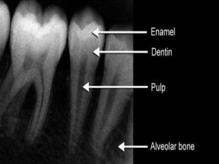
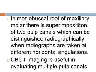
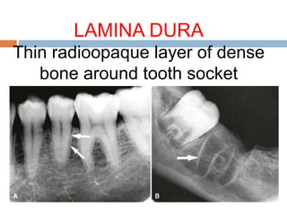
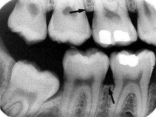
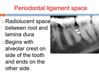
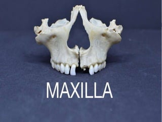
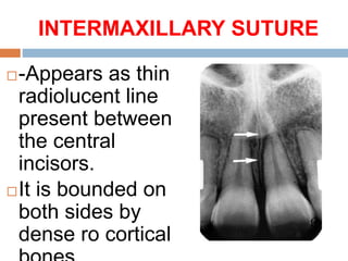
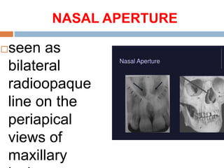
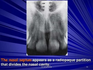
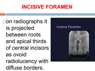
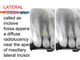
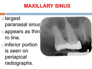
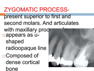
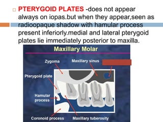
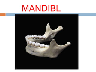
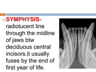
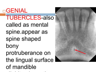
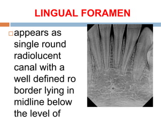
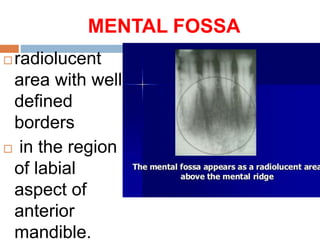
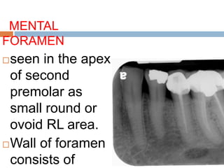
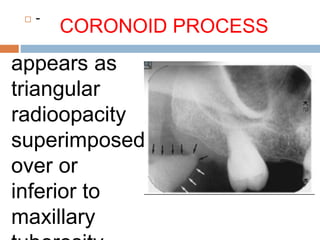
NORMAL
RADIOGRAPHIC
LANDMARKS
AAKRITI
AGGARWAL
BDS
BATCH 2016
CONTENTS
TEETH
SUPPORTING
DENTOALVEOLAR
STRUCTURES
MAXILLA AND MIDFACIAL
BONES
MANDIBLE
TEETH
ENAMEL -outermost
radioopaque layer of the
crown of tooth.
DENTIN-Radioopaque but less
than enamel
Found beneath enamel layer
and surrounds pulp cavity.it
comprises most of the tooth
CEMENTUM-Usually not
apparent radiographically
because the contrast between it
and dentin is low and also
because it is a very thin layer
PULP CHAMBER- it consists of
pulp chamber and root canal.it
appears radiolucent.
In mesiobuccal root of maxillary
molar there is superimposititon
of two pulp canals which can be
distinguished radiographically
when radiographs are taken at
different horizontal angulations.
CBCT imaging is useful in
evaluating multiple pulp canals
SUPPORTING
DENTOALVEOLAR
STRUCTURES
LAMINA DURA
Thin radioopaque layer of dense
bone around tooth socket
CORTICAL BONE
 Dense outer layer of bone
 More radioopaque than adjacent
cancellous bone.
CANCELLOUS BONE
Also called as trabecular bone
Lies between cortical plates in
both jaws
Appear as thin radioopaque
plates and rods surrounding
many small radiolucent
pockets of marrow.
ALVEOLAR CREST
 Appears as a radioopaque line
between two teeth
 Normal level-0.5 -2mm apical to
cej of adjacent tooth
 Level recedes with age or any
periodontal disease
 Continuous with lamina dura and
forms a sharp angle with it
Periodontal ligament space
 Radiolucent space
between root and
lamina dura
 Begins with
alveolar crest on
side of the tooth
and ends on the
other side
INTERMAXILLARY SUTURE
-Appears as thin
radiolucent line
present between
the central
incisors.
It is bounded on
both sides by
dense ro cortical
NASAL APERTURE
seen as
bilateral
radioopaque
line on the
periapical
views of
maxillary
INCISIVE FORAMEN
on radiographs it
is projected
between roots
and apical thirds
of central incisors
as ovoid
radiolucency with
diffuse borders.
 LATERAL
FOSSA-also
called as
incisive
fossa.apperas
a diffuse
radiolucency
near the apex
of maxillary
lateral incisor.
MAXILLARY SINUS
 largest
paranasal sinus
 appears as thin
ro line.
 inferior portion
is seen on
periapical
radiographs.
ZYGOMATIC PROCESS-
present superior to first and
second molars. And articulates
with maxillary process of zygoma.
appears as u-
shaped
radioopaque line
Composed of
dense cortical
bone
 PTERYGOID PLATES -does not appear
always on iopas.but when they appear,seen as
radioopaque shadow with hamular process
present inferiorly.medial and lateral pterygoid
plates lie immediately posterior to maxilla.
MANDIBL
E
SYMPHYSIS-
radiolucent line
through the midline
of jaws btw
deciduous central
incisors.it usually
fuses by the end of
first year of life.
GENIAL
TUBERCLES-also
called as mental
spine.appear as
spine shaped
bony
protruberance on
the lingual surface
of mandible
LINGUAL FORAMEN
appears as
single round
radiolucent
canal with a
well defined ro
border lying in
midline below
the level of
MENTAL RIDGE
 on iopa of
mand CI ,seen
as two
radioopaque
lines sweeping
bilaterally
forward and
upward
towards the
MENTAL FOSSA
 radiolucent
area with well
defined
borders
 in the region
of labial
aspect of
anterior
mandible.
MENTAL
FORAMEN
seen in the apex
of second
premolar as
small round or
ovoid RL area.
Wall of foramen
consists of
CORONOID PROCESS
 -
appears as
triangular
radioopacity
superimposed
over or
inferior to
maxillary
RESTORATIVE MATERIALS
 Amalgam-completely
radioopaque
 Gold-radioopaque
 Stainless steel pins-highly
radioopaque
 Composite rest.-radiolucent
over radioopaque metal
coping.
REFERENCES
WHITE AND PHAROAH’S
ORAL RADIOLOGY
C ANAND KUMAR
AAKRITI AGGARWAL
BDS-2016-01

NORMAL RADIOGRAPHIC LANDMARKS