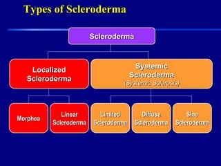
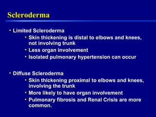
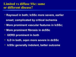
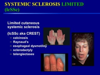
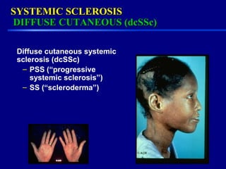
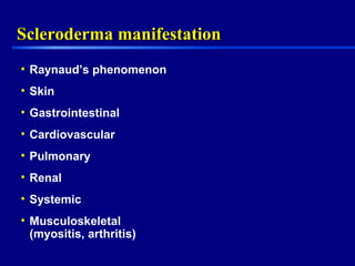
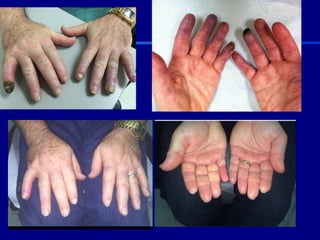
Overview of SclerodermaOverview of Scleroderma
Scleroderma Patient Education Conference
Apr 22, 2017
Nadera J Sweiss, MD, FACR
Associate Professor of Medicine
Director, Bernie Mac STAR Center and Cold Hand Clinic
Section of Rheumatology, Section of Pulmonary Medicine
DisclosureDisclosure
•None
•I will discuss non FDA approved
therapies
The Mission of the Scleroderma FoundationThe Mission of the Scleroderma Foundation
Making life easier for patients and making strides toward a
cure by focusing on three critical goals:
•SUPPORT: To help patients and their families cope with
scleroderma through mutual support programs, peer
counseling, physician referrals, and educational information.
•EDUCATION: To promote public awareness and education
through patient and health professional seminars, literature,
and publicity campaigns.
•RESEARCH: To stimulate and support research to improve
treatment and ultimately find the cause of and cure for
scleroderma.
ObjectivesObjectives
•Scleroderma overview
•Update
• Future directions
SclerodermaScleroderma
• Multisystem disorder
• Unknown etiology
• Thickening of skin caused by accumulation of
connective tissue (collagen types I and III)
• Involvement of visceral organs
What is Scleroderma?What is Scleroderma?
• The word scleroderma comes from Greek, meaning hard
skin.
• Early symptoms of the disease often include cold hands,
tightening of the skin, changes in facial features, joint pain,
heartburn, shortness of breath and fatigue.
• Patient awareness about scleroderma is important. It gives a
better chance for early intervention, treatment and a return to
normal routines and activities. Also, primary care physicians
awareness of the disease allows them to make more referrals
to specialists at earlier stages.
Classification of SclerodermaClassification of Scleroderma
• Systemic sclerosis, limited and diffuse
• Localized sclerdoerma
• Mimicks
Types of SclerodermaTypes of Scleroderma
SclerodermaSclerodermaSclerodermaScleroderma
LocalizedLocalized
SclerodermaScleroderma
LocalizedLocalized
SclerodermaScleroderma
SystemicSystemic
SclerodermaScleroderma
(Systemic Sclerosis)(Systemic Sclerosis)
SystemicSystemic
SclerodermaScleroderma
(Systemic Sclerosis)(Systemic Sclerosis)
MorpheaMorpheaMorpheaMorphea
LinearLinear
SclerodermaScleroderma
LinearLinear
SclerodermaScleroderma
LimitedLimited
SclerodermaScleroderma
LimitedLimited
SclerodermaScleroderma
DiffuseDiffuse
SclerodermaScleroderma
DiffuseDiffuse
SclerodermaScleroderma
SineSine
SclerodermaScleroderma
SineSine
SclerodermaScleroderma
Limited and Diffuse SSc—
Skin Involvement
Limited Diffuse
Overview of SclerodermaOverview of Scleroderma
• Peak age range: 35-64
• Younger age in women and with diffuse
disease.
• Female:Male = 3:1
• 8:1 in child bearing years
• Incidence: 20/million per year in US
• Prevalence: 240/million in US.
SclerodermaScleroderma
• Limited Scleroderma
• Skin thickening is distal to elbows and knees,
not involving trunk
• Less organ involvement
• Isolated pulmonary hypertension can occur
• Diffuse Scleroderma
• Skin thickening proximal to elbows and knees,
involving the trunk
• More likely to have organ involvement
• Pulmonary fibrosis and Renal Crisis are more
common.
Limited vs diffuse SSc: sameLimited vs diffuse SSc: same
or different disease?or different disease?
• Raynaud in both; lcSSc more severe, earlier
onset; complicated by critical ischemia
• More prominent vascular features in lcSSc;
• More prominent fibrosis in dcSSc
• GERD prominent in both
• ILD in both, oqen more severe in dcSSc
• lcSSc generally indolent, better outcome
SYSTEMIC SCLEROSISSYSTEMIC SCLEROSIS LIMITEDLIMITED
(lcSSc)(lcSSc)
Limited cutaneous
systemic sclerosis
(lcSSc aka CREST)
– calcinosis
– Raynaud’s
– esophageal dysmotility
– sclerodactyly
– telangiectases
SYSTEMIC SCLEROSISSYSTEMIC SCLEROSIS
DIFFUSE CUTANEOUS (dcSSc)DIFFUSE CUTANEOUS (dcSSc)
Diffuse cutaneous systemic
sclerosis (dcSSc)
– PSS (“progressive
systemic sclerosis”)
– SS (“scleroderma”)
Scleroderma manifestationScleroderma manifestation
• Raynaud’s phenomenon
• Skin
• Gastrointestinal
• Cardiovascular
• Pulmonary
• Renal
• Systemic
• Musculoskeletal
(myositis, arthritis)
Skin ScoringSkin Scoring
Scleroderma AutoantibodiesScleroderma Autoantibodies
Antigen ANA
Pattern
Frequency Clinical
Associations
Organs Involved
Scl-70
(topoisomerase 1)
Speckled 10-40 dcSSC Lung fibrosis
RNA Polymerase III Speck/Nuc 4-25 dcSSC Renal,
Pulmonary HTN
Centromere Centromere 15-40 lcSSc, CREST Pulmonary HTN
Esophageal
U1-RNP Speckled 5-35 lcSSC, MCTD Muscle
U3 RNP (fibrillarin) Nucleolar 1-5 dcSSC, poor prognosis Muscle
Pulmonary HTN
PM-SCL Nucleolar 3-6 Overlap, mixed Muscle
Th/To Nucleolar 1-7 lcSSc Pulmonary HTN,
Lung fibrosis,
Small bowel
Anti U11/U12 Nucleolar 1-5 lcSSc & dcSSC Lung Fibrosis
Anti-Ku 1-3 Overlap Ssc Muscle, Joint,
SLE overlap
Adapted from: Nihtyanova SI, Denton CP. Nat Rev Rheumatol 2010; 6:112
2013 ACR Diagnostic Criteria2013 ACR Diagnostic Criteria
Scleroderma ChallengesScleroderma Challenges
• While there is no cure for scleroderma, with early
intervention, it’s often possible to manage the symptoms
and offer a lifetime of hope. With early diagnosis of the
disease, better treatment can be offered to patients.
• Scleroderma can also happen in men, infants and seniors.
It happens with women 4 times greater than with men.
• Scleroderma continues to be a challenging disease, that
requires:
finding a cure, and/or at the very least,
finding a better treatment for patients so that they can
live a better life.
Cardiac InvolvementCardiac Involvement
Adapted from Desai, et al; Curr Opin Rheumatol 2011m 23:545-554
Cardiac
Manifestation
Prevalence Diagnosis Treatment
Myocarditis Rare Cardiac MRI, Biopsy Cytoxan + steroids
Pericardial effusion 5-16% Echocardiogram None; NSAIDs if
symptomatic
Microvascular CAD > 60% MRI/nuclear
medicine
Calcium channel
blockers
Macrovascular CAD 25% Coronary
Angiogram
Stenting/medical tx
Bradyarrhythmias Rare EKG/Holter Pacemaker
Tachyarrhythmias 15% EKG/Holter Diltiazem, ablation,
defibrillator
Digital Ischemia/ Ulcers in Systemic SclerosisDigital Ischemia/ Ulcers in Systemic Sclerosis
Clinical PresentationClinical Presentation
Digital Ischemia/ Ulcers in Systemic Sclerosis andDigital Ischemia/ Ulcers in Systemic Sclerosis and
MimicsMimics
Digital Ischemia/ Ulcers in Systemic SclerosisDigital Ischemia/ Ulcers in Systemic Sclerosis
Therapeutic OptionsTherapeutic Options
• AVOID THE COLD
• Vasodilator
• Target the endothelium
• Target circulating cells
• Inhibit mediators of
vascular disease
• Treat consequences of
ischemia- reperfusion
tissue injury
26Fat Transfer in Raynaud’s
Treatment
Ca
Channel
Blockers
ARB
Protective
Measures
Protective
Measures PDE-I
Clopidogre
l
Endothelin
Receptor
Blockers
Alpha
Blockers
Topical
Nitrates
ACE-I
ASA
Prostanoid
s
27Fat Transfer in Raynaud’s
Treatment
Ca
Channel
Blockers
ARB
Protective
Measures
Protective
Measures
PDE-I
Clopidogre
l
Endothelin
Receptor
Blockers
Alpha
Blockers
Topical
Nitrates
ACE-I
ASA
Prostanoid
s
Invasive
Modalities
Surgical
Sympathec
tomy
Neuro-
modulators
Fat
Transfer
Fat
Transfer
Periarterial Symapthectomy in thePeriarterial Symapthectomy in the
Treatment of Digital IschemiaTreatment of Digital Ischemia
29Fat Transfer in Raynaud’s
Technique
30Fat Transfer in Raynaud’s
Quantitative Flow Measurement of Digital Arteries at 3 TeslaQuantitative Flow Measurement of Digital Arteries at 3 Tesla
Guanzhong Liu, Kezhou Wang, Xinjian Du, Yi Sui1, Michael P Flannery,Guanzhong Liu, Kezhou Wang, Xinjian Du, Yi Sui1, Michael P Flannery,
Fady Charbel, Nadera Sweiss, and X. Joe ZhouFady Charbel, Nadera Sweiss, and X. Joe Zhou
a
)
b
)
A 3D angiogram showing all proper
palmer digital arteries from a
representative female volunteer. (b) A
zoomed view of PPDA2 showing the
automatically determined perpendicular
plane for the subsequent 2D PC
acquisition
ConclusionConclusion
• Scleroderma is multisystem heterogeneous disiease;
individualized therapy is warranted
• Outcome measures need to be defined
• Translational team approach will be helpful to identify responders
Thank You

Overview of Scleroderma

  • 1.
    Overview of SclerodermaOverviewof Scleroderma Scleroderma Patient Education Conference Apr 22, 2017 Nadera J Sweiss, MD, FACR Associate Professor of Medicine Director, Bernie Mac STAR Center and Cold Hand Clinic Section of Rheumatology, Section of Pulmonary Medicine
  • 2.
  • 3.
    The Mission ofthe Scleroderma FoundationThe Mission of the Scleroderma Foundation Making life easier for patients and making strides toward a cure by focusing on three critical goals: •SUPPORT: To help patients and their families cope with scleroderma through mutual support programs, peer counseling, physician referrals, and educational information. •EDUCATION: To promote public awareness and education through patient and health professional seminars, literature, and publicity campaigns. •RESEARCH: To stimulate and support research to improve treatment and ultimately find the cause of and cure for scleroderma.
  • 4.
  • 5.
    SclerodermaScleroderma • Multisystem disorder •Unknown etiology • Thickening of skin caused by accumulation of connective tissue (collagen types I and III) • Involvement of visceral organs
  • 6.
    What is Scleroderma?Whatis Scleroderma? • The word scleroderma comes from Greek, meaning hard skin. • Early symptoms of the disease often include cold hands, tightening of the skin, changes in facial features, joint pain, heartburn, shortness of breath and fatigue. • Patient awareness about scleroderma is important. It gives a better chance for early intervention, treatment and a return to normal routines and activities. Also, primary care physicians awareness of the disease allows them to make more referrals to specialists at earlier stages.
  • 7.
    Classification of SclerodermaClassificationof Scleroderma • Systemic sclerosis, limited and diffuse • Localized sclerdoerma • Mimicks
  • 8.
    Types of SclerodermaTypesof Scleroderma SclerodermaSclerodermaSclerodermaScleroderma LocalizedLocalized SclerodermaScleroderma LocalizedLocalized SclerodermaScleroderma SystemicSystemic SclerodermaScleroderma (Systemic Sclerosis)(Systemic Sclerosis) SystemicSystemic SclerodermaScleroderma (Systemic Sclerosis)(Systemic Sclerosis) MorpheaMorpheaMorpheaMorphea LinearLinear SclerodermaScleroderma LinearLinear SclerodermaScleroderma LimitedLimited SclerodermaScleroderma LimitedLimited SclerodermaScleroderma DiffuseDiffuse SclerodermaScleroderma DiffuseDiffuse SclerodermaScleroderma SineSine SclerodermaScleroderma SineSine SclerodermaScleroderma
  • 9.
    Limited and DiffuseSSc— Skin Involvement Limited Diffuse
  • 10.
    Overview of SclerodermaOverviewof Scleroderma • Peak age range: 35-64 • Younger age in women and with diffuse disease. • Female:Male = 3:1 • 8:1 in child bearing years • Incidence: 20/million per year in US • Prevalence: 240/million in US.
  • 11.
    SclerodermaScleroderma • Limited Scleroderma •Skin thickening is distal to elbows and knees, not involving trunk • Less organ involvement • Isolated pulmonary hypertension can occur • Diffuse Scleroderma • Skin thickening proximal to elbows and knees, involving the trunk • More likely to have organ involvement • Pulmonary fibrosis and Renal Crisis are more common.
  • 12.
    Limited vs diffuseSSc: sameLimited vs diffuse SSc: same or different disease?or different disease? • Raynaud in both; lcSSc more severe, earlier onset; complicated by critical ischemia • More prominent vascular features in lcSSc; • More prominent fibrosis in dcSSc • GERD prominent in both • ILD in both, oqen more severe in dcSSc • lcSSc generally indolent, better outcome
  • 13.
    SYSTEMIC SCLEROSISSYSTEMIC SCLEROSISLIMITEDLIMITED (lcSSc)(lcSSc) Limited cutaneous systemic sclerosis (lcSSc aka CREST) – calcinosis – Raynaud’s – esophageal dysmotility – sclerodactyly – telangiectases
  • 14.
    SYSTEMIC SCLEROSISSYSTEMIC SCLEROSIS DIFFUSECUTANEOUS (dcSSc)DIFFUSE CUTANEOUS (dcSSc) Diffuse cutaneous systemic sclerosis (dcSSc) – PSS (“progressive systemic sclerosis”) – SS (“scleroderma”)
  • 15.
    Scleroderma manifestationScleroderma manifestation •Raynaud’s phenomenon • Skin • Gastrointestinal • Cardiovascular • Pulmonary • Renal • Systemic • Musculoskeletal (myositis, arthritis)
  • 16.
  • 18.
    Scleroderma AutoantibodiesScleroderma Autoantibodies AntigenANA Pattern Frequency Clinical Associations Organs Involved Scl-70 (topoisomerase 1) Speckled 10-40 dcSSC Lung fibrosis RNA Polymerase III Speck/Nuc 4-25 dcSSC Renal, Pulmonary HTN Centromere Centromere 15-40 lcSSc, CREST Pulmonary HTN Esophageal U1-RNP Speckled 5-35 lcSSC, MCTD Muscle U3 RNP (fibrillarin) Nucleolar 1-5 dcSSC, poor prognosis Muscle Pulmonary HTN PM-SCL Nucleolar 3-6 Overlap, mixed Muscle Th/To Nucleolar 1-7 lcSSc Pulmonary HTN, Lung fibrosis, Small bowel Anti U11/U12 Nucleolar 1-5 lcSSc & dcSSC Lung Fibrosis Anti-Ku 1-3 Overlap Ssc Muscle, Joint, SLE overlap Adapted from: Nihtyanova SI, Denton CP. Nat Rev Rheumatol 2010; 6:112
  • 20.
    2013 ACR DiagnosticCriteria2013 ACR Diagnostic Criteria
  • 21.
    Scleroderma ChallengesScleroderma Challenges •While there is no cure for scleroderma, with early intervention, it’s often possible to manage the symptoms and offer a lifetime of hope. With early diagnosis of the disease, better treatment can be offered to patients. • Scleroderma can also happen in men, infants and seniors. It happens with women 4 times greater than with men. • Scleroderma continues to be a challenging disease, that requires: finding a cure, and/or at the very least, finding a better treatment for patients so that they can live a better life.
  • 22.
    Cardiac InvolvementCardiac Involvement Adaptedfrom Desai, et al; Curr Opin Rheumatol 2011m 23:545-554 Cardiac Manifestation Prevalence Diagnosis Treatment Myocarditis Rare Cardiac MRI, Biopsy Cytoxan + steroids Pericardial effusion 5-16% Echocardiogram None; NSAIDs if symptomatic Microvascular CAD > 60% MRI/nuclear medicine Calcium channel blockers Macrovascular CAD 25% Coronary Angiogram Stenting/medical tx Bradyarrhythmias Rare EKG/Holter Pacemaker Tachyarrhythmias 15% EKG/Holter Diltiazem, ablation, defibrillator
  • 23.
    Digital Ischemia/ Ulcersin Systemic SclerosisDigital Ischemia/ Ulcers in Systemic Sclerosis Clinical PresentationClinical Presentation
  • 24.
    Clinical PresentationClinical Presentation DigitalIschemia/ Ulcers in Systemic Sclerosis andDigital Ischemia/ Ulcers in Systemic Sclerosis and MimicsMimics
  • 25.
    Digital Ischemia/ Ulcersin Systemic SclerosisDigital Ischemia/ Ulcers in Systemic Sclerosis Therapeutic OptionsTherapeutic Options • AVOID THE COLD • Vasodilator • Target the endothelium • Target circulating cells • Inhibit mediators of vascular disease • Treat consequences of ischemia- reperfusion tissue injury
  • 26.
    26Fat Transfer inRaynaud’s Treatment Ca Channel Blockers ARB Protective Measures Protective Measures PDE-I Clopidogre l Endothelin Receptor Blockers Alpha Blockers Topical Nitrates ACE-I ASA Prostanoid s
  • 27.
    27Fat Transfer inRaynaud’s Treatment Ca Channel Blockers ARB Protective Measures Protective Measures PDE-I Clopidogre l Endothelin Receptor Blockers Alpha Blockers Topical Nitrates ACE-I ASA Prostanoid s Invasive Modalities Surgical Sympathec tomy Neuro- modulators Fat Transfer Fat Transfer
  • 28.
    Periarterial Symapthectomy inthePeriarterial Symapthectomy in the Treatment of Digital IschemiaTreatment of Digital Ischemia
  • 29.
    29Fat Transfer inRaynaud’s Technique
  • 30.
    30Fat Transfer inRaynaud’s
  • 32.
    Quantitative Flow Measurementof Digital Arteries at 3 TeslaQuantitative Flow Measurement of Digital Arteries at 3 Tesla Guanzhong Liu, Kezhou Wang, Xinjian Du, Yi Sui1, Michael P Flannery,Guanzhong Liu, Kezhou Wang, Xinjian Du, Yi Sui1, Michael P Flannery, Fady Charbel, Nadera Sweiss, and X. Joe ZhouFady Charbel, Nadera Sweiss, and X. Joe Zhou a ) b ) A 3D angiogram showing all proper palmer digital arteries from a representative female volunteer. (b) A zoomed view of PPDA2 showing the automatically determined perpendicular plane for the subsequent 2D PC acquisition
  • 33.
    ConclusionConclusion • Scleroderma ismultisystem heterogeneous disiease; individualized therapy is warranted • Outcome measures need to be defined • Translational team approach will be helpful to identify responders
  • 34.

Editor's Notes

  • #27 Initial treatment is preventative – with gloves and cold avoidance But many patients progress to require medical treatment with calcium channel blockers and topical nitrates And there is a whole host of other medications aimed at vasodilation and prevention of arterial thrombosis
  • #28 Despite all of these options, A portion of these patients develop severe, refractory symptoms And this is where the hand surgeon comes in to play, invoking invasive modalities With injection of neuromodulators such as botulinum toxin – which has shown to be effective, but short lived, and can be an arduous undertaking for the patient and surgeon when done in the clinic setting Another option would be surgical sympathectomy of the hands of affected feet – this is not a trivial undertaking, with inherent procedural risks, and requires somewhat specialized training and my not be long-lived with the regrowth of the stripped adventitia The intervention we propose he today is autologous fat grafting into the affected extremities
  • #30 Here you can see the fat being injected while withdrawing the cannula
  • #31 An example of fat transfer to the feet