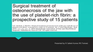
This study evaluated the outcome of surgical treatment of osteonecrosis of the jaw (ONJ) with the additional use of autologous platelet-rich fibrin (PRF) membranes. 15 patients underwent surgical resection of necrotic bone followed by placement of multiple PRF membrane layers over the bone. At follow-up between 7-20 months post-op, 14 of 15 patients (93%) showed complete mucosal healing with no symptoms or bone exposure, indicating the PRF membranes aided in wound healing. One patient had recurrence. The study concluded PRF membrane use provides multilayer closure and benefits patients with reduced complications and better healing.
Osteoradionecrosis is bone necrosis that occurs in the radiation treatment volume months after treatment. It is caused by loss of vasculature due to radiation damage. Risk factors include radiation dose over 6500 cGy, chemotherapy, brachytherapy, and post-radiation dental extractions. Advanced cases can lead to fistulas, fractures, and discontinuity defects impacting functions like speech and swallowing.
BRONJ bisphosphonates osteonecrosis of jawWeam Faroun
This document discusses bisphosphonate-related osteonecrosis of the jaws (BRONJ) and provides guidance on dental management of patients taking bisphosphonates. It notes that bisphosphonates are commonly used to treat osteoporosis and bone metastases but can increase the risk of BRONJ, especially after dental surgery. The document recommends conservative dental treatment and antibiotic prophylaxis for high-risk patients. It identifies risk factors for BRONJ and advises monitoring of bone turnover markers like CTX to further assess surgical risk. The goal is to minimize but not eliminate the low risk of BRONJ from oral bisphosphonate use through preventative dental care and cautious management of invasive procedures.
This document discusses osteoradionecrosis of the jaws, providing definitions, epidemiology, staging systems, clinical and radiological findings, risk factors, diagnosis, and radiation techniques. It defines ORN as exposed irradiated bone that fails to heal over 3 months without evidence of tumor. Risk factors include high radiation dose, oral cavity primary site, and tooth extractions after radiation. Diagnosis requires previous irradiation, no recurrent tumor, mucosal breakdown exposing bone for over 3 months. Later radiation techniques like IMRT may reduce risks by more precisely targeting tumors and minimizing dose to surrounding tissues.
Osteoradionecrosis is a serious complication of radiation therapy for cancer where exposed irradiated bone fails to heal. It is caused by hypoxia, hypocellularity, and hypovascularity due to radiation therapy damaging blood vessels and reducing oxygen levels in tissues. Common risk factors include radiation doses over 50Gy, history of dental extractions post-radiation, and location in the mandible which has a richer blood supply. Treatment focuses on controlling infection with antibiotics and debriding necrotic tissue. Hyperbaric oxygen therapy helps by increasing oxygen levels in tissues and promoting healing. Free omental transfers are also used to improve blood flow in severe cases.
This document discusses oroantral communication, which is a communication between the maxillary sinus and oral cavity that can occur after extraction of upper molars. It describes the etiology, signs and symptoms, patients at high risk, diagnosis, prevention, management, and complications of oroantral communication. The management depends on the size of the opening and may involve ensuring a blood clot forms, suturing, antibiotics, or surgical closure of larger openings using buccal or palatal flaps. Complications include postoperative maxillary sinusitis or formation of a chronic oroantral fistula.
Osteoradionecrosis is an inflammatory bone condition that occurs after exposure to therapeutic radiation doses, usually for head and neck cancers. It is characterized by exposed bone for at least 3 months after radiation therapy. The mandible is most commonly affected due to its anatomy and low vascularity. Management includes conservative approaches like antibiotics and debridement to maintain bone integrity and prevent infection, as well as hyperbaric oxygen therapy to reduce hypoxia and improve healing. Prevention involves dental work before radiation and careful oral hygiene during and after treatment.
This document discusses osteoradionecrosis of the jaws, which is bone necrosis caused by radiation therapy for head and neck cancer. It defines osteoradionecrosis and describes its classification, types, risk factors, and theories of pathophysiology. The document outlines protocols for preventing and treating osteoradionecrosis, including conservative management, hyperbaric oxygen therapy, pentoxifyllin and tocopherol supplements, and surgical interventions. Hyperbaric oxygen therapy is described as an adjuvant treatment involving intermittent high-pressure oxygen sessions to promote revascularization and healing.
This document discusses osteoradionecrosis (ORN), a serious complication of radiation therapy where exposed irradiated bone fails to heal. It can occur spontaneously or after trauma. The mandible is most commonly affected due to its tenuous blood supply. Pathophysiology involves hypoxic, hypocellular tissue with impaired healing ability. Management includes controlling infection, supportive care, and hyperbaric oxygen therapy (HBO) as an adjuvant to surgery to improve tissue oxygenation and revasculation. The Marx protocol uses HBO followed by surgical resection of necrotic bone in stages depending on severity.
The document discusses osteomyelitis, which is an inflammatory condition of bone that begins as an infection of the medullary cavity and spreads to involve the periosteum. It can be acute or chronic, and is caused by bacteria or fungi entering via trauma or a blood-borne route. Symptoms include pain, swelling, and pus drainage. Diagnosis involves medical imaging and biopsy. Treatment involves antibiotics, drainage of pus, debridement of infected tissue, and sometimes surgery. Chronic osteomyelitis can be difficult to treat and may require repeated surgeries. Risk factors include reduced blood supply to bone from conditions like diabetes.
This document discusses the anatomy and pathology of the maxillary sinus and oroantral communications. It describes the location and drainage of the maxillary sinus and causes of sinusitis. Oroantral communications are defined as pathological connections between the oral cavity and maxillary sinus that can form due to dental procedures or trauma. Signs, testing methods, prevention, and management strategies are outlined for both acute communications and oroantral fistulas. Surgical techniques for repair include local soft tissue flaps, grafts, and use of the buccal fat pad flap. Immediate closure of communications less than 3 weeks old has a high success rate, while delayed or recurrent fistulas require surgical intervention.
This document provides information about the surgical procedure of apicoectomy. It begins with an introduction defining apicoectomy as the surgical resection and removal of the root tip and pathological periapical tissues. It then lists the indications and contraindications for the procedure. The rest of the document details the armamentarium, surgical technique including designing the flap, localizing and exposing the apex, resection of the apex, retrograde filling if needed, and wound closure. The surgical technique section provides step-by-step details of each part of the procedure.
Dental extractions in irradiated patientsUjwal Gautam
Dental extractions in patients undergoing radiotherapy carry risks of osteoradionecrosis and impaired wound healing due to radiation damage to vasculature, bone marrow, and fibroblasts. Extraction after radiotherapy requires preventive measures like antibiotics and atraumatic technique. Hyperbaric oxygen therapy has been used preventively for extractions, though its effectiveness is less than 100%. Where possible, extractions in irradiated patients should be avoided or meticulous preventive measures undertaken due to osteoradionecrosis risk.
Mandibular angle fractures account for 23-42% of facial fractures and are commonly caused by motor vehicle accidents and assaults. The angle is prone to fractures due to its thin cross-section and presence of impacted third molars. Fractures are classified as vertically or horizontally favorable/unfavorable based on the direction of the fracture line and effect of muscle forces. Traditionally, rigid plate fixation and intermaxillary fixation were used but caused complications. Currently, semi-rigid fixation using a single miniplate placed along the superior border based on Champy's lines of osteosynthesis is the standard approach, allowing early function with low complications.
Different flap designs used for the management of impacted wisdommohamedamr94
This document discusses different flap designs used for surgical removal of impacted third molars. It describes several types of mucosal and mucoperiosteal flaps including envelope, triangular, modified triangular, comma-shaped, and Szmyd flaps. Key principles for flap design are outlined such as ensuring adequate access and blood supply while avoiding vital structures. Factors like bone exposure, flap position, and limitations of different techniques are compared. The literature review evaluates studies on primary wound healing comparing conventional versus modified flap designs.
Vestibuloplasty is a surgical procedure to deepen the oral vestibule by changing the attachments of the soft tissue. There are several types of vestibuloplasty procedures, including mucosal advancement, secondary epithelization, and grafting. Mucosal advancement involves undermining and advancing the oral mucosa, while secondary epithelization uses the oral mucosa to line one side and allows the other side to heal through epithelization. Grafting can use skin, mucosa, or dermis grafts to line the extended vestibule. The document discusses techniques for each type of vestibuloplasty procedure.
Wisdom teeth are the third and last molars on each side of the upper and lower jaws. They are also the final teeth to erupt; they usually appear when a person is in their late teens or early twenties
This document discusses various pre-prosthetic hard tissue procedures including: recontouring alveolar ridges through alveoloplasty and Dean's alveoloplasty; reducing maxillary tuberosities, palatal exostoses, mylohyoid ridges, and genial tubercles; removing tori and bone augmentation of atrophic maxillary and mandibular ridges through onlay grafts, sinus lifts, and hydroxyapatite grafts. The goal is to modify oral anatomy and eliminate undercuts/protuberances to facilitate dental prosthesis placement through reshaping bony areas.
This document discusses infections that can occur in the five masticatory spaces. It describes the boundaries, contents, causes, clinical features, and treatment for infections of the pterygomandibular space, submasseteric space, and the three temporal spaces (superficial, infratemporal, and deep). Infections in these spaces can spread between adjacent spaces and present with symptoms like trismus, swelling, and pain. Treatment involves incision and drainage through intraoral, extraoral, or combined approaches depending on the specific infected space.
Corticosteroids - Role in Oral and Maxillofacial Surgeryanchalag8
Corticosteroids have many uses in oral and maxillofacial surgery. They can be used to treat TMJ disorders by reducing pain and inflammation via intracapsular injections. Topical steroids can treat oral ulcers and lesions. Intralesional injections are used to treat keloids, hypertrophic scars, and central giant cell granulomas. Oral steroids are the standard treatment for Bell's palsy and can reduce postoperative morbidities from surgeries like wisdom tooth extraction and orthognathic surgery by blocking excessive inflammation. While corticosteroids have benefits, their use requires weighing risks and using the minimum dose and least potent type needed for the therapeutic effect.
This document discusses angle fractures of the mandible, including signs and symptoms, diagnostic aids, treatment options, and principles for selecting treatment. Signs include pain, swelling, restricted movement, and malocclusion. Diagnostic aids include panoramic x-rays, CT scans, and 3D CT scans. Treatment options range from closed reduction with maxillomandibular fixation to open reduction with plates, screws, or wiring depending on the fracture characteristics and direction of forces. The goal is to select treatment that resists muscular and masticatory forces at the fracture site.
The document provides information on internal derangement of the temporomandibular joint (TMJ). It begins with definitions of internal derangement and Wilkes classification system for stages of derangement. It then discusses etiology, including trauma as a common cause. Physical findings and non-surgical and surgical treatment procedures are outlined. Non-surgical options include splint therapy, medications, acupuncture and others aimed at reducing pain and improving joint function.
This document discusses oral submucous fibrosis (OSF), a chronic disease caused by chewing betel quid that results in fibrosis of the oral cavity and inability to open the mouth fully. It describes the epidemiology, pathogenesis, clinical features and classification system for OSF. Surgical management is often needed for advanced OSF cases. One approach described is resection of fibrotic bands and reconstruction of the surgical defect using a buccal fat pad graft, which provides a well-vascularized tissue that reliably epithelializes the area. The document includes a case report demonstrating successful application of this technique.
This document provides an overview of Lefort I osteotomy, including:
- A brief history describing the development of maxillary osteotomy techniques.
- Anatomical considerations and the biologic basis for maxillary osteotomies, which have shown adequate blood supply can be maintained.
- Indications for Lefort I osteotomy include altering vertical dimension, anteroposterior movements, and surgical expansion of the maxilla.
- Types of Lefort I osteotomies are described, including classic, quadrangular, and segmental variations. Postoperative management and potential complications are also outlined.
Local & systemic Complications of Local AnesthesiaIAU Dent
This document discusses local anesthesia (LA), including its mechanism of action, factors influencing injection discomfort and techniques to reduce discomfort, testing the success of LA, causes and management of failed LA, complications of LA including local and systemic complications, and management of specific complications like needle breakage, pain/burning on injection, persistent anesthesia, and trismus. It provides anatomical and technical details related to achieving successful LA and avoiding complications.
The document discusses temporomandibular joint ankylosis, including its causes, clinical features, diagnosis using radiographs, and various treatment methods. Key points include: TMJ ankylosis is the fusion of the mandibular condyle with the glenoid fossa, immobilizing the mandible. Common causes are trauma, infection, and inflammation. Treatment involves surgical resection of the ankylotic mass with coronoidectomy and interpositional arthroplasty using grafts to prevent re-ankylosis, followed by aggressive physiotherapy. Complications can include recurrence if physiotherapy is not continued long-term.
This document discusses tooth impaction, which occurs when a tooth is unerupted or malpositioned against another tooth, bone, or soft tissue beyond its normal eruption time. The most common impacted teeth are mandibular and maxillary third molars, followed by maxillary canines. Causes of impaction include genetics, small jaw size, dental anomalies, systemic conditions, and local factors. Impacted teeth are classified based on their position, relationship to other teeth, and degree of bone coverage. Surgical removal may be needed to address related issues like pericoronitis or to allow proper eruption of other teeth.
The document describes the submandibular and retromandibular surgical approaches. The submandibular approach involves making a 1.5-2 cm incision inferior to the mandible and dissecting through the layers of the skin, subcutaneous tissue, platysma muscle, and pterygomasseteric sling. The retromandibular approach uses a vertical incision 2 cm posterior to the mandibular ramus and dissects through the same layers to the pterygomasseteric sling. Both approaches give access below the mandible for surgical procedures.
Mahendra Azad et al. GAINT ODONTOGENIC KERATOCYST OF MANDIBLE OPERATED UNDER LOCAL ANESTHESIA- A CASE REPORT. JOURNAL OF DENTAL HEALTH & RESEARCH (VOL. 1, ISSUE 2, JUL - DEC 2020): 24-2
This document describes a two-stage technique for treating chronic osteomyelitis of long bones. In stage one, all infected and compromised bone and soft tissue is radically debrided. Healthy soft tissue coverage is then provided, either directly or with a muscle flap. Stage two occurs 3-6 weeks later, where any remaining bone defects are grafted with cancellous bone grafts. This technique was used to treat 37 patients with chronic osteomyelitis of the tibia, femur, radius or humerus. Infection was eradicated in 34 patients, with no patients requiring amputation.
A comparative study on the clinical and functional outcome of limb salvage su...NAAR Journal
The aim of this study was to analyze the survival, recurrence, complications as well as the quality of life (QOL) in tibial osteosarcoma (OSA) patients managed by limb salvage surgery (LSS), either by a prosthesis, resection or graft or by amputation. 106 tibial osteosarcoma patients were enrolled where 39 had custom-designed endoprosthetic arthroplasty (LSS1), 36 underwent resection and bone graft (LSS2) while only 31 underwent amputation. A Comparison was done based on post-operative survival rates, postoperative recurrence, and complications. The impact of the patient’s QOL was also evaluated.
This document provides a history of distraction osteogenesis. It began in 1905 with lengthening of long bones, was pioneered for the maxillofacial region by Ilizarov in the 1950s using gradual traction to regenerate bone, and was first applied to the human mandible by McCarthy in 1989. Since then, distraction techniques have been used increasingly as alternatives to orthognathic surgery, with applications to the maxilla beginning in the 1990s and advances in device design improving three-dimensional control and outcomes. The biology of distraction osteogenesis involves regeneration of new bone between segments separated by gradual traction applied during the distraction phase.
Dr. Rahul VC Tiwari - Fellowship In Orthognathic Surgery - Jubilee Mission Medical College Hospital and Research Center, Thrissur, Kerala - 20TH PUBLICATION - IJADS
Rapid canine retraction and orthodontic treatment with dentoalveolar distract...Dr Mujtaba Ashraf
This document presents a study on a new technique called dentoalveolar distraction (DAD) to rapidly retract canines during orthodontic treatment. The study involved 10 patients where the maxillary first premolars were extracted and a distractor device was used to move the canines into the extraction sites at a rate of 0.8 mm per day. Full canine retraction was achieved in an average of 10 days with minimal anchorage loss. The canines tipped and translated distally on average 13 degrees. No complications were observed. The DAD technique reduces orthodontic treatment time by nearly 50% with no adverse effects on surrounding structures.
Role of Adjuvant Therapy in Osteoradionecrosis (Orn) and Bisphosphonate Induc...inventionjournals
This study evaluated the effectiveness of a treatment protocol using pentoxifylline and alpha-tocoferol as an adjuvant therapy for 13 cases of osteoradionecrosis (ORN) and bisphosphonate-induced osteonecrosis of the jaw (BRONJ). The protocol was prescribed for 2 months pre- and post-operatively along with standard antimicrobial therapy and chlorhexidine rinses. Results found a decrease in symptoms for all cases before and after surgery. All patients were pain-free with no purulence or erythema post-operatively. The therapy was concluded to be highly beneficial for ORN and BRONJ without adverse effects.
Management of posttraumatic malocclusion caused by condylar process fractureDr. SHEETAL KAPSE
This study evaluated the treatment of 21 patients with post-traumatic malocclusions caused by condylar process fractures. For asymmetric malocclusions from unilateral fractures (n=15), patients underwent unilateral or bilateral mandibular ramus osteotomies. For anterior open bites from bilateral fractures (n=6), patients underwent either Le Fort I osteotomies (n=5) or bilateral ramus osteotomies (n=1). All patients had stable dental and skeletal results after 1+ years except one treated with bilateral ramus osteotomies. The authors conclude that osteotomies of the affected jaw are effective for treating post-traumatic malocclusions from condylar fractures.
An Evaluation of Short Term Success and Survival Rate of Implants Placed in F...DrHeena tiwari
An Evaluation of Short Term Success and Survival Rate of Implants Placed in Fresh Extraction Socket Post Prosthetic Rehabilitation- A Prospective Study
The use of low level laser in periodontal diseaseJan Tunér
This study investigated the effects of low-level laser therapy (LLLT) as an adjunct to scaling and root planing (SRP) for the treatment of chronic periodontitis. 16 patients received SRP on one side of their mouth and SRP plus 10 sessions of LLLT on the other side. Sites treated with SRP plus LLLT showed greater reductions in pocket depth at 5 weeks and 3 months compared to SRP alone. SRP plus LLLT sites also had a statistically significant increase in mean radiographic bone density from baseline to 12 months. However, LLLT did not significantly affect gingival index, plaque index, or levels of the inflammatory marker IL-1β in gingival crevicular fluid
Dr. Rahul VC Tiwari - Fellowship In Orthognathic Surgery - Jubilee Mission Medical College Hospital and Research Center, Thrissur, Kerala - 31st publication IJAR 1st name
This document provides a synopsis for a study comparing the use of temporalis fascia with and without platelet rich fibrin (PRF) for myringoplasty. The study aims to evaluate the success rates and benefits of PRF in improving hearing outcomes when applied to the temporalis fascia graft. It is a prospective study that will recruit 100 patients with inactive chronic otitis media requiring myringoplasty, who will be divided into two groups: one receiving temporalis fascia alone and the other receiving PRF applied to the fascia graft. Patients will be followed for 12 weeks to assess graft healing and post-operative hearing.
1) The document discusses the "All-on-4" technique for placing four dental implants (two tilted posteriorly and two anterior) to support a fixed prosthesis in edentulous maxilla or mandible.
2) Previous studies have shown this technique to be predictable and successful with high implant survival rates and minimal bone loss.
3) Tilting the posterior implants provides benefits like avoiding anatomical structures and improving prosthesis support.
Journal Club on Autologous blood injection for the treatment of recurrent tmj...Dr Bhavik Miyani
The document summarizes a journal club presentation on a study evaluating the effectiveness of autologous blood injection for the treatment of recurrent mandibular dislocation. The study included 11 patients with recurrent dislocation who underwent injection of their own blood into the temporomandibular joint. After a follow up period ranging from 24 to 35 months, 8 of the 11 patients (72.7%) did not experience further dislocation episodes. Autologous blood injection was found to be a simple, minimally invasive procedure for treating recurrent mandibular dislocation. However, more research with larger sample sizes and longer follow up periods is still needed.
A Prospective comparative study of Local anaesthesia & Spinal anaesthesia for...iosrjce
IOSR Journal of Dental and Medical Sciences is one of the speciality Journal in Dental Science and Medical Science published by International Organization of Scientific Research (IOSR). The Journal publishes papers of the highest scientific merit and widest possible scope work in all areas related to medical and dental science. The Journal welcome review articles, leading medical and clinical research articles, technical notes, case reports and others.
The document discusses reconstruction of the temporomandibular joint (TMJ) following ankylosis. It describes a study using hydroxyapatite/collagen scaffolds combined with bone marrow aspirate to regenerate condylar bone in 7 pediatric patients with TMJ ankylosis. Postoperative outcomes showed significantly improved mouth opening, chewing, and quality of life scores at 6 months and 1 year follow up. The technique aims to maintain ramal height for craniofacial growth and stability in young patients, using a simple, cost-effective method without donor site morbidity.
open versus closed reduction of adult condylar fracturesailesh kumar
This document reviews the evolution of treatment approaches for adult mandibular condyle fractures. It summarizes literature comparing outcomes of open reduction with internal fixation (ORIF) versus closed reduction with maxillomandibular fixation (CRMMF). While CRMMF was traditionally preferred, more recent studies show higher complication rates with CRMMF, including malocclusion, asymmetry and limited mobility. ORIF is associated with better anatomic reconstruction and outcomes. The document outlines classification systems for condyle fractures and indications for different treatment methods based on displacement and stability. It also reviews techniques used for ORIF and concludes that based on reviewed literature, ORIF provides better outcomes and is the preferred approach for treating condyle fractures.
The document discusses the anatomy of the temporomandibular joint, labeling structures like the articular eminence, condyle, and glenoid fossa. It notes that synovial fluid in the joint is derived from plasma by dialysis and secretion from synoviocytes, and that the upper compartment can hold approximately 1.2 ml of fluid without pressure while the lower compartment holds around 0.5 ml.
Low Level LASER therapy in impaction socketsailesh kumar
The document discusses the history and applications of low-level laser therapy (LLLT). It summarizes three studies that examined the use of LLLT to reduce pain, swelling, and trismus following surgical removal of impacted third molars. The first study applied laser intraorally and extraorally immediately and 24 hours after surgery. It found reduced pain, swelling, and increased interincisal opening with laser. The second study used a zonal laser technique and found reductions in pain and swelling, though not statistically significant. The third study applied intraoral laser once and found significantly reduced pain compared to medication alone. Overall, LLLT shows potential for improving outcomes following third molar surgery, but standardized power settings are needed
Differential diagnosis of orofacial painsailesh kumar
Differential diagnosis of orofacial pain can be divided into acute and chronic categories. Acute pain includes dental, periodontal, sinus, and salivary gland issues and is usually inflammatory in origin. Chronic pain includes myofascial pain, TMJ disorders, migraines, and various neuropathies. Neuropathic pain includes trigeminal neuralgia, postherpetic neuralgia, glossopharyngeal neuralgia, and complex regional pain syndrome. Management depends on the underlying cause and includes medications, physical therapy, and occasionally surgery.
1) NOE fractures involve the nose, orbit, ethmoids, and frontal sinus floor, including the medial canthal tendon attachment area.
2) Classification systems include the Markowitz system of Types I-III based on medial canthal tendon involvement and displacement.
3) Treatment involves open reduction and internal fixation to restore anatomy, including medial canthal tendon reconstruction using transnasal wiring or plating.
Late mandibular fracture occurring in the postoperative periodsailesh kumar
This systematic review analyzed 124 cases of late mandibular fractures occurring after surgical removal of lower third molars. It found that fractures most often occurred on the left side, involved teeth in positions II and C according to classification systems, and were of the mesioangular or vertical variety. Common causes of fracture included mastication and most fractures presented with crackling sounds and pain within 2-3 weeks of extraction. Management often involved non-surgical treatment such as a soft diet. The review concluded that risk of late fracture is associated with excessive bone removal during surgery and that further prospective studies are needed.
Role of radiotherapy in oral ca ppt for csmsailesh kumar
Radiotherapy plays an important role in the management of oral cancer. It uses ionizing radiation to deliver tumoricidal doses to cancer while limiting dose to surrounding normal tissues. There are several techniques of radiotherapy including external beam therapy and brachytherapy. Factors like total radiation dose, chemotherapy combination, treatment delays and interruptions can influence effectiveness. Complications include both early side effects like mucositis and late effects like osteoradionecrosis. Advances in radiotherapy techniques aim to improve targeting accuracy and reduce side effects.
Osteomyelitis is an inflammatory condition of bone that usually begins as an infection of the bone marrow. It can spread rapidly through the bone tissue. There are several classifications of osteomyelitis including acute vs chronic forms and suppurative (pus-forming) vs non-suppurative. Common causes include spread from nearby infected tissues, trauma or surgery, or hematogenous spread from other infections. Staphylococcus aureus is a common cause. Imaging like x-rays, CT and MRI can help identify bone changes. Treatment involves antibiotics, sometimes implanted directly into the bone, along with surgical drainage or debridement of infected tissues.
Test bank for karp s cell and molecular biology 9th edition by gerald karp.pdfrightmanforbloodline
Test bank for karp s cell and molecular biology 9th edition by gerald karp.pdf
Travel Clinic Cardiff: Health Advice for International TravelersNX Healthcare
Travel Clinic Cardiff offers comprehensive travel health services, including vaccinations, travel advice, and preventive care for international travelers. Our expert team ensures you are well-prepared and protected for your journey, providing personalized consultations tailored to your destination. Conveniently located in Cardiff, we help you travel with confidence and peace of mind. Visit us: www.nxhealthcare.co.uk
Histololgy of Female Reproductive System.pptxAyeshaZaid1
Dive into an in-depth exploration of the histological structure of female reproductive system with this comprehensive lecture. Presented by Dr. Ayesha Irfan, Assistant Professor of Anatomy, this presentation covers the Gross anatomy and functional histology of the female reproductive organs. Ideal for students, educators, and anyone interested in medical science, this lecture provides clear explanations, detailed diagrams, and valuable insights into female reproductive system. Enhance your knowledge and understanding of this essential aspect of human biology.
Cell Therapy Expansion and Challenges in Autoimmune DiseaseHealth Advances
There is increasing confidence that cell therapies will soon play a role in the treatment of autoimmune disorders, but the extent of this impact remains to be seen. Early readouts on autologous CAR-Ts in lupus are encouraging, but manufacturing and cost limitations are likely to restrict access to highly refractory patients. Allogeneic CAR-Ts have the potential to broaden access to earlier lines of treatment due to their inherent cost benefits, however they will need to demonstrate comparable or improved efficacy to established modalities.
In addition to infrastructure and capacity constraints, CAR-Ts face a very different risk-benefit dynamic in autoimmune compared to oncology, highlighting the need for tolerable therapies with low adverse event risk. CAR-NK and Treg-based therapies are also being developed in certain autoimmune disorders and may demonstrate favorable safety profiles. Several novel non-cell therapies such as bispecific antibodies, nanobodies, and RNAi drugs, may also offer future alternative competitive solutions with variable value propositions.
Widespread adoption of cell therapies will not only require strong efficacy and safety data, but also adapted pricing and access strategies. At oncology-based price points, CAR-Ts are unlikely to achieve broad market access in autoimmune disorders, with eligible patient populations that are potentially orders of magnitude greater than the number of currently addressable cancer patients. Developers have made strides towards reducing cell therapy COGS while improving manufacturing efficiency, but payors will inevitably restrict access until more sustainable pricing is achieved.
Despite these headwinds, industry leaders and investors remain confident that cell therapies are poised to address significant unmet need in patients suffering from autoimmune disorders. However, the extent of this impact on the treatment landscape remains to be seen, as the industry rapidly approaches an inflection point.
Lecture 6 -- Memory 2015.pptlearning occurs when a stimulus (unconditioned st...AyushGadhvi1
learning occurs when a stimulus (unconditioned stimulus) eliciting a response (unconditioned response) • is paired with another stimulus (conditioned stimulus)
NAVIGATING THE HORIZONS OF TIME LAPSE EMBRYO MONITORING.pdfRahul Sen
Time-lapse embryo monitoring is an advanced imaging technique used in IVF to continuously observe embryo development. It captures high-resolution images at regular intervals, allowing embryologists to select the most viable embryos for transfer based on detailed growth patterns. This technology enhances embryo selection, potentially increasing pregnancy success rates.
- Video recording of this lecture in English language: https://youtu.be/Pt1nA32sdHQ
- Video recording of this lecture in Arabic language: https://youtu.be/uFdc9F0rlP0
- Link to download the book free: https://nephrotube.blogspot.com/p/nephrotube-nephrology-books.html
- Link to NephroTube website: www.NephroTube.com
- Link to NephroTube social media accounts: https://nephrotube.blogspot.com/p/join-nephrotube-on-social-media.html
How to Control Your Asthma Tips by gokuldas hospital.Gokuldas Hospital
Respiratory issues like asthma are the most sensitive issue that is affecting millions worldwide. It hampers the daily activities leaving the body tired and breathless.
The key to a good grip on asthma is proper knowledge and management strategies. Understanding the patient-specific symptoms and carving out an effective treatment likewise is the best way to keep asthma under control.
Co-Chairs, Val J. Lowe, MD, and Cyrus A. Raji, MD, PhD, prepared useful Practice Aids pertaining to Alzheimer’s disease for this CME/AAPA activity titled “Alzheimer’s Disease Case Conference: Gearing Up for the Expanding Role of Neuroradiology in Diagnosis and Treatment.” For the full presentation, downloadable Practice Aids, and complete CME/AAPA information, and to apply for credit, please visit us at https://bit.ly/3PvVY25. CME/AAPA credit will be available until June 28, 2025.
DECLARATION OF HELSINKI - History and principlesanaghabharat01
This SlideShare presentation provides a comprehensive overview of the Declaration of Helsinki, a foundational document outlining ethical guidelines for conducting medical research involving human subjects.
The skin is the largest organ and its health plays a vital role among the other sense organs. The skin concerns like acne breakout, psoriasis, or anything similar along the lines, finding a qualified and experienced dermatologist becomes paramount.
2. Definition of Osteoradionecrosis
‘Irradiated bone becomes devitalised
and exposed through the overlying
skin or mucosa, persisting without
healing for 3 months in the absence
of tumor recurrence’
(Harris M. The conservative management of osteoradionecrosis of the mandible with ultrasound therapy.
Br J Oral Maxillofac Surg 1992;30:313e318)
3. Definition
“When bone in the radiation field is exposed for at least 2 months in the absence of
local neoplastic disease”, (BEUMER)
“An area greater than 1 cm of exposed bone in a field of irradiation that fails to show
any evidence of healing for at least 6 months” (MARX)
“An area of exposed mandible present for longer than 2 months in a previously
irradiated field, in the absence of recurrent tumor” (HUTCHINSON)
Ulceration of the mucous membrane with exposure of necrotic bone (EPSTEIN)
4. History
Regaud published the first report on ORN of the jaw after radiotherapy
in 1922
Ewing reported in 1926 on the bone changes associated with radiation
therapy and described this disease state as “radiation osteitis”
5. ORN has also been described as
Radiation osteitis,
Radio-osteonecrosis,
Radiation osteomyelitis,
Osteomyelitis of irradiated bone,
Osteonecrosis,
Radio-osteomyelitis,
Septic osteoradionecrosis,
Post-radiotherapy osteonecrosis
6. Clinical Features of ORN
Symptoms
• Pain
• Trismus
• Dysesthesia.
Clinical signs
• Ulceration and/or necrosis of the oral mucosa
• Exposure of underlying bone
• Malodor
• In advanced stages
• Ulceration of overlying skin
• Pathologic fracture
7. Hypotheses for the development of ORN
1. Watson and Scarboroughfirst described the sequence of radiation exposure, local injury, and infection as a
possible cause, and this hypothesis was further popularized by Meyer
2. Marx (1983)described the “Three-H” hypothesis: wherein the area shows a
Hypocellular, Hypoxic, and Hypovascular state
3. Suppression of osteoclast mediated bone turnover, wherein irradiation-induced loss of osteoclast
function results in the clinical features described earlier.
4. Delanian and Lefaix proposed a fourth hypothesis of fibroatrophic bone change in 2004
10. MARX
(1983)
Staging ORN based on response to treatment
STAGE DESCRIPTION
I Exposed alveolar bone without pathologic fracture,
which responds to hyperbaric oxygen therapy
II Disease does not respond to HBOT, and requires
sequestrectomy and saucerization
III Full thickness bone damage or pathologic fracture, usually requires
complete resection and reconstruction with free tissue
12. NOTANI et al (2003)
Notani K, Yamazaki Y, Kitada H, Sakakibara N, Fukuda H, Omori K, Nakamura M. Management of mandibular osteroradionecrosis corresponding to
the severity of osteoradionecrosis and the method of radiotherapy. Head Neck 2003: 25: 181–186.
GRADE DESCRIPTION
I ORN confined to alveolar bone
II ORN limited to the alveolar bone and/or mandible above
the level of the inferior alveolar canal
III ORN involving the mandible below the level of the inferior
alveolar canal and/or skin fistula and/or pathological fracture
13. Characteristics:
• Irradiated bone becomes devitalized and
exposed through the overlying skin or
mucosa without healing for 3months,
without recurrence of tumor
• Most case happen in mandible
• 70-94% of cases developed within the
first 3 years after radiotherapy
14. Risk Factors
• Hyperfractionated irradiation regimen - High total
dose (6000-7000cGy)
• Recent reports have suggested that when
chemotherapy is added to radiotherapy the incidence
of ORN may be increased
• Pre-irradiation and post-irradiation dental extractions
• Poor oral hygien with periodontal disease
• Tobacco and alcohol use
17. Objective
To evaluate the outcome of the surgical treatment of
osteonecrosis of the jaw (ONJ) with the additional use of
autologous membranes of platelet-rich fibrin (PRF).
18. The study used leucocyte-rich and platelet-rich fibrin (L-PRF), which is prepared
without the addition of chemicals.
It can be prepared in the form of membranes with physical properties that
allow it to be handled and layered to cover the bone
This study was done to evaluate the outcome of surgical treatment of ONJ with
the use of L-PRF
19. Platelet rich fibrin (PRF)
Platelet rich fibrin (PRF) is a fibrin
matrix in which platelet cytokines,
growth factors, and cells are trapped
and may be released after a certain
time and that can serve as a
resorbable membrane
PDGF, TGF-B1, VEGF, EGF, IGF-1 are
growth factors released from PRF (in 5
hours of placement)
20. • Promote wound healing,
• Bone regeneration,
• Graft stabilization,
• Wound sealing,
• Hemostasis.
Uses of
PRF
21. Advantages of PRF
It is an autogenous material with an inherent strength to support growth
factors for timely and optimum release.
It is user-friendly and economical, and has huge potential to be used
routinely to reduce postoperative discomfort.
22. Advantages of PRF
It may also be used to hasten natural healing in immuno-compromised
patients, those taking drugs that interfere with natural healing, and those
with a history of radiotherapy.
As minimal cost is involved, it can be used for all types of patients.
23. Diagnosis of ONJ and planning of treatment
Patient’s history and physical and oral examinations, as well as the
necessary radiological examination (OPG, CBCT)
Detailed information was obtained on the patients’ medical history
including anti-resorptive drug treatment and any concomitant
medications.
24. Treatment Plan
Oral or IV antibiotics were administered based on LA / GA administration,
the day before the surgery
The standard antibiotic regime was
2 MIU(Million International Units) / 1200mg penicillin and 1 g metronidazole
preoperatively,
Followed by 1 MIU/ 600mg penicillin four times a day for 4 weeks and 0.5 g
metronidazole twice a day for 5 days.
Clindamycin 600 mg three times a day was used in the case of an allergy to penicillin.
25. PRF collection method
The L-PRF was prepared from blood samples collected before
surgery from the cubital vein in 10-ml tubes with no
anticoagulants
The samples were centrifuged at 1300 rpm for 14 min using
the L-PRF
Centrifuge immediately - avoids the natural coagulation
process.
26. When using eight tubes, centrifugation was started once the first four
tubes had been filled and was restarted after the last four tubes had
been obtained.
After centrifugation, the tubes were placed vertically in a rack to
allowing the blood to clot for 10–15 min.
27. Lastly, the fibrin clots in the middle of the tubes were
transferred to the surgical table to be put under gentle
pressure for a few minutes, after which they were ready for
use as membranes with a size of approximately 10 mm x 20
mm x 2 mm
28. Surgical Procedure
The surgical procedure included elevation of a mucoperiosteal flap mobilized
to facilitate tension-free closure.
Necrotic bone was removed with a piezoelectric device or with rotating burs
and the bone surface was smoothened to remove any sharp edges.
The extent of the resection was based on the preoperative radiological findings
and perioperative appearance of the bone at the resected surface.
31. Multiple layers of the PRF membranes - used to cover the bony surfaces.
A buccal fat pad was mobilized to help cover the bony defect in five patients.
The mucoperiosteal flap was adapted and a tension-free closure with 5–0
resorbable sutures was performed.
32. Postoperatively, the use of any dentures was not allowed for the first 2
weeks
A soft diet was prescribed for 2 weeks and mouth rinse with 0.12%
chlorhexidine was used for 2 weeks.
Follow up to be done for - 6 months postoperatively
Outcome - complete mucosal healing and no symptoms from the jaw
33. Discussion
The use of PRF membranes seemed to work as an adjunct measure in
the present study, providing multilayered coverage of the bone surface
and strengthening the efforts made to obtain an intact mucosa.
In addition to the physical properties of the membranes, PRF has been
shown to contain growth factors and leukocytes, which tend to stimulate
the healing process.
34. The buccal fat pad is an attractive pedicled flap for use in covering
defects in the posterior part of the maxilla or mandible.
However, the use of this flap is not always applicable, and other
techniques including mobilization of the soft tissue, local flaps, and tissue
transfer to ensure a complete soft tissue cover have been described
35. Result of the Study
TOTAL sample : 15 patients (11 females and 4 males)
Mean age - 68.5 years (range 54–83 years).
Follow-up - 7 to 20 months.
Site : Maxilla- 11 patients
Mandible- 3, both- 1
36. The development of ONJ –
Dental extraction in 11 patients,
Pressure from a prosthesis in 3 patients, and
Spontaneous in 1 patient.
A buccal fat pad was mobilized to add a layer in the coverage of the
bony lesion in five patients; three of these lesions were in the maxilla and
two in the posterior mandible.
37. Eight patients had malignant disease and were treated with high-dose
anti-resorptive drugs; seven patients with osteoporosis were treated with
low-dose anti-resorptive drugs.
The mean duration of high-dose anti-resorptive drug treatment was 34
months (range 15–73 months); for low-dose treatment, the mean duration
was 126 months (range 48–240 months).
38. Outcome of treatment
The outcome of the surgical treatment was successful in 14 of the 15 patients (93%).
One patient had recurrence of the exposed bone.
This patient had been treated with high-dose anti-resorptive drugs and had bilateral
involvement of the mandible.
At the latest follow-up, the bone was exposed but without signs of infection.
The patient had cancer and died 14 months after the jaw resection.
39. Merits
Benefits of this approach are
The relief from symptoms such as pain, sharp edges, odour, swelling, etc.,
The avoidance of recurrent infections, and the prevention of a more
extensive osteonecrotic lesion
Usage of L-PRF is economical , can be taken from patient.
40. Critical Analysis
The design of the present study does not allow for conclusive statements
regarding the association between the use of PRF membranes and a successful
outcome;
A study with a randomized design is required to elucidate this further.
Comparitive study analysis with other modes of management of ORN would
provide details of which treatment is better
41. Conclusion of the Study
The surgical treatment of ONJ with the use of PRF membranes provides
multilayered closure.
Availability of PRF is easy and economical.
Benefits the patient with reduced post operative complications and
better healing
42. Radiation therapy and Dental Extractions
Atraumatic extraction is preferred in irradiated patients; its done by limited
mucoperiosteal disruption and minimal bone injury.
If one has to consider dental extraction after R/T,
An extraction time less than 6 months after R/T or during the period of head and neck R/T
and
Extraction tooth number fewer than 5 teeth
These considerations would significantly lower the ORNJ prevalence.
Jaw osteoradionecrosis and dental extraction after head and neck radiotherapy: A nationwide population-based retrospective study in
Taiwan , Tsu-Jen Kuo et al, Oral Oncology 56 (2016) 71–77
43. References
Nørholt SE, Hartlev J. Surgical treatment of osteonecrosis of the jaw with the use of platelet-rich
fibrin: a prospective study of 15 patients, Int J Oral Maxillofac Surg (2016)
Brad.W. Neville, Carl M.Allen et al Textbook of Oral & maxillofacial pathology 2nd edition (2002)
Pharmacologic Modalities in the Treatment of Osteoradionecrosis of the Jaw James Anthony
McCaul, PhD, FRCS(OMFS), FRCS, FDSRCPS. Oral Maxillofacial Surg Clin N Am 26 (2014) 247–252
Kumar YR, et al. Platelet-rich fibrin: the benefits. Br J Oral Maxillofac Surg (2015),
Jaw osteoradionecrosis and dental extraction after head and neck radiotherapy: A nationwide
population-based retrospective study in Taiwan , Tsu-Jen Kuo et al, Oral Oncology 56 (2016) 71–
77