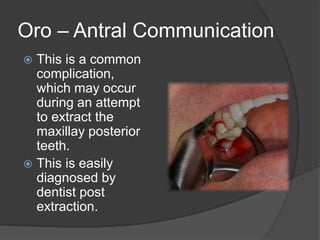
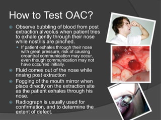
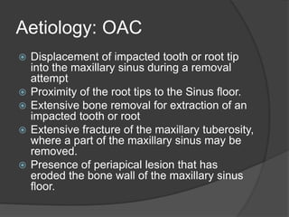
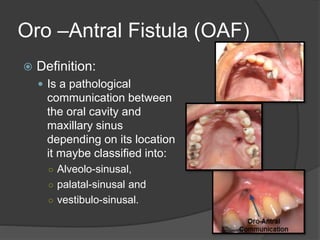
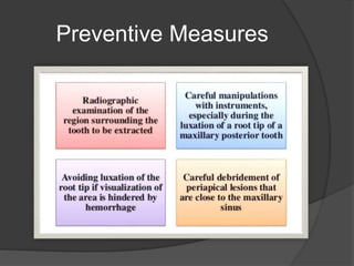
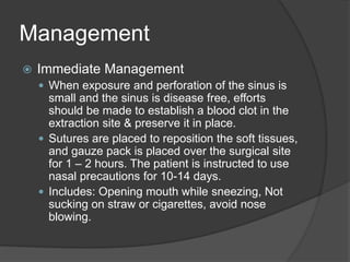
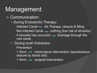
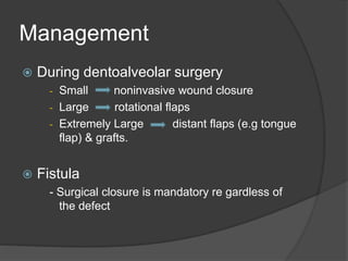
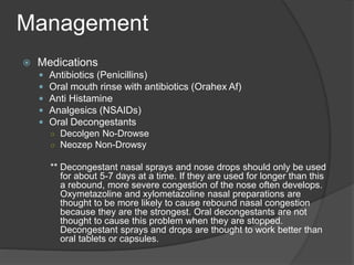
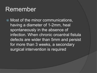
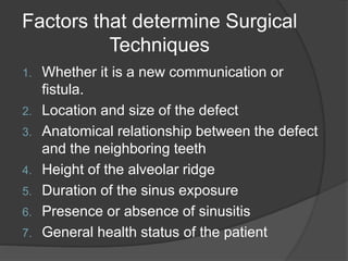
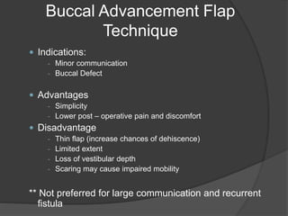
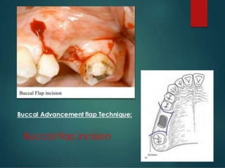
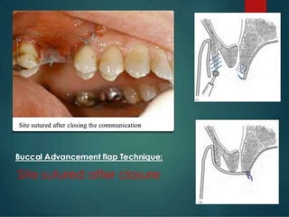
Arrantxa Danielle M. Sumabat, DMD
Resident, Department of Oral & Maxillo Facial Surgery
Craniofacial Foundation of the Philippines
Maxillary Sinus
 The maxillary sinuses are
located beneath the cheeks,
above the teeth and on
either sides of the nose.
 It drains into the nose
through a hole located
about half way up the side
of the sinus wall called the
ostia.
 If the maxillary sinuses
become blocked they fill up
with liquid that often gets
infected (sinusitis). This
could cause toothache or a
dull aching pain under your
cheeks.
Oro – Antral Communication
 This is a common
complication,
which may occur
during an attempt
to extract the
maxillay posterior
teeth.
 This is easily
diagnosed by
dentist post
extraction.
 Observe bubbling of blood from post
extraction alveolus when patient tries
to exhale gently through their nose
while nostrils are pinched.
 If patient exhales through their nose
with great pressure, risk of causing
oroantral communication may occur,
even though communication may not
have occurred initially.
 Fluid comes out of the nose while
rinsing post extraction
 Fogging of the mouth mirror when
place directly on the extraction site
as the patient exhales through his
nose.
 Radiograph is usually used for
confirmation, and to determine the
extent of defect.
How to Test OAC?
Aetiology: OAC
 Displacement of impacted tooth or root tip
into the maxillary sinus during a removal
attempt
 Proximity of the root tips to the Sinus floor.
 Extensive bone removal for extraction of an
impacted tooth or root
 Extensive fracture of the maxillary tuberosity,
where a part of the maxillary sinus may be
removed.
 Presence of periapical lesion that has
eroded the bone wall of the maxillary sinus
floor.
Oro –Antral Fistula (OAF)
 Definition:
 Is a pathological
communication between
the oral cavity and
maxillary sinus
depending on its location
it maybe classified into:
○ Alveolo-sinusal,
○ palatal-sinusal and
○ vestibulo-sinusal.
OROANTRAL FITULA
 The term OAF is meant to indicate a canal
lined by epithelium that may be filled by
granulation tissue or by polyposis of the sinus
membrane, most frequently due to iatrogenic
oroantral communication.
 It must be emphasized that unlike the oro-
antral communication (OAC), OAF is
characterized by the presence of epithelium
arising from the oral mucosa and/or from the
antral sinus mucosa that, if not removed, could
inhibit spontaneous healing.
Aetiology
 OAF could be caused by dental infection,
osteomyelitis, radiation therapy, trauma or
following removal of maxillary cysts or
tumors.
 The extraction of maxillary posterior teeth
represents the most common etiology of
OAF due to the proximity of the bicuspid
apices and molars to the antrum.
 Alternatively, OAF might arise during
preparation of bone for insertion of a dental
implant as a consequence of poor surgical
planning.
Signs and Symptoms
 Unpleasant tasting discharge and odor
 Reflux of fluids and food into the nose
from the mouth
 Leakage of air
 Some patients may be asymptomatic
Preventive Measures
Management
 Immediate Management
 When exposure and perforation of the sinus is
small and the sinus is disease free, efforts
should be made to establish a blood clot in the
extraction site & preserve it in place.
 Sutures are placed to reposition the soft tissues,
and gauze pack is placed over the surgical site
for 1 – 2 hours. The patient is instructed to use
nasal precautions for 10-14 days.
 Includes: Opening mouth while sneezing, Not
sucking on straw or cigarettes, avoid nose
blowing.
Management
 Communication:
 During Endodontic Therapy:
- Infected Canal Ab Therapy, closure & filling
- Not infected Canal nothing (low risk of sinusitis)
• If sinusitis has occurred drainage through the
root canal.
 During tooth Extraction
- Prevention
- < 5mm noninvasive intervention (spontaneous
closure by blood clot)
- > 5mm surgical intervention
Management
 During dentoalveolar surgery
- Small noninvasive wound closure
- Large rotational flaps
- Extremely Large distant flaps (e.g tongue
flap) & grafts.
 Fistula
- Surgical closure is mandatory re gardless of
the defect
 Medications
 Antibiotics (Penicillins)
 Oral mouth rinse with antibiotics (Orahex Af)
 Anti Histamine
 Analgesics (NSAIDs)
 Oral Decongestants
○ Decolgen No-Drowse
○ Neozep Non-Drowsy
** Decongestant nasal sprays and nose drops should only be used
for about 5-7 days at a time. If they are used for longer than this
a rebound, more severe congestion of the nose often develops.
Oxymetazoline and xylometazoline nasal preparations are
thought to be more likely to cause rebound nasal congestion
because they are the strongest. Oral decongestants are not
thought to cause this problem when they are stopped.
Decongestant sprays and drops are thought to work better than
oral tablets or capsules.
Management
Remember
 Most of the minor communications,
having a diameter of 1-2mm, heal
spontaneously in the absence of
infection. When chronic oroantral fistula
defects are wider than 5mm and persist
for more than 3 weeks, a secondary
surgical intervention is required
Immediate Management
Factors that determine Surgical
Techniques
1. Whether it is a new communication or
fistula.
2. Location and size of the defect
3. Anatomical relationship between the defect
and the neighboring teeth
4. Height of the alveolar ridge
5. Duration of the sinus exposure
6. Presence or absence of sinusitis
7. General health status of the patient
Buccal Advancement Flap
Technique
 Indications:
- Minor communication
- Buccal Defect
 Advantages
- Simplicity
- Lower post – operative pain and discomfort
 Disadvantage
- Thin flap (increase chances of dehiscence)
- Limited extent
- Loss of vestibular depth
- Scaring may cause impaired mobility
** Not preferred for large communication and recurrent
fistula
Buccal Advancement Flap
Techniques
Palatal Flap Technique
 Advantage
- More tissue attachment without
tension
- Firmer and more resistant to trauma
& infection
- Could be used with large defect
- Preserve the buccal vestibular depth
- Good vascularization
 Disadvantage
- Denudation of the palatal surface
- Greater post-op pain
- More complicated technique
- Appearance of roughness at the
donor site (epithelialization)
- Possible flap necrosis
- Interfere with wearing partial denture
for covering the hard palate
Palatal Pedicle Flap
Buccal & Palatal Flap
Buccal Fat Pad Flap
 The use of this type of flap has
limited clinical usage and for
many years has been
considered a risky procedure
due to the possibility of
traumatizing the pterygo-
maxillary space.
 This is commonly used for
OAF due to its location which
is anatomically favorable, easy
and minimal dissection with
which it can be harvested and
mobilized. The fat pad
provides a good rate of
epithelialization and low rate of
failure.
Buccal Fat Pad
 Is a biconvex disc of vascularized fat
lying behind the zygomatic arch.
 There are four processes, the buccal
process, the pterygoid process, the
superficial process and the deep
temporal process. These extend from
the body to the surrounding tissue
spaces such as the
pterygomandibular space and the
infratemporal space.
 6 The arterial supply to the BFP
depends on small branches of the
maxillary, superficial temporal and
facial arteries.
 The size of the BFP is fairly constant
among individuals, regardless of the
overall body weight and fat
distribution.
How to reach BFP?
 In order to reach the BFP an incision of the posterior
mucosa must be made in the area of the zygomatic
buttress, followed by a light in incision of the periosteum
and the fascial envelope of the buccal pad.
 A gentle dissection with fine curved artery forceps
exposes the yellowish-colored buccal fat. The buccal fat
pad flap, preferably of the pedicled type, has been used
most commonly for the closure of the OAF. This is due
to the location of the buccal fat pad which is
anatomically favorable, to the easy and minimal
dissection with which it can be harvested and
mobilized. The fat pad provides a good rate of
epithelialization and a low rate of failure
Buccal Fat Pad Flap Techniques
Points to Remember
 According to the literature and to the author’s clinical
experience, any communication between the maxillary
sinus and the oral cavity lasting for more than three weeks
should be surgically closed in order to avoid further
medical problems.
 Immediate repairs of the acute oroantral defect have a
uniformly high success rate approaching 95% that
decreases to 67% in cases of delayed closure. An
important role in the healing process is played by the
presence of sinus diseases.
 An important role in the healing process is played by the
presence of sinus diseases. In these cases the advice of a
specialist will help to deal with complications.
Points to Remember
 Treatment modalities to repair the oroantral
fistula include local or free soft tissue flaps,
with or with or without autogenous grafts or
alloplastic implants.
 The closure of an oroantal communication
of any origin, can be achieved by different
techniques. Particular emphasis should be
made in choosing most appropriate
surgical technique to use.
References
 Use of the buccal fat pad in maxillary and sinus grafting of
the severely atrophic maxilla preparatory to implant
reconstruction of the partially or completely edentulous
patient: technical note.Liversedge RL, Wong K
Int J Oral Maxillofac Implants. 2002 May-Jun; 17(3):424-8.
 Andrea Enrico Borgonovo, Frederick Valerio
Berardinelli.2012. Surgical options in Oroantral Fistula
Treatment. The Open Dentistry Journal.2013
 Closure of Oroantral Communications: A Review of the
Literature. Susan H. Visscher, Baucke van Minnen,.2010,
Journal of Oral and Maxillofacial Surgery.
 Hupp, J.R, Edward Ellis III and Tucker, M.R.(2009) –
CONTEMPORARY Oral and Maxillofacial Surgery,
Missouri: Mosby
 Fragiskos, D.F. (1965) – Oral Surery, Heidelberg: Springer
- Verlag
Oro – antral  communication

Oro – antral communication

  • 1.
    Arrantxa Danielle M.Sumabat, DMD Resident, Department of Oral & Maxillo Facial Surgery Craniofacial Foundation of the Philippines
  • 2.
    Maxillary Sinus  Themaxillary sinuses are located beneath the cheeks, above the teeth and on either sides of the nose.  It drains into the nose through a hole located about half way up the side of the sinus wall called the ostia.  If the maxillary sinuses become blocked they fill up with liquid that often gets infected (sinusitis). This could cause toothache or a dull aching pain under your cheeks.
  • 3.
    Oro – AntralCommunication  This is a common complication, which may occur during an attempt to extract the maxillay posterior teeth.  This is easily diagnosed by dentist post extraction.
  • 4.
     Observe bubblingof blood from post extraction alveolus when patient tries to exhale gently through their nose while nostrils are pinched.  If patient exhales through their nose with great pressure, risk of causing oroantral communication may occur, even though communication may not have occurred initially.  Fluid comes out of the nose while rinsing post extraction  Fogging of the mouth mirror when place directly on the extraction site as the patient exhales through his nose.  Radiograph is usually used for confirmation, and to determine the extent of defect. How to Test OAC?
  • 5.
    Aetiology: OAC  Displacementof impacted tooth or root tip into the maxillary sinus during a removal attempt  Proximity of the root tips to the Sinus floor.  Extensive bone removal for extraction of an impacted tooth or root  Extensive fracture of the maxillary tuberosity, where a part of the maxillary sinus may be removed.  Presence of periapical lesion that has eroded the bone wall of the maxillary sinus floor.
  • 6.
    Oro –Antral Fistula(OAF)  Definition:  Is a pathological communication between the oral cavity and maxillary sinus depending on its location it maybe classified into: ○ Alveolo-sinusal, ○ palatal-sinusal and ○ vestibulo-sinusal.
  • 7.
    OROANTRAL FITULA  Theterm OAF is meant to indicate a canal lined by epithelium that may be filled by granulation tissue or by polyposis of the sinus membrane, most frequently due to iatrogenic oroantral communication.  It must be emphasized that unlike the oro- antral communication (OAC), OAF is characterized by the presence of epithelium arising from the oral mucosa and/or from the antral sinus mucosa that, if not removed, could inhibit spontaneous healing.
  • 8.
    Aetiology  OAF couldbe caused by dental infection, osteomyelitis, radiation therapy, trauma or following removal of maxillary cysts or tumors.  The extraction of maxillary posterior teeth represents the most common etiology of OAF due to the proximity of the bicuspid apices and molars to the antrum.  Alternatively, OAF might arise during preparation of bone for insertion of a dental implant as a consequence of poor surgical planning.
  • 9.
    Signs and Symptoms Unpleasant tasting discharge and odor  Reflux of fluids and food into the nose from the mouth  Leakage of air  Some patients may be asymptomatic
  • 10.
  • 11.
    Management  Immediate Management When exposure and perforation of the sinus is small and the sinus is disease free, efforts should be made to establish a blood clot in the extraction site & preserve it in place.  Sutures are placed to reposition the soft tissues, and gauze pack is placed over the surgical site for 1 – 2 hours. The patient is instructed to use nasal precautions for 10-14 days.  Includes: Opening mouth while sneezing, Not sucking on straw or cigarettes, avoid nose blowing.
  • 12.
    Management  Communication:  DuringEndodontic Therapy: - Infected Canal Ab Therapy, closure & filling - Not infected Canal nothing (low risk of sinusitis) • If sinusitis has occurred drainage through the root canal.  During tooth Extraction - Prevention - < 5mm noninvasive intervention (spontaneous closure by blood clot) - > 5mm surgical intervention
  • 13.
    Management  During dentoalveolarsurgery - Small noninvasive wound closure - Large rotational flaps - Extremely Large distant flaps (e.g tongue flap) & grafts.  Fistula - Surgical closure is mandatory re gardless of the defect
  • 14.
     Medications  Antibiotics(Penicillins)  Oral mouth rinse with antibiotics (Orahex Af)  Anti Histamine  Analgesics (NSAIDs)  Oral Decongestants ○ Decolgen No-Drowse ○ Neozep Non-Drowsy ** Decongestant nasal sprays and nose drops should only be used for about 5-7 days at a time. If they are used for longer than this a rebound, more severe congestion of the nose often develops. Oxymetazoline and xylometazoline nasal preparations are thought to be more likely to cause rebound nasal congestion because they are the strongest. Oral decongestants are not thought to cause this problem when they are stopped. Decongestant sprays and drops are thought to work better than oral tablets or capsules. Management
  • 15.
    Remember  Most ofthe minor communications, having a diameter of 1-2mm, heal spontaneously in the absence of infection. When chronic oroantral fistula defects are wider than 5mm and persist for more than 3 weeks, a secondary surgical intervention is required
  • 16.
  • 17.
    Factors that determineSurgical Techniques 1. Whether it is a new communication or fistula. 2. Location and size of the defect 3. Anatomical relationship between the defect and the neighboring teeth 4. Height of the alveolar ridge 5. Duration of the sinus exposure 6. Presence or absence of sinusitis 7. General health status of the patient
  • 18.
    Buccal Advancement Flap Technique Indications: - Minor communication - Buccal Defect  Advantages - Simplicity - Lower post – operative pain and discomfort  Disadvantage - Thin flap (increase chances of dehiscence) - Limited extent - Loss of vestibular depth - Scaring may cause impaired mobility ** Not preferred for large communication and recurrent fistula
  • 21.
  • 22.
    Palatal Flap Technique Advantage - More tissue attachment without tension - Firmer and more resistant to trauma & infection - Could be used with large defect - Preserve the buccal vestibular depth - Good vascularization  Disadvantage - Denudation of the palatal surface - Greater post-op pain - More complicated technique - Appearance of roughness at the donor site (epithelialization) - Possible flap necrosis - Interfere with wearing partial denture for covering the hard palate
  • 23.
  • 24.
  • 25.
    Buccal Fat PadFlap  The use of this type of flap has limited clinical usage and for many years has been considered a risky procedure due to the possibility of traumatizing the pterygo- maxillary space.  This is commonly used for OAF due to its location which is anatomically favorable, easy and minimal dissection with which it can be harvested and mobilized. The fat pad provides a good rate of epithelialization and low rate of failure.
  • 26.
    Buccal Fat Pad Is a biconvex disc of vascularized fat lying behind the zygomatic arch.  There are four processes, the buccal process, the pterygoid process, the superficial process and the deep temporal process. These extend from the body to the surrounding tissue spaces such as the pterygomandibular space and the infratemporal space.  6 The arterial supply to the BFP depends on small branches of the maxillary, superficial temporal and facial arteries.  The size of the BFP is fairly constant among individuals, regardless of the overall body weight and fat distribution.
  • 27.
    How to reachBFP?  In order to reach the BFP an incision of the posterior mucosa must be made in the area of the zygomatic buttress, followed by a light in incision of the periosteum and the fascial envelope of the buccal pad.  A gentle dissection with fine curved artery forceps exposes the yellowish-colored buccal fat. The buccal fat pad flap, preferably of the pedicled type, has been used most commonly for the closure of the OAF. This is due to the location of the buccal fat pad which is anatomically favorable, to the easy and minimal dissection with which it can be harvested and mobilized. The fat pad provides a good rate of epithelialization and a low rate of failure
  • 28.
    Buccal Fat PadFlap Techniques
  • 29.
    Points to Remember According to the literature and to the author’s clinical experience, any communication between the maxillary sinus and the oral cavity lasting for more than three weeks should be surgically closed in order to avoid further medical problems.  Immediate repairs of the acute oroantral defect have a uniformly high success rate approaching 95% that decreases to 67% in cases of delayed closure. An important role in the healing process is played by the presence of sinus diseases.  An important role in the healing process is played by the presence of sinus diseases. In these cases the advice of a specialist will help to deal with complications.
  • 30.
    Points to Remember Treatment modalities to repair the oroantral fistula include local or free soft tissue flaps, with or with or without autogenous grafts or alloplastic implants.  The closure of an oroantal communication of any origin, can be achieved by different techniques. Particular emphasis should be made in choosing most appropriate surgical technique to use.
  • 31.
    References  Use ofthe buccal fat pad in maxillary and sinus grafting of the severely atrophic maxilla preparatory to implant reconstruction of the partially or completely edentulous patient: technical note.Liversedge RL, Wong K Int J Oral Maxillofac Implants. 2002 May-Jun; 17(3):424-8.  Andrea Enrico Borgonovo, Frederick Valerio Berardinelli.2012. Surgical options in Oroantral Fistula Treatment. The Open Dentistry Journal.2013  Closure of Oroantral Communications: A Review of the Literature. Susan H. Visscher, Baucke van Minnen,.2010, Journal of Oral and Maxillofacial Surgery.  Hupp, J.R, Edward Ellis III and Tucker, M.R.(2009) – CONTEMPORARY Oral and Maxillofacial Surgery, Missouri: Mosby  Fragiskos, D.F. (1965) – Oral Surery, Heidelberg: Springer - Verlag

Editor's Notes

  • #2 The most common dental complication of oral surgical procedures that subsequently involve the maxillary sinus include displacement of teeth, roots, or instrument fragments into the sinus or the communication between the oral cavity and the sinus during posterior maxilla surgery
  • #5 Probing is generally not recommended, could cause perforation.
  • #6 Proximity to sinus floor = in this case the bony portion above the root tip is very thin or may even be absent, where upon oroantral communication is inevitable during the extraction of the tooth, especially in the alveolus is debrided unnecessarily.
  • #8 This type of OAF will develop an epithelium similar to the pseudo stratified ciliated columnar respiratory cells of the maxillary antrum and to those of the squamous epithelium of the oral mucosa. The fistula must be quickly closed since its persistence increases the possible inflammation of the sinus through contamination of the oral cavity. It is important to establish whether or not sinus infection has occurred [2] Sinus infection can occur with any size and duration of fistula canal.
  • #12 The best treatment of a potential sinus exposure is aoiding the problem through careful observation and treatment planning.
  • #15 How do decongestants work? They help reduce swelling in the passageways of your nose, which relieves the feeling of pressure and improves the flow of air. You'll be able to breathe a whole lot better. Decongestants come in pill form or nasal sprays. Don't use the sprays for more than 3 days, or you may get more stuffed up. - The vast majority of decongestants act via enhancing norepinephrine (noradrenaline) and epinephrine(adrenaline) or adrenergic activity by stimulating the α-adrenergic receptors. This inducesvasoconstriction of the blood vessels in the nose, throat, and paranasal sinuses, which results in reducedinflammation (swelling) and mucus formation in these areas. How Do Antihistamines Work? Some types of them can help relieve your runny nose and sneezing when you have a cold. They block a chemical your body makes called histamine that makes the tissues in your nose itch and swell. Most experts say that histamine isn't the major cause of a runny nose when you have a cold. Even so, some of the older antihistamines, such as brompheniramine and chlorpheniramine, can bring relief.