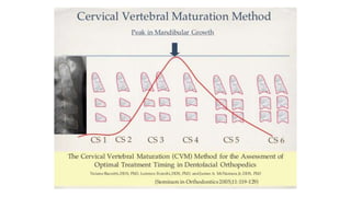
Cervical Vertebral
Maturation
Aslam Alkadhimi
Aims
• Overview of CVM
• Critique Szemraj et al 2018
Introduction
• The timing of the treatment onset may be as critical as the selection
of the specific treatment protocol
• “Timing is the fourth dimension in orthodontics (transverse, sagittal ,
vertical)”
• The issue of optimal timing for dentofacial orthopedics is linked
intimately to the identification of periods of accelerated growth that
can contribute significantly to the correction of skeletal imbalances in
the individual patient.
Skeletal maturity
Individual skeletal maturity can be assessed by means of several
biologic indicators:
• increase in body height
• skeletal maturation of the hand and wrist
• dental development and eruption
• menarche or voice changes
• cervical vertebral maturation
Biologic indicator
• The reliability and efficiency of a biologic indicator can be evaluated
with respect to several fundamental requisites:
• Efficacy in detecting the peak in mandibular growth.
• No need for additional x-ray exposure.
• Ease in recording.
• Consistency in the interpretation of the data.
• Usefulness for the anticipation of the occurrence of the peak.
History of CVM
• The cervical vertebrae undergo a series of well-defined ossification
changes, which correlate with different stages of hand–wrist
development
• Given that the upper cervical vertebrae are identifiable on a
cephalometric radiograph, this precludes the need for additional
radiation
• Lamparski 1972 Franchi 2000 Baccetti 2002 Baccetti 2005
• The method is based upon morphology of the second (C2-odontoid
process), third (C3) and fourth (C4) cervical vertebrae bodies
History of CVM
• A total sample (706) that comprises the cephalometric files of the
University of Michigan Elementary and Secondary School Growth
Study was evaluated.
• Total mandibular length (Co-Gn) was measured on the longitudinal
sets of lateral cephalograms at yearly intervals.
• The maximum increase in Co-Gn between two consecutive annual
cephalograms was used to define the peak in mandibular growth at
puberty.
• The morphology of the bodies of C2, C3 and C4 were analysed in the
six consecutive annual observations (T1 through T6).
Mandibular length
Development of CVM
Mandibular length changes:
• CS1CS2 2.5mm pre-peak
• CS2CS3 2.5mm
• CS3CS4 5.4mm peak
• CS4CS5 1.6mm
• CS5CS6 2.1mm post-peak
CVM stages
McNamara and Franchi 2018
-Concavity of lower borders C2
C3 C4
-Shape of bodies C3 C4
Trapezoid=least mature
Rect. Vertica=adult life
Uncertain cases?
Anterior spikes v. posterior spikes
Depth of concavity > 0.8mm
If uncertain choose earlier stage
Clinical application
• Class II treatment is most effective when it includes the peak in mandibular growth; CS3 and 4
• Class III treatment with maxillary expansion and protraction is effective in the maxilla only when
it is performed before the peak (CS1 or CS2), whereas it is effective in the mandible during both
prepubertal and pubertal stages;
• Skeletal effects of rapid maxillary expansion for the correction of transverse maxillary deficiency
are greater at prepubertal stages, while pubertal or postpubertal use of the rapid maxillary
expander entails more dentoalveolar effects;
• Deficiency of mandibular ramus height can be enhanced significantly in subjects with increased
vertical facial dimension when orthopedic treatment is performed at the peak in mandibular
growth (CS3).
To summarize, effects of therapies aimed to enhance/restrict mandibular growth appear to be of
greater magnitude at the circumpubertal period during which the growth spurt occurs in
comparison to earlier intervention, while effects of therapies aimed to alter the maxilla
orthopedically (maxillary protraction/maxillary expansion) are greater at prepubertal
Evidence
• Mellion et al, 2013: utilized the archive at the Bolton-Brush Growth
Study Centre in Cleveland, Ohio, and investigated patterns of facial
skeletal growth and their relationship to various common indices of
maturation.
• Serial records of 100 children (50 males and 50 females) were
selected to encompass onset and peak of the adolescent facial
growth spurt. This investigation has some interesting findings:
• In terms of growth prediction, hand–wrist radiographs provided the
best indication that maturation had reached the peak velocity stage,
and whilst chronological age was nearly as good, CMV was
consistently the worst.
Evidence
Evidence
Evidence
Evidence
Evidence
Aim
TO:
• Assess the usefulness of the CVM method
• Verify the assumption, according to which the CVM method modified
by Baccetti et al. may replace the method for the assessment of
skeletal maturation based on hand-wrist maturation (HWM) method.
Materials and methods
• Review of literature from 2006-2016
• Exclusion criteria: articles related to patients with prior orthodontic
treatment, patients with systemic diseases, cleft palate and other
skeletal deformities
• 9055214810 articles included
• No restriction on language
Materials and methods
• The largest group consisted of 709 and the smallest of 30
• One hand-wrist X-ray and one Ceph were analysed in each patient. In most articles, the
results were presented with no gender division
• In all of the articles the bone age was assessed on Cephs using the CVM method,
according to Baccetti, Franchi and McNamara.
• However, the methods of determining the skeletal maturation of the hand and wrist
were different:
3 articles used Bjork’s method
2 used Fishman method
1 used Grave and Brown method
1 used National Taiwan University Hospital Skeletal Maturation Index
1 used Greulich and Pyle method
1 used Martins method
Results
• Not possible to carry out meta analysis due to the inherent
heterogeneity of the studies
• All of the studies presented a high level of correlation between the
examined methods.
• In eight articles the researchers concluded that the CVM classification
could replace the HWM method, known as the “gold standard”.
• The lowest correlation coefficient was 0.616 and the highest 0.937.
Conclusion
Cvm

Cvm

  • 1.
  • 2.
    Aims • Overview ofCVM • Critique Szemraj et al 2018
  • 3.
    Introduction • The timingof the treatment onset may be as critical as the selection of the specific treatment protocol • “Timing is the fourth dimension in orthodontics (transverse, sagittal , vertical)” • The issue of optimal timing for dentofacial orthopedics is linked intimately to the identification of periods of accelerated growth that can contribute significantly to the correction of skeletal imbalances in the individual patient.
  • 4.
    Skeletal maturity Individual skeletalmaturity can be assessed by means of several biologic indicators: • increase in body height • skeletal maturation of the hand and wrist • dental development and eruption • menarche or voice changes • cervical vertebral maturation
  • 5.
    Biologic indicator • Thereliability and efficiency of a biologic indicator can be evaluated with respect to several fundamental requisites: • Efficacy in detecting the peak in mandibular growth. • No need for additional x-ray exposure. • Ease in recording. • Consistency in the interpretation of the data. • Usefulness for the anticipation of the occurrence of the peak.
  • 6.
    History of CVM •The cervical vertebrae undergo a series of well-defined ossification changes, which correlate with different stages of hand–wrist development • Given that the upper cervical vertebrae are identifiable on a cephalometric radiograph, this precludes the need for additional radiation • Lamparski 1972 Franchi 2000 Baccetti 2002 Baccetti 2005 • The method is based upon morphology of the second (C2-odontoid process), third (C3) and fourth (C4) cervical vertebrae bodies
  • 7.
    History of CVM •A total sample (706) that comprises the cephalometric files of the University of Michigan Elementary and Secondary School Growth Study was evaluated. • Total mandibular length (Co-Gn) was measured on the longitudinal sets of lateral cephalograms at yearly intervals. • The maximum increase in Co-Gn between two consecutive annual cephalograms was used to define the peak in mandibular growth at puberty. • The morphology of the bodies of C2, C3 and C4 were analysed in the six consecutive annual observations (T1 through T6).
  • 8.
  • 9.
    Development of CVM Mandibularlength changes: • CS1CS2 2.5mm pre-peak • CS2CS3 2.5mm • CS3CS4 5.4mm peak • CS4CS5 1.6mm • CS5CS6 2.1mm post-peak
  • 10.
    CVM stages McNamara andFranchi 2018 -Concavity of lower borders C2 C3 C4 -Shape of bodies C3 C4 Trapezoid=least mature Rect. Vertica=adult life
  • 12.
    Uncertain cases? Anterior spikesv. posterior spikes Depth of concavity > 0.8mm If uncertain choose earlier stage
  • 14.
    Clinical application • ClassII treatment is most effective when it includes the peak in mandibular growth; CS3 and 4 • Class III treatment with maxillary expansion and protraction is effective in the maxilla only when it is performed before the peak (CS1 or CS2), whereas it is effective in the mandible during both prepubertal and pubertal stages; • Skeletal effects of rapid maxillary expansion for the correction of transverse maxillary deficiency are greater at prepubertal stages, while pubertal or postpubertal use of the rapid maxillary expander entails more dentoalveolar effects; • Deficiency of mandibular ramus height can be enhanced significantly in subjects with increased vertical facial dimension when orthopedic treatment is performed at the peak in mandibular growth (CS3). To summarize, effects of therapies aimed to enhance/restrict mandibular growth appear to be of greater magnitude at the circumpubertal period during which the growth spurt occurs in comparison to earlier intervention, while effects of therapies aimed to alter the maxilla orthopedically (maxillary protraction/maxillary expansion) are greater at prepubertal
  • 15.
    Evidence • Mellion etal, 2013: utilized the archive at the Bolton-Brush Growth Study Centre in Cleveland, Ohio, and investigated patterns of facial skeletal growth and their relationship to various common indices of maturation. • Serial records of 100 children (50 males and 50 females) were selected to encompass onset and peak of the adolescent facial growth spurt. This investigation has some interesting findings: • In terms of growth prediction, hand–wrist radiographs provided the best indication that maturation had reached the peak velocity stage, and whilst chronological age was nearly as good, CMV was consistently the worst.
  • 16.
  • 17.
  • 18.
  • 19.
  • 20.
  • 22.
    Aim TO: • Assess theusefulness of the CVM method • Verify the assumption, according to which the CVM method modified by Baccetti et al. may replace the method for the assessment of skeletal maturation based on hand-wrist maturation (HWM) method.
  • 23.
    Materials and methods •Review of literature from 2006-2016 • Exclusion criteria: articles related to patients with prior orthodontic treatment, patients with systemic diseases, cleft palate and other skeletal deformities • 9055214810 articles included • No restriction on language
  • 26.
    Materials and methods •The largest group consisted of 709 and the smallest of 30 • One hand-wrist X-ray and one Ceph were analysed in each patient. In most articles, the results were presented with no gender division • In all of the articles the bone age was assessed on Cephs using the CVM method, according to Baccetti, Franchi and McNamara. • However, the methods of determining the skeletal maturation of the hand and wrist were different: 3 articles used Bjork’s method 2 used Fishman method 1 used Grave and Brown method 1 used National Taiwan University Hospital Skeletal Maturation Index 1 used Greulich and Pyle method 1 used Martins method
  • 27.
    Results • Not possibleto carry out meta analysis due to the inherent heterogeneity of the studies • All of the studies presented a high level of correlation between the examined methods. • In eight articles the researchers concluded that the CVM classification could replace the HWM method, known as the “gold standard”. • The lowest correlation coefficient was 0.616 and the highest 0.937.
  • 28.