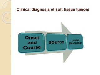
This document provides a 3-step algorithm for diagnosing oral exophytic lesions:
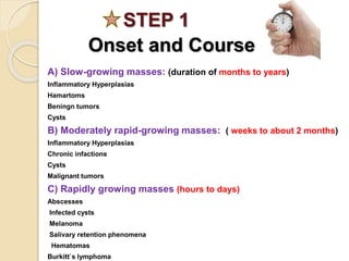
1. Determine the onset and course of the lesion which can indicate inflammatory hyperplasias, benign or malignant tumors, or infections.
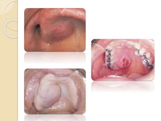
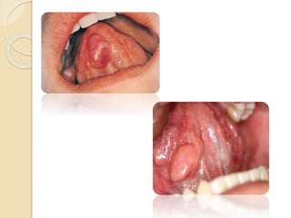
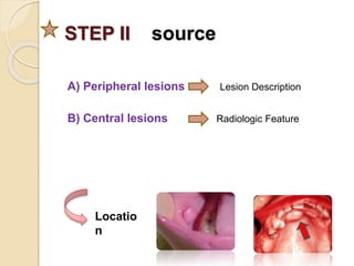
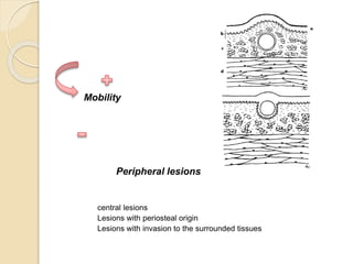
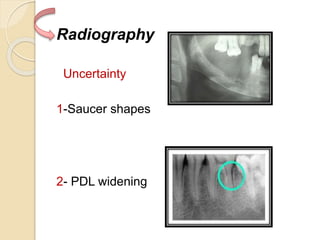
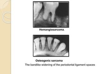
2. Examine the location, mobility, and radiographic features of the lesion which can provide clues to whether it is peripheral or central in location and if there are signs of periosteal involvement or tissue invasion.
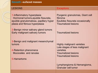
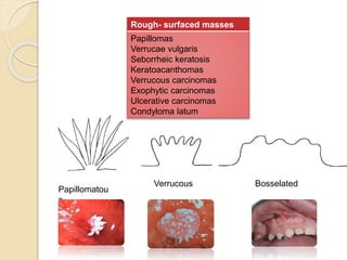
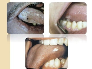
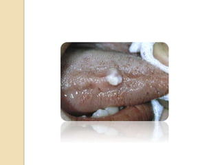
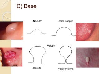
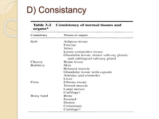
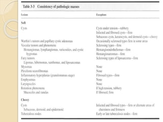
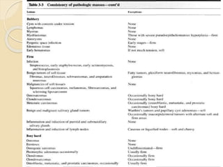
3. Evaluate the characteristics of the lesion such as its surface, base, and consistency which when combined with location and features can indicate possibilities like inflammatory hyperplasias, benign or malignant tumors, cysts, or infections.