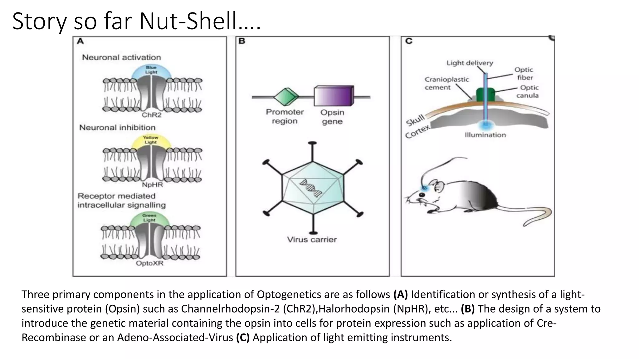
The basics for symbiosis of Optics and Genetics have been explained in this presentation. " How light can change the very way of life?" .This question has been addressed using relevant web content, consultations from book and through nature videos. This presentation was awarded the highest score in PHM805 at Dayalbagh Educational Institute, Agra.
Optogenetics is a technique that uses light to control neurons that have been genetically modified to express light-sensitive ion channels. It allows scientists to precisely stimulate or silence neural activity by exposing specific neurons to light. The first demonstration of optogenetics in mammalian neurons used channelrhodopsin, a light-activated ion channel from algae, to activate neurons with light. Optogenetics holds promise for advancing understanding of brain function and developing new treatments for neurological disorders like Parkinson's disease, epilepsy, and blindness through targeted neuromodulation with light. Challenges include improving light-sensitive tools and light sources to target deeper brain regions.
Optogenetics integrates optics and genetics to control neural activity with light. It involves introducing light-sensitive proteins into neurons using viruses. When exposed to light, these proteins cause neurons to become active or inactive. This allows controlling and studying neural circuits with high spatial and temporal precision. Optogenetics was pioneered in 2005 and has since been applied to study various neural systems and behaviors.
Optogenetics is a technique developed in 2006 that uses light to control neurons by inserting light-sensitive ion channel genes into brain cells. It works by using channelrhodopsin genes to excite neurons or halorhodopsin genes to inhibit neurons when light hits the nerve cell. This allows precise activation or stopping of specific neuron groups with high temporal resolution. Optogenetics offers potential treatment for conditions like anxiety, addiction, chronic pain, and sleep disorders by providing a less invasive way to stimulate or inhibit neurons compared to electrical methods. However, challenges include introducing foreign genes into the brain and technical issues like fiber optics posing infection risks or requiring heavy batteries.
The document summarizes the Encyclopedia of DNA Elements (ENCODE) project. It describes ENCODE as a follow-up to the Human Genome Project that aims to identify all functional elements in the human genome, including regions that regulate genes. The document outlines the phases of the project and some of the high-throughput techniques used, such as ChIP-seq, DNase-seq, and MNase-seq. It also discusses how the data from ENCODE is being utilized and the future plans to expand the project.
The document summarizes several genome and brain mapping projects that followed the completion of the Human Genome Project in 2003. It describes the objectives and outcomes of the HapMap Project, ENCODE Project, Human Proteome Project, European Commission's Human Brain Project, and U.S. Brain Mapping Project. All of these projects aimed to further understand the human genome and proteome, characterize gene functions, and map the structure and diseases of the human brain. The research generated vast amounts of freely available data and furthered knowledge in human biology, disease research, and brain-inspired technologies.
The document discusses the brain as a complex network and introduces the concept of the connectome. It describes how the brain exhibits both segregation into specialized areas and integration through connections between areas. Mapping the structural and functional connectivity between brain regions using tools from network science and graph theory provides a powerful way to quantitatively describe the topological organization of the brain. Analyzing the human connectome has revealed hierarchical modular organization and the presence of connector hubs and rich clubs that facilitate integration and efficient communication in the brain network. Understanding disruptions to functional and structural connectivity may help explain neurological and psychiatric disorders.
Genomics is the study of whole genomes. In the 1980s, scientists determined sequences of important genes. In the 1990s, the genome of H. influenzae was fully sequenced. The Human Genome Project, begun in 1990, fully sequenced the human genome ahead of schedule in 2003. The human genome contains 3.2 billion DNA base pairs and 30,000-40,000 genes. While genomics provides medical benefits, it also raises safety, ethical, and privacy concerns that remain open questions.
EEG has relatively low sensitivity (25–56%) for diagnosing epilepsy but high specificity (78–98%). Epileptiform discharges are seen in 0.5% of healthy adults on routine EEG but 10-30% in those with brain pathologies. Factors like the location of the epileptogenic zone, seizure frequency, and timing of the EEG affect whether a patient shows interictal epileptiform discharges. Intraoperative electrocorticography directly records cortical potentials during epilepsy surgery to localize the irritative zone, map brain functions, and predict surgical outcomes. It has higher spatial resolution than scalp EEG but recordings are limited by anesthesia effects and duration.
The nervous system receives millions of bits of sensory information per minute and integrates them to determine responses. The basic functional unit of the brain is neurons, which communicate via electrical signals. An electroencephalogram (EEG) records these electrical signals from the scalp using electrodes. During an EEG test, electrodes are placed on the scalp to detect brain waves which are displayed as wavy lines. EEGs can help diagnose conditions affecting brain function and electrical activity such as epilepsy, brain tumors, and sleep disorders.
The document discusses electroencephalography (EEG), which records the electrical activity of the brain through electrodes placed on the scalp. It describes how Hans Berger first recorded human EEG in 1929. It then covers the different types of brain waves seen on EEG (alpha, beta, theta, delta), how they are generated and what they indicate. The document discusses how EEG is used to study epilepsy, sleep disorders and brain function. It outlines the procedure for performing an EEG and analyzing the results.
An electroencephalogram (EEG) is a test that tracks and records brain wave patterns using small metal discs placed on the scalp. EEGs are used to detect problems with brain electrical activity and locate areas of damage. Brain waves are categorized into four main groups - beta, alpha, theta, and delta - based on their frequency. EEGs have various applications like monitoring alertness, locating seizures, and investigating sleep disorders. The EEG methodology involves non-invasive electrodes, amplifiers to strengthen microvolt signals, an analog to digital converter, and computer for storage and display.
Overview of epigenetics and its role in diseaseGarry D. Lasaga
Epigenetics is the study of heritable changes in gene expression (active versus inactive genes) that do not involve changes to the underlying DNA sequence — a change in phenotype without a change in genotype — which in turn affects how cells read the genes.
Neuro-technology aims to restore or improve human nervous system function through electronics. Neuromotor prostheses (NMPs) extract signals from the nervous system to control devices. The goal of NMPs is to convey motor control intent from the central nervous system to drive multi-degree of freedom prosthetic devices for amputees or paralyzed patients. Key challenges are developing neural interfaces that last a lifetime and providing dexterous, natural control of prosthetics. NMP systems involve neural implants to record brain signals, decoding software to translate signals into motor commands, and output devices like prosthetics. Technological advances now allow basic NMP control but further progress is still needed.
The document discusses three fluorescence microscopy techniques: FRET, FRAP, and TIRF microscopy. It explains the principles, instrumentation, sample preparation, applications, and future aspects of each technique. FRET involves energy transfer between fluorophores and is used to study molecular interactions and structure. FRAP examines fluorophore diffusion by photobleaching a region and monitoring recovery. It provides information about molecular mobility. TIRF microscopy uses evanescent waves to selectively excite fluorophores very near a surface and is applied to studies of cellular processes at membranes. All three techniques provide insights into biological phenomena at the molecular level.
Alzheimer's disease is a progressive brain disorder that causes memory loss and cognitive decline. The main pathological features are amyloid plaques and neurofibrillary tangles in the brain. Risk factors include age and family history. Symptoms include memory loss, problems with thinking and language, and behavioral changes. A diagnosis is made based on medical history, cognitive tests, and brain imaging. Currently, treatment focuses on managing symptoms with medications and lifestyle changes. Research continues on developing disease-modifying therapies to slow or stop progression.
This document provides information about zinc finger proteins. It begins with an introduction to zinc finger motifs, which are protein structural domains characterized by the coordination of zinc ions. The document then discusses the history of zinc finger discovery, functions, and families. It provides details on the most common Cys2His2 zinc finger proteins and their role in DNA recognition and transcriptional regulation. The document also examines uses of zinc finger nucleases for genome editing and their mechanism of action involving creating double-strand breaks in DNA.
Epigenetics is the study of inherited changes in gene expression and phenotype that are not caused by changes to DNA sequence. Epigenetic "tags" on DNA and histone proteins can be passed from parents to offspring and influence gene expression and traits. For example, smoking can increase the likelihood that children will also smoke due to epigenetic inheritance. Methyl groups are one way cells determine which genes to express by binding differently in various cell types like skin or eye cells. Histone proteins also control gene expression by tightly or loosely winding DNA around them.
Artifacts in EEG - Recognition and differentiationRahul Kumar
This Presentation discusses the variously commonly seen artifacts in EEG, and how to recognize them. In EEG interpretation, it is often more important to identify an artifact than to identify true pathology. Once all the artifacts are ruled out, one is sure that what one is dealing with represents disease/abnormality
New neurons are generated from neural stem cells located in the subventricular zone and subgranular zone of the adult brain. These stem cells differentiate into precursor cells, then neuroblasts, and finally mature neurons. Neuroblasts migrate through glial tubes made by astrocytes, with those from the subventricular zone traveling to the olfactory bulb along the rostral migratory stream. BrdU and tritiated thymidine are used to track the generation of new neurons by labeling cells during DNA synthesis. Adult neurogenesis is regulated by various environmental factors that can increase or decrease the proliferation and survival of new neurons.
Epigenetics is the study, in the field of genetics, of cellular and physiological phenotypic trait variations that are caused by external or environmental factors that switch genes on and off and affect how cells read genes instead of being caused by changes in the DNA sequence. -Wikipedia
This document discusses protein sequence databases and their role in storing protein data generated from genome projects and new proteomics technologies. It describes several types of protein databases, including universal repositories like GenPept that store sequences with little annotation, and expertly curated databases like Swiss-Prot that enrich sequence data with additional validation and integration. Specialized databases also exist that focus on specific protein families, organisms, structures like SCOP, or classifications like CATH.
Histone Modification: Acetylation n MethylationSomanna AN
This document discusses histone modifications through acetylation and methylation. It explains that DNA wraps around histone proteins to form nucleosomes. Histone tails can be modified through acetylation and methylation, which are regulated by specific enzyme complexes. These modifications affect chromatin structure and transcription by altering the interaction of histone tails with DNA and recruiting other proteins. The combination of modifications on histone tails is referred to as the "histone code", which is read by protein complexes to influence gene expression.
Enzyme replacement therapy in neurological disordersNeurologyKota
This document discusses enzyme replacement therapy (ERT) for lysosomal storage disorders. It provides details on ERT including its development, mechanisms, available products, dosing and costs. Challenges with ERT include limited blood-brain barrier penetration and immunogenicity. Alternative therapies discussed include substrate reduction therapy, pharmacological chaperones, and direct delivery of enzymes into the cerebrospinal fluid. ERT remains the standard treatment but has limitations for treating neurological manifestations of lysosomal storage disorders.
The patch clamp technique allows for high-resolution recording of ion channel currents. It involves using a glass pipette to form a high-resistance seal with a cell membrane, isolating a small portion of the membrane. This enables measurement of the electric current passing through individual or small groups of ion channels as voltages are varied. The technique was developed in the late 1970s and has provided insights into the functions of many ion channels in excitable cells like neurons.
Bacteriorhodopsin (BR) is a light-driven proton pump found in halophilic archaea that uses energy from light to transport protons across cell membranes. The study discovered a similar protein encoded in the genome of an uncultured gamma-proteobacterium. This bacterial rhodopsin was functionally expressed in E. coli where it formed an active light-driven proton pump, demonstrating that archaeal-like rhodopsins are more broadly distributed than previously thought. BR is a seven-helix transmembrane protein with a bound retinal molecule that undergoes structural changes upon light absorption to transport protons from the extracellular to cytoplasmic side of the cell membrane.
This document provides a summary of a presentation on electrodiagnosis and somatosensory evoked potentials (SEPs). It discusses the historical aspects, anatomical basis, nomenclature, instrumentation, waveforms, techniques, and clinical usage of SEPs. Specific topics covered include electrode placement, stimulus parameters, montages for upper and lower limb SEPs, waveform characteristics, factors affecting SEPs such as age, medication, and temperature.
Optical propagation of blue LED light in brain tissue and Parylene-C used in ...Manjunath Pujar
Understanding the propagation of LED light in the brain tissue can facilitate the advanced development of LED based neuroprosthetic devices for optogenetic applications. The attenuation coefficient of blue LED light in thin tissue slices and Parylene-C films were quantified, which is 19.9 cm-1 and 1.70 cm-1, respectively. Optical simulations in TracePro show good agreement with the experiments.
As a revolutionary neuromodulation technology, optogenetics offers remote manipulation on neural activities of genetically-targeted neural cells with millisecond temporal precision through light illumination. Compared to electrical stimulation, optogenetics has unique benefits including specificity control of neural cell types as well as minimal artifacts and instrumental interferences with electrophysiological recording. Application of optogenetics in neuroscience studies has created an increasing need for the development of light sources and the instruments for light delivery. Among various light sources, micro-light-emitting diodes (μ-LEDs) are favored for its high power efficiency, low cost, and capability of complex system integration. Successful in-vivo optogenetic stimulation on neural cells with the employment of μ-LEDs has been widely reported.
This seminar presentation summarizes research on chemoarchitectonics and neural growth factors. It discusses chemoarchitectonics as the study of brain anatomy and function through staining specific chemicals to identify neurotransmitter patterns. The presentation reviews a study on lungfish brains that identified neuronal populations and tracts using calbindin and calretinin staining. It also discusses Brodmann's cytoarchitectonic mapping of the cerebral cortex and Nissl staining technique. Finally, it provides an overview of neurotrophins like NGF, their discovery, biogenesis, functions in neuron survival and differentiation, and signaling mechanisms through neurotrophin receptors.
Microdialysis is an integral part of preclinical research to determine extracellular fluid and blood concentrations of metabolites, hormones, drugs, etc, and is often used in quantifying the biochemistry of brain and peripheral tissues. However, it is a molecular-only technique and other imaging modalities are needed to provide the researcher with functional and anatomical information of the animal in vivo.
This document describes an experiment that used near-infrared spectroscopy (NIRS) to noninvasively measure the optical properties of a songbird's brain. Researchers placed optical fibers on the head of anesthetized zebra finches to transmit laser light and collect the light after it passed through the brain tissue. They were able to measure the absorption and scattering coefficients of the caudal nidopallium region of the brain in vivo. This technique could help monitor brain activity and oxygenation levels in songbirds.
This document discusses the use of fluorescent proteins in current biological research. It begins with an overview of the development of optical microscopy and fluorescence techniques. It then focuses on the green fluorescent protein (GFP) and how it has been used as a molecular tag to study protein expression and interactions in living cells through techniques like gene delivery, transfection, viral infection, FRET, and optogenetics. The document concludes that fluorescent proteins have revolutionized cell biology by enabling the real-time visualization and control of molecular pathways and signaling processes in living systems.
Employing Electrophysiology and Optogenetics to Measure and Manipulate Neuron...InsideScientific
In this webinar, Dr. Tahl Holtzman, Founder of Cambridge NeuroTech, describes a new generation of silicon neural probes offering dozens of recording channels in precisely spaced, high-resolution arrays, built using sophisticated fabrication techniques borrowed from the electronics industry, along with simple-to-follow surgical implantation schemes for both acute and chronic animals.
Watch to learn how to take advantage of ultra-small chronic drives to open up scalability to span multiple brain areas in parallel and to achieve excellent chronic stability. In addition, Dr. Holtzman demonstrates integration of novel probes and drives offered by Cambridge NeuroTech with optogenetics that thereby enable your experiments to have the combined capability for measurement AND manipulation of neuronal activity in both acute and freely behaving settings.
This webinar will benefit both established electrophysiologists who wish to increase their data yield and experimental reach as well as those investigators whose expertise is centred in and around the animal behavioural, neuropharmacological, and optogenetics arenas. Viewers will learn what silicon neural probes are and how to use them in both acute and chronic experiments, best-practice techniques for surgical implantation in species ranging from mice to monkeys and how to integrate fibre optic cannulas with your probes to enable simultaneous opto-electrophysiology.
Studying Epilepsy in Awake Head-Fixed Mice Using Microscopy, Electrophysiolog...InsideScientific
Epilepsy research employs sophisticated research methods such as fluorescence optical imaging and optogenetics, as well as novel electrophysiological techniques, to address unresolved questions about seizure generation and propagation on the cellular and circuitry levels. Since epilepsy research is most relevant when performed in non-anesthetized mice, it requires specialized tools that ensure stable head fixation during high-precision imaging and recordings.
In this webinar, Dr. Anthony Umpierre (Prof. LongJun Wu group, Mayo Clinic, USA) and Prof. Rob Wykes (UCL, UK) present their research on microglial calcium signaling and epileptic networks carried out in awake head-fixed mice. In addition to sharing exciting new findings, the presenters address the challenges of working with awake mice.
Key topics will include…
- Mesoscopic investigations of seizure dynamics and propagation using widefield calcium imaging
- Generating full-bandwidth electrophysiological recordings enabled by graphene micro-transistors to detect spreading depolarizations and seizures
- On-demand optogenetic induction of spreading depolarizations to investigate pharmacological suppression in the awake brain
- The impact of acute versus chronic window preparations on microglial calcium activity
- The use of genetically encoded calcium indicators to study calcium dynamics in microglia
- The effects of bi-directional shifts in neuronal activity caused by kainate-triggered status epilepticus and isoflurane anesthesia on microglial calcium
This document discusses the use of nanotechnology in enzyme technology. It defines nanotechnology as manipulating matter at the atomic scale. Some key points discussed include immobilizing enzymes onto nanomaterials like nanoparticles to improve biocatalytic efficiency for applications. Nanoparticles are also shown to enhance enzyme activity and thermostability. Single enzyme nanoparticles are created by caging individual enzymes within nanostructures to increase their longevity. New techniques like EnzMet use enzymes with metallographic substrates to provide high clarity staining for applications in immunohistochemistry and electron microscopy. Harnessing firefly bioluminescence using luciferase enzymes attached to nanorods is also summarized.
Computational neuropharmacology drug designingRevathi Boyina
This document discusses computational neuropharmacology, which uses computational modeling approaches from neuroscience and dynamical systems theory integrated with traditional neuropharmacological methods to study drug effects on the brain and behavior. It describes how computational models are used in neuroscience to simulate neurons, neural circuits, and brain regions. It suggests computational neuropharmacology could help integrate molecular and systems-level descriptions of the nervous system to analyze drug effects on neural activity patterns and behavioral states. This may provide strategies for molecular screening of drugs and searching for target-specific drugs to shift pathological brain dynamics to normal patterns.
This presentation shows a basic overview of all aspects of Fluorescence Microscopy including its description, history, mechanism, applications, advantages, limitations, and some examples of studies that used this technique.
Epi-Fluorescence Microscopy: Explore Its Amazing Powers and Uses | The Lifesc...The Lifesciences Magazine
Epi-fluorescence microscopy, also known as epifluorescence microscopy, is a specialized imaging technique that utilizes fluorescence to illuminate specimens of interest.
Monitoring live cell viability Comparative studyWerden Keeler
This document compares three live cell imaging techniques: fluorescence microscopy, oblique incidence reflection microscopy, and phase contrast microscopy. It finds that oblique incidence reflection microscopy is the simplest, least expensive, and least phototoxic method, causing the least damage to live cells during long-term monitoring of cell viability. The document describes the equipment and cell lines used, including normal and cancerous cell lines tagged with fluorescent proteins or unlabeled, to evaluate the stresses induced by different illumination techniques.
White Paper: In vivo Fiberoptic Fluorescence Microscopy in freely behaving miceFUJIFILM VisualSonics Inc.
Fiberoptic fluorescence microscopy (FFM) employs optical fibers as small as 300 micrometers in diameter and offers the ability to image cellular and subcellular processes in deep brain structures including the Ventral Tegmental Area (VTA) and the substantia nigra (Sn).
This document discusses the history and development of nanotechnology. It describes how the field originated from Feynman's 1959 talk where he first proposed the concept of nanotechnology. It then discusses how the term was introduced by Professor Taniguchi in 1974 and promoted by Dr. K. Eric Drexler in the 1980s. The development of cluster science and the scanning tunneling microscope in the 1980s helped mature the field. The document outlines several applications of nanotechnology in areas like medicine, materials science, and engineering.
Immunofluorescence and fluoroscence microscopyManjubala Us
This document provides information about immunofluorescence and fluorescence microscopy techniques. It discusses immunofluorescence, which uses fluorescent-labeled antibodies to detect target antigens. It describes direct and indirect immunofluorescence methods. It also discusses fluorescence microscopy, including different types of fluorescent dyes, fluorescence microscopes, applications such as visualizing viral plaques and detecting proteins in cells, and considerations for effective immunofluorescence applications. Flow cytometry is also summarized, which uses fluorescence to examine particles like cells that are passed through a flow chamber.
dokumen.tips_immunofluorescence-and-fluoroscence-microscopy.pdfBassem Ahmed
Immunofluorescence is a technique that uses fluorescent-labeled antibodies to detect specific target antigens in cells or tissues. It allows visualization of the target under a fluorescence microscope. There are two main methods - direct immunofluorescence, which uses pre-labeled antibodies, and indirect immunofluorescence, which uses a secondary antibody labeled with a fluorophore. Immunofluorescence is widely used in research and clinical diagnosis to study the distribution of proteins, glycoproteins and other molecules in cells and tissues.
This document summarizes various molecular imaging techniques including magnetic resonance imaging (MRI), positron emission tomography (PET), single-photon emission computed tomography (SPECT), fluorescence resonance energy transfer (FRET), fluorescence, and bioluminescence. It then discusses specific applications of these techniques such as measuring molecular distances, conformational changes, and enzymatic activities. The document also describes challenges with using fluorescent proteins as sensors and the need for new sensor technologies.
Neurochemistry involves the study of neurochemicals that influence neuronal function and neural networks. Key developments include the discovery that messages are sent chemically between neurons rather than electrically. Molecular neuroscience applies concepts of molecular biology to the nervous system. Main components of neuroanatomy include the brain, neurons, synapses, and neuroglia. Neurons have a cell body, dendrites that receive inputs, and an axon that transmits signals. Synapses allow communication between neurons. Neurotransmitters are the chemical messengers released at synapses to affect another neuron.
Main Java[All of the Base Concepts}.docxadhitya5119
This is part 1 of my Java Learning Journey. This Contains Custom methods, classes, constructors, packages, multithreading , try- catch block, finally block and more.
Strategies for Effective Upskilling is a presentation by Chinwendu Peace in a Your Skill Boost Masterclass organisation by the Excellence Foundation for South Sudan on 08th and 09th June 2024 from 1 PM to 3 PM on each day.
How to Make a Field Mandatory in Odoo 17Celine George
In Odoo, making a field required can be done through both Python code and XML views. When you set the required attribute to True in Python code, it makes the field required across all views where it's used. Conversely, when you set the required attribute in XML views, it makes the field required only in the context of that particular view.
How to Add Chatter in the odoo 17 ERP ModuleCeline George
In Odoo, the chatter is like a chat tool that helps you work together on records. You can leave notes and track things, making it easier to talk with your team and partners. Inside chatter, all communication history, activity, and changes will be displayed.
How to Build a Module in Odoo 17 Using the Scaffold MethodCeline George
Odoo provides an option for creating a module by using a single line command. By using this command the user can make a whole structure of a module. It is very easy for a beginner to make a module. There is no need to make each file manually. This slide will show how to create a module using the scaffold method.
ISO/IEC 27001, ISO/IEC 42001, and GDPR: Best Practices for Implementation and...PECB
Denis is a dynamic and results-driven Chief Information Officer (CIO) with a distinguished career spanning information systems analysis and technical project management. With a proven track record of spearheading the design and delivery of cutting-edge Information Management solutions, he has consistently elevated business operations, streamlined reporting functions, and maximized process efficiency.
Certified as an ISO/IEC 27001: Information Security Management Systems (ISMS) Lead Implementer, Data Protection Officer, and Cyber Risks Analyst, Denis brings a heightened focus on data security, privacy, and cyber resilience to every endeavor.
His expertise extends across a diverse spectrum of reporting, database, and web development applications, underpinned by an exceptional grasp of data storage and virtualization technologies. His proficiency in application testing, database administration, and data cleansing ensures seamless execution of complex projects.
What sets Denis apart is his comprehensive understanding of Business and Systems Analysis technologies, honed through involvement in all phases of the Software Development Lifecycle (SDLC). From meticulous requirements gathering to precise analysis, innovative design, rigorous development, thorough testing, and successful implementation, he has consistently delivered exceptional results.
Throughout his career, he has taken on multifaceted roles, from leading technical project management teams to owning solutions that drive operational excellence. His conscientious and proactive approach is unwavering, whether he is working independently or collaboratively within a team. His ability to connect with colleagues on a personal level underscores his commitment to fostering a harmonious and productive workplace environment.
Date: May 29, 2024
Tags: Information Security, ISO/IEC 27001, ISO/IEC 42001, Artificial Intelligence, GDPR
-------------------------------------------------------------------------------
Find out more about ISO training and certification services
Training: ISO/IEC 27001 Information Security Management System - EN | PECB
ISO/IEC 42001 Artificial Intelligence Management System - EN | PECB
General Data Protection Regulation (GDPR) - Training Courses - EN | PECB
Webinars: https://pecb.com/webinars
Article: https://pecb.com/article
-------------------------------------------------------------------------------
For more information about PECB:
Website: https://pecb.com/
LinkedIn: https://www.linkedin.com/company/pecb/
Facebook: https://www.facebook.com/PECBInternational/
Slideshare: http://www.slideshare.net/PECBCERTIFICATION
The simplified electron and muon model, Oscillating Spacetime: The Foundation...RitikBhardwaj56
Discover the Simplified Electron and Muon Model: A New Wave-Based Approach to Understanding Particles delves into a groundbreaking theory that presents electrons and muons as rotating soliton waves within oscillating spacetime. Geared towards students, researchers, and science buffs, this book breaks down complex ideas into simple explanations. It covers topics such as electron waves, temporal dynamics, and the implications of this model on particle physics. With clear illustrations and easy-to-follow explanations, readers will gain a new outlook on the universe's fundamental nature.
हिंदी वर्णमाला पीपीटी, hindi alphabet PPT presentation, hindi varnamala PPT, Hindi Varnamala pdf, हिंदी स्वर, हिंदी व्यंजन, sikhiye hindi varnmala, dr. mulla adam ali, hindi language and literature, hindi alphabet with drawing, hindi alphabet pdf, hindi varnamala for childrens, hindi language, hindi varnamala practice for kids, https://www.drmullaadamali.com
4. Introduction…
What is Optogenetics?
Optogenetics is a biological technique which involves the use
of light to control cells in living tissue, typically neurons, that
have been genetically modified to express light-sensitive ion
channels.
5. • It is a neuromodulation method employed in neuroscience
that uses a combination of techniques from optics and
genetics to control and monitor the activities of individual
neurons in living tissue—even within freely-moving
animals—and to precisely measure these manipulation
effects.
6. • The key reagents used in Optogenetics are light-sensitive
proteins.
• Neuronal control is achieved using optogenetic actuators like
channel rhodopsin, halorhodopsin, and archaerhodopsin.
• Optical recording of neuronal activities can be made with the
help of optogenetic sensors for calcium, vesicular release
(synapto-pHluorin), Neurotransmitter (GluSnFRs), or membrane
voltage .
• Control or recording is confined to genetically defined neurons
and performed in a spatiotemporal-specific manner by light.
How it is done??
7. Understanding Opsin And Rhodopsin…..
Opsins:
Opsins are a group of light-sensitive proteins found in photoreceptor
cells of the retina. Opsins are involved in vision, mediating the
conversion of a photon of light into an electrochemical signal.
8. Rhodopsins:
Rhodopsin (also known as visual purple) is a light-
sensitive receptor protein involved in visual
phototransduction. It is named after ancient
Greek ῥόδον (rhódon) for “rose”, due to its
pinkish color, and ὄψις (ópsis) for “sight”.
Rhodopsin is a biological pigment found in the
rods of the retina. Rhodopsin is extremely
sensitive to light, and thus enables vision in low-
light conditions. When rhodopsin is exposed to
light, it immediately photobleaches.
10. History…
• The "far-fetched" possibility of using light for selectively controlling precise
neural activity (action potential) patterns within subtypes of cells in the
brain was thought of by Francis Crick in his Kuffler Lectures at the University
of California in San Diego in 1999.
• An earlier use of light to activate neurons was carried out by Richard Fork,
who demonstrated laser activation of neurons within intact tissue, although
not in a genetically-targeted manner.
• The earliest genetically targeted method, which used light to control
genetically-sensitised neurons, was reported in January 2002 by Boris
Zemelman and Gero Miesenböck, who employed rhodopsin photoreceptors
for controlling neural activity in cultured mammalian neurons.
• In 2003 Zemelman and Miesenböck developed a second method for light-
dependent activation of neurons in which single ionotropic channels were
gated by caged ligands in response to light.
11. History…
• Beginning in 2004, the Kramer and Isacoff groups developed organic
photoswitches or "reversibly caged" compounds in collaboration with the
Trauner group that could interact with genetically introduced ion channels.
However, these earlier approaches were not applied outside the original
laboratories, likely because of technical challenges in delivering the multiple
component parts required.
• Dr. Zhuo-Hua Pan of Wayne State University, researching on restore sight to
blindness, thought about using channelrhodopsin when it came out in late 2003.
• By February 2004, he was trying channelrhodopsin out in ganglion cells — the
neurons in our eyes that connect directly to the brain — that he had cultured in
a dish.
• They became electrically active in response to light. Over the moon with
excitement, Pan applied for a grant from the National Institutes of Health. The
NIH awarded him $300,000, with the comment that his research was "quite an
unprecedented, highly innovative proposal, bordering on the unknown."
Continued…
12. History….
• In August 2005, Karl Deisseroth's laboratory in the Bioengineering
Department at Stanford including graduate students Ed Boyden and
Feng Zhang (both now at MIT) published the first demonstration of a
single-component optogenetic system, beginning in cultured
mammalian neurons using channelrhodopsin, a single-component
light-activated cation channel from unicellular algae.
15. Channelrhodopsin-2 (ChR2) induces temporally precise blue
light-driven activity in rat prelimbic prefrontal cortical
neurons.
a) In vitro schematic
(left) showing blue light delivery and whole-cell patch-clamp
recording of light-evoked activity from a fluorescent
CaMKllα::ChR2-EYFP expressing pyramidal neuron
(right) in an acute brain slice.
b) In vivo schematic
(left) showing blue light (473 nm) delivery and single-unit
recording.
(bottom left) Coronal brain slice showing expression of
CaMKllα::ChR2-EYFP in the prelimbic region. Light blue arrow
shows tip of the optical fiber; black arrow shows tip of the
recording electrode (left). White bar, 100 µm.
(bottom right) In vivo light recording of prefrontal cortical
neuron in a transduced CaMKllα::ChR2-EYFP rat showing
light-evoked spiking to 20 Hz delivery of blue light pulses
(right). Inset, representative light-evoked single-unit response.
Implementation….
16.
17. Well one more clip to get the feel of….
A nematode expressing ChR2
in its gubernacular-oblique
muscle group responding to
stimulation by blue light. Blue
light stimulation causes the
gubernacular-oblique muscles
to repeatedly contract,
causing repetitive thrusts of
the spicule, as would be seen
naturally during copulation
19. • The technique of using optogenetics is flexible and adaptable to the experimenter's
needs. For starters, experimenters genetically engineer a microbial opsin based on
the gating properties (rate of excitability, refractory period, etc..) required for the
experiment.
• There is a challenge in introducing the microbial opsin, an optogenetic actuator,
into a specific region of the organism in question. A rudimentary approach is to
introduce an engineered viral vector that contains the optogenetic actuator gene
attached to a recognizable promoter such as CAMKIIα. This allows for some level of
specificity as cells that already contain and can translate the given promoter will be
infected with the viral vector and hopefully express the optogenetic actuator gene.
• Another approach is the creation of transgenic mice where the optogenetic
actuator gene is introduced into mice zygotes with a given promoter, most
commonly Thy1. Introduction of the optogenetic actuator at an early stage allows
for a larger genetic code to be incorporated and as a result, increases the specificity
of cells to be infected.
20. • A third and rather novel approach that has been developed is creating transgenic
mice with Cre-Recombinase, an enzyme that catalyzes recombination between two
lox-P sites. Then by introducing an engineered viral vector containing the
optogenetic actuator gene in between two lox-P sites, only the cells containing the
Cre-Recombinase will express the microbial opsin. This last technique has allowed
for multiple modified optogenetic actuators to be used without the need to create a
whole line of transgenic animals every time a new microbial opsin is needed.
• After the introduction and expression of the microbial opsin, depending on the type
of analysis being performed, application of light can be placed at the terminal ends
or the main region where the infected cells are situated. Light stimulation can be
performed with a vast array of instruments from light emitting diodes (LEDs) or
diode-pumped solid state (DPSS). These light sources are most commonly connected
to a computer through a fiber optic cable. Recent advances include the advent of
wireless head-mounted devices that also apply LED to targeted areas and as a result
give the animal more freedom of mobility to reproduce in vivo results
21. Story so far Nut-Shell….
Three primary components in the application of Optogenetics are as follows (A) Identification or synthesis of a light-
sensitive protein (Opsin) such as Channelrhodopsin-2 (ChR2),Halorhodopsin (NpHR), etc... (B) The design of a system to
introduce the genetic material containing the opsin into cells for protein expression such as application of Cre-
Recombinase or an Adeno-Associated-Virus (C) Application of light emitting instruments.
23. Applications…
• The field of optogenetics has furthered the fundamental scientific
understanding of how specific cell types contribute to the function of
biological tissues such as neural circuits in vivo.
• Moreover, on the clinical side, optogenetics-driven research has led
to insights into Parkinson's disease and other neurological and
psychiatric disorders.
• Indeed, optogenetics papers in 2009 have also provided insight into
neural codes relevant to autism, Schizophrenia, drug abuse, anxiety,
and depression.
24.
25. Examples :
Amygdala
• Optogenetic approaches have been used
to map neural circuits in the amygdala
that contribute to fear conditioning.
• One such example of a neural circuit is
the connection made from the
basolateral amygdala to the dorsal-
medial prefrontal cortex where neuronal
oscillations of 4 Hz have been observed in
correlation to fear induced freezing
behaviours in mice.
26. Examples:
Olfactory Bulb
• Optogenetic activation of olfactory sensory neurons was critical for
demonstrating timing in odour processing and for mechanism of
neuromodulatory mediated olfactory guided behaviours (e.g.
aggression, mating). In addition, with the aid of optogenetics,
evidence has been reproduced to show that the "afterimage" of
odours is concentrated more centrally around the olfactory bulb
rather than on the periphery where the olfactory receptor neurons
would be located
27. Example:
Heart
• Optogenetics was applied on atrial cardiomyocytes to end spiral wave
irregular heartbeats, found to occur in irregular heart beat, with light.
• This method is still in the development stage. A recent study explored
the possibilities of optogenetics as a method to correct for
arrhythmias and resynchronize cardiac pacing.
28. Example:
Spiral ganglion
• Optogenetic stimulation of the spiral ganglion in deaf mice restored auditory
activity.Optogenetic application onto the cochlear region allows for the stimulation or
inhibition of the spiral ganglion cells (SGN).
• In addition, due to the characteristics of the resting potentials of SGN's, different variants
of the protein Channelrhodopsin-2 have been employed such as Chronos and CatCh.
• Chronos and CatCh variants are particularly useful in that they have less time spent in
their deactivated states, which allow for more activity with less bursts of blue light
emitted.
• The result being that the LED producing the light would require less energy and the idea
of cochlear prosthetics in association with photo-stimulation, would be more feasible.
30. Summary
In this presentation We learnt-
• Basic idea of Optogenetics.
• Historical development.
• How is it implemented.
• Techniques involved.
• Real Life Examples.
32. Thank You
Special Thanks to :
Prof. Sukhdev Roy
Dept. of Physics and Comp Sci.
DEI
Dr. K.S. Daya
Dept. of Physics and Comp Sci.
DEI
Motivated by talks of Prof. Vibha R. Satsangi