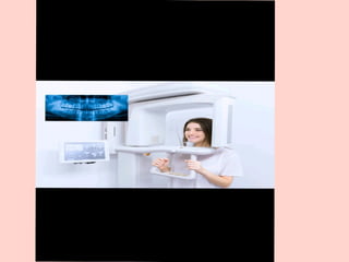
OPG (Orthopantomogram) ImagingSystem
OPG (Orthopantomogram) is a panoramic dental X-
ray imaging system that provides a single wide view
of:
1. Upper and lower teeth
2. Maxilla and mandible
3. Temporomandibular joints (TMJ)
4. Surrounding jaw structures
It is a 2D imaging technique but covers the entire jaw
in one image.
4.
Working Principle ofOPG:
It works on the principle of panoramic
tomography .
The X-ray tube and image receptor rotate
around the patient’s head.
A narrow, slit-shaped X-ray beam is
used.
Only the structures located in the focal
trough (image layer) appear sharp.
Structures outside this layer appear
blurred.
Continuous low-dose X-ray exposure is
used during rotation.
5.
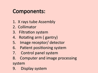
Components:
1. X raystube Assembly
2. Collimator
3. Filtration system
4. Rotating arm ( gantry)
5. Image receptor/ detector
6. Patient positioning system
7. Control panel system
8. Computer and image processing
system
9. Display system
6.
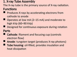
1. X-ray TubeAssembly
The X-ray tube is the primary source of X-ray radiation.
Functions
Produces X-rays by accelerating electrons from
cathode to anode
Operates at low mA (2–15 mA) and moderate to
high kVp (60–90 kVp)
Designed for continuous exposure during rotation
Parts
Cathode: filament and focusing cup (controls
electron flow)
Anode: tungsten target (produces X-ray photons)
Tube housing: oil-filled, provides insulation and
heat dissipation
7.
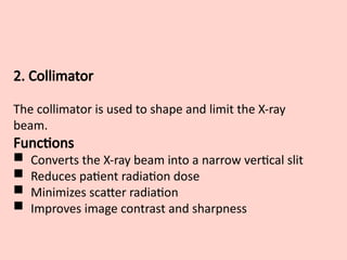
2. Collimator
The collimatoris used to shape and limit the X-ray
beam.
Functions
Converts the X-ray beam into a narrow vertical slit
Reduces patient radiation dose
Minimizes scatter radiation
Improves image contrast and sharpness
8.
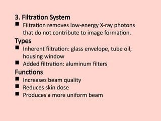
3. Filtration System
Filtration removes low-energy X-ray photons
that do not contribute to image formation.
Types
Inherent filtration: glass envelope, tube oil,
housing window
Added filtration: aluminum filters
Functions
Increases beam quality
Reduces skin dose
Produces a more uniform beam
9.
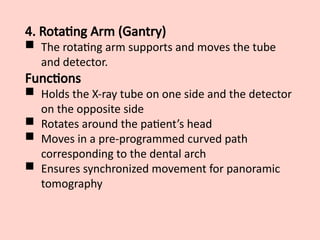
4. Rotating Arm(Gantry)
The rotating arm supports and moves the tube
and detector.
Functions
Holds the X-ray tube on one side and the detector
on the opposite side
Rotates around the patient’s head
Moves in a pre-programmed curved path
corresponding to the dental arch
Ensures synchronized movement for panoramic
tomography
10.
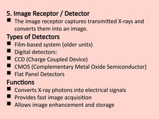
5. Image Receptor/ Detector
The image receptor captures transmitted X-rays and
converts them into an image.
Types of Detectors
Film-based system (older units)
Digital detectors:
CCD (Charge Coupled Device)
CMOS (Complementary Metal Oxide Semiconductor)
Flat Panel Detectors
Functions
Converts X-ray photons into electrical signals
Provides fast image acquisition
Allows image enhancement and storage
11.
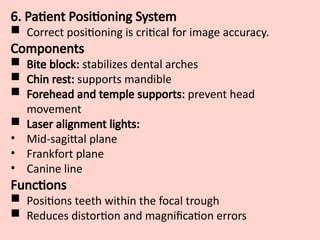
6. Patient PositioningSystem
Correct positioning is critical for image accuracy.
Components
Bite block: stabilizes dental arches
Chin rest: supports mandible
Forehead and temple supports: prevent head
movement
Laser alignment lights:
• Mid-sagittal plane
• Frankfort plane
• Canine line
Functions
Positions teeth within the focal trough
Reduces distortion and magnification errors
12.
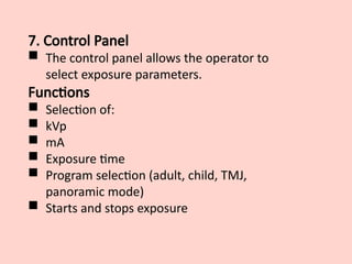
7. Control Panel
The control panel allows the operator to
select exposure parameters.
Functions
Selection of:
kVp
mA
Exposure time
Program selection (adult, child, TMJ,
panoramic mode)
Starts and stops exposure
13.
8. Computer andImage Processing Unit
This unit processes and displays the image.
Functions
Analog-to-digital conversion (ADC)
Image reconstruction
Image enhancement (contrast, brightness, zoom)
Noise reduction
Storage and transfer (PACS, DICOM)
9. Display Monitor
Displays the final panoramic image.
Functions
High-resolution visualization
Allows diagnostic interpretation
Image manipulation by the operator
14.
Protective and SafetyComponents
These ensure patient and operator safety.
Components
Lead shielding
Emergency stop switch
Exposure indicator lights
Radiation warning labels
15.
Clinical Indications ofOPG
Impacted teeth (especially third molars)
Jaw fractures
Dental caries (advanced cases)
TMJ disorders
Cysts and tumors of the jaw
Orthodontic treatment planning
Pre-operative dental assessment
16.
Advantages of OPG
Large field of view
Entire maxilla and mandible in one image
Low radiation dose
Fast image acquisition
Patient friendly and comfortable
Useful screening tool
Detects impacted teeth
Helps in orthodontic planning
Digital image storage and processing
17.
Disadvantages
Low imageresolution
Image magnification and distortion
Positioning errors affect image quality
No 3D information
Poor soft tissue visualization
Not suitable for early caries detection
Superimposition of structures
Artifacts from metallic objects
TOMOGRAPHY
Definition
Tomography is animaging technique used to
obtain cross-sectional (slice) images of the body by
moving the X-ray tube and detector in a
synchronized manner so that structures at a
particular depth are in focus, while structures
above and below that plane are blurred.
👉 The word comes from:
Tomo = slice
Graphy = recording
21.
Principle of Tomography
Whenthe X-ray tube and film/detector move in
opposite directions around a pivot point (fulcrum),
Structures lying in the focal plane (layer of
interest) remain sharp,
Structures outside this plane move relative to the
detector and become blurred.
📌 This selective blurring helps visualize a specific
anatomical layer.
22.
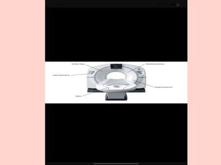
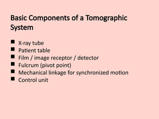
Basic Components ofa Tomographic
System
X-ray tube
Patient table
Film / image receptor / detector
Fulcrum (pivot point)
Mechanical linkage for synchronized motion
Control unit
23.
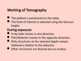
Working of Tomography
The patient is positioned on the table.
The level of interest is selected using the fulcrum
height.
During exposure:
X-ray tube moves in one direction,
Film/detector moves in the opposite direction.
Only structures at the selected depth remain
stationary relative to the detector.
Other structures are blurred due to motion.
24.
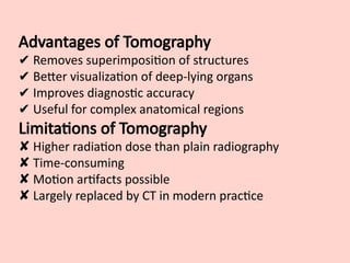
Advantages of Tomography
✔Removes superimposition of structures
✔ Better visualization of deep-lying organs
✔ Improves diagnostic accuracy
✔ Useful for complex anatomical regions
Limitations of Tomography
✘ Higher radiation dose than plain radiography
✘ Time-consuming
✘ Motion artifacts possible
✘ Largely replaced by CT in modern practice