Download to read offline
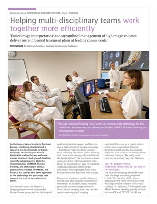


The Norwegian Radium Hospital in Oslo is a leading cancer center in Northern Europe known for new cancer treatments and research. It has implemented IMPAX Volume Viewing to help radiologists and oncology teams better visualize and interpret large volumes of MRI, CT, and PET CT images. Volume Viewing provides advanced 3D visualization tools and allows fusion of image sets for efficient comparison over time. Its integration with the hospital's IMPAX PACS allows seamless usage and improved workflow during daily tumor board meetings where radiologists demonstrate patient images and pathology to clinician groups. The hospital anticipates future technologies will further aid in managing and analyzing the large and growing volumes of complex patient image data.


