SlideShare a Scribd company logo
1 of 41
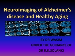
Neuroimaging of Alzheimer’s
disease and Healthy Aging
BY DR WASIM
UNDER THE GUIDANCE OF
DR R.K.SOLANKI
ANATOMICAL BRAIN IMAGING
CT – cerebral tomography
MRI – magnetic resonance imaging
FUNCTIONAL BRAIN IMAGING
SPECT – single photon emission computed tomography
PET – FDG – Positron emission tomography
BRAIN CHEMISTRY MEASUREMENT
MRS (spectroscopy – NAA/Cr: estimate neuronal volume)
BRAIN PATHOLOGY IMAGING
FDDNP – neurofibrillary pathology
BRAIN SCANNING TECHNIQUES
Evolution of Neuroimaging in AD
• Computed Tomography
• MRI
• Volumetric MRI
• Functional MRI
• FDG Glucose PET
• Amyloid Imaging
FDG Glucose PET
Lab of Neuro Imaging UCLA School of Medicine. www.loni.ucla.edu/~thompson/AD_4D/dynamic.html.
Helmuth L. Science.
2002;297:1260-1262.
Alzheimer Disease Forum.
http://www.alzforum.org/new/det
ail.asp?id=948.
MRI-guided SPECT data: (a) rostral anterior
cingulate (blue), caudal anterior cingulate
(green) and posterior cingulate (red); (b)
temporal horn (purple), hippocampus (blue),
and entorhinal cortex (orange); (c) basal
forebrain (blue) and amygdala (yellow); (d)
banks of the superior temporal sulcus (green).
CT Scan
• The initial criteria for CT scan diagnosis of Alzheimer
disease includes diffuse cerebral atrophy with
enlargement of the cortical sulci and increased size
of the ventricles.
• This concept was soon challenged because cerebral
atrophy can be present in elderly and healthy
persons, and some patients with dementia may have
no cerebral atrophy, at least in the early stages.
Rate of change of brain atrophy-Changes in the rate of atrophy
progression can be useful in diagnosing Alzheimer disease.
Longitudinal changes in brain size are associated with longitudinal
progression of cognitive loss and enlargement of the third and lateral
ventricles is greater in patients with Alzheimer disease than in control
subjects.
Changes in brain structure-Diffuse cerebral atrophy with widened sulci
and dilatation of the lateral ventricles can be observed.
Disproportionate atrophy of the medial temporal lobe, particularly of
the volume of the hippocampal formations (< 50%), can be seen.
• Dilatation of the perihippocampal fissure is a useful radiologic marker
for the initial diagnosis of Alzheimer disease (predictive accuracy of
91%)
• The hippocampal fissure is surrounded laterally by the hippocampus,
superiorly by the dentate gyrus and inferiorly by the subiculum.
• These structures are all involved in the early development of
Alzheimer disease and explain the enlargement in the early stages.
• At the medial aspect the fissure communicates with the
ambient cistern and its enlargement on CT scans is often
seen as hippocampal lucency or hypoattenuation in the
temporal area medial to the temporal horn.
• The temporal horns of the lateral ventricles may be
enlarged.
• Prominence of the choroid and hippocampal fissures and
enlargement of the sylvian fissure may be noted.
• White matter attenuation is not a feature of Alzheimer
disease.
• Degree of confidence-CT scan indices of hippocampal atrophy
are highly associated with Alzheimer disease but the
specificity is not well established.
• Use of a nonquantitative rating scale showed a sensitivity of
81% and a specificity of 67% in differentiating 21 patients with
Alzheimer disease with moderate dementia from 21 age-
matched control subjects.
• Hippocampal volumes in a sample of similar size permitted
correct classification of 85% of control subjects.
• Many studies have shown that cerebral atrophy is significantly
greater in patients with Alzheimer disease than in persons
without it. However, the variability of atrophy in the normal aging
process makes it difficult to use MRI as a definitive diagnostic
technique. Alzheimer disease. Brain image reveals hippocampal
atrophy
MRI
Axial, T2-weighted magnetic
resonance imaging (MRI) scan
shows dilated sylvian fissure
On structural MRI, atrophy of the entorhinal cortex is already present in
MCI.
MRI measurements of the hippocampus, amygdala, cingulate gyrus,
head of the caudate nucleus, temporal horn, lateral ventricles, third
ventricle and basal forebrain yield a prediction rate of 77% for
conversion to Alzheimer disease from questionable Alzheimer disease.
Functional MRI (fMRI) techniques can be used to measure cerebral
perfusion.
• Studies have been performed using MRI with echo-planar imaging
and signal targeting with attenuation radiofrequency (EPISTAR) in
patients with Alzheimer disease.
• Focal areas of hypoperfusion were in the posterior
temporoparietooccipital regions.
• On fMRI paradigms activate a larger area of parietotemporal
association cortex in persons at high risk for Alzheimer disease
than in others.
• The entorhinal cortex activation is relatively low in MCI.
Degree of confidence-MRI findings of hippocampal atrophy are highly
associated with Alzheimer disease (Alzheimer's disease), but the
specificity is not well established.
In patients with Alzheimer disease and moderate dementia
hippocampal volumes permitted correct classification in 85% of
patients.
In Alzheimer disease and mild dementia, sensitivity was 77% and
specificity, 80%.
Hippocampal volume was the best discriminator, although a number of
medical temporal-lobe structures were studied, including the amygdala
and the parahippocampal gyrus.
Bilateral medial temporal lobe atrophy (right hippocampus illustrated with
arrows) in the same subject with Alzheimer’s disease demonstrated on
coronal images acquired with: (A) 64 detector row computed tomography
scanning; (B) 1.5 tesla MRI volumetric T1 weighted sequence
ANATOMICAL BRAIN IMAGING
CT – cerebral tomography
MRI – magnetic resonance imaging
Coronal T1WI of the hippocampus demonstrating progressive atrophy in familial AD
NORMAL ALZHEIMER
Hippocampus outline Entorhinal Cortex outline
PET Scanning-
PET scan with fluorodeoxyglucose used for
1. Early diagnosis
2. Differentiation of Alzheimer disease from other types of dementia.
In Alzheimer's low activity is mostly in the back part (parietal,
posterior temporal and posterior cingulate cortices) of the brain; in
FTD low activity is mostly in the front of the brain.
3. Detection of persons at risk for Alzheimer disease even before the
onset of symptoms
It is likely to be caused by a combination of neuronal cell loss and
decreased synaptic activity.
Individuals at high risk for Alzheimer disease (asymptomatic carriers of
the APOE*E4 allele) exhibit a pattern of glucose hypometabolism similar
to that of patients with Alzheimer disease.
PET with ligand PK11195 labeled with11 C showed increased binding in
the entorhinal, temporoparietal and cingulate cortices.
This finding corresponded to the postmortem distribution of Alzheimer
disease pathology
Degree of confidence- PET scanning is more sensitive than SPECT
scanning.
FDG-PET has a sensitivity of 94% and a specificity of 73%.
Courtesy of S. Minoshima, University of Washington
FDG-PET in AD and MCI
Different amyloid-binding PET scan agents—Pittsburgh Compound-B
and FDDNP ‘’2-(1-(6-[(2-[18F]fluoroethyl)(methyl)amino]-2-
naphthyl)ethylidene)malononitrile’’ amyloid imaging agents may be
useful in the diagnosis of early onset dementia
Fibrillar A
Amyloid Imaging with
Positron Emission Tomography (PET)
PET Imaging
Amyloid Plaques
Alzheimer’s Disease
Normal Aging (Amyloid Negative)
Normal Aging (Amyloid Positive)
Amyloid PET Imaging in Aging
30% of normal older
people are amyloid
positive
Single-photon emission computed tomography- Early SPECT studies of
blood flow replicated findings of functional reductions in the posterior
tempoparietal cortex.
The severity of temporoparietal hypofunction has been correlated with
the severity of dementia in a number of studies.
Reductions of blood flow and oxygen use can be found in the
temporoparietal cortex in patients with Alzheimer disease and
moderate to severe symptoms.
Early reductions of glucose metabolism are seen in the posterior
cingulate cortex.
SPECT
Degree of confidence validated SPECT scan studies showing differences
between patients with Alzheimer disease (Alzheimer's disease) and
control subjects reveal high sensitivities and specificities of 80-90%.
Magnetic resonance spectroscopy (MRS) is a means
of noninvasive physiologic imaging of the brain that
measures relative levels of various tissue
metabolites
Decrease NAA/Cr
Decrease NAA/ Cho
Increase Myo/NAA
MRS in Alzheimer Disease Axial T2-weighted images from an AD patient (L, left) and
a healthy control (R, right). These images show the left elevated NAA, Cr/PCr, Cho
containing compounds, Glu and mI. Most authors have opted for following up AD .
Reduced NAA and NAA / Cr (reduction of neuronal population); Increased mI and
mI / Cr (presence of glial repairers phenomena); The reason mI / NAA is considered
the most reliable in the assessment of metabolites in Alzheimer's disease.
Magnetic resonance spectroscopy (MRS) in Alzheimer's disease.
•T1W image shows
reduction in the volume of
the hippocampus.
•Proton MRS in
hippocampal region shows
MI peak, decreased NAA
and elevated MI/Cr ratio
•Dx - Alzheimer’s Disease
BRAIN PATHOLOGY IMAGING
FDDNP – neurofibrillary pathology
FDDNP-PET scans in the parietal region (top) and the temporal region (bottom) in
one control subject and
one subject with mild cognitive impairment who was reclassified on follow-up as
having Alzheimer's disease.
Scans of the subject with mild cognitive impairment, who was reclassified as having
Alzheimer's disease,
showed increased binding in the frontal (8.6%), parietal (8.9%), and lateral temporal
(6.6%) regions.
Red and yellow areas correspond to high FDDNP binding values.
Thank You.

More Related Content

What's hot

Apraxia examination and evaluation
Apraxia examination and evaluation Apraxia examination and evaluation
Apraxia examination and evaluation NeurologyKota
 
Mri imaging finding in wilson disease
Mri imaging finding in wilson diseaseMri imaging finding in wilson disease
Mri imaging finding in wilson diseasegulabsoni
 
Muscle MRI in Neuromuscular Disease
Muscle MRI in Neuromuscular DiseaseMuscle MRI in Neuromuscular Disease
Muscle MRI in Neuromuscular DiseaseDr. Arghya Deb
 
Presentation1, radiological imaging of leigh disease.
Presentation1, radiological imaging of leigh disease.Presentation1, radiological imaging of leigh disease.
Presentation1, radiological imaging of leigh disease.Abdellah Nazeer
 
Presentation1, radiological imaging of pediatric leukodystrophy.
Presentation1, radiological imaging of pediatric leukodystrophy.Presentation1, radiological imaging of pediatric leukodystrophy.
Presentation1, radiological imaging of pediatric leukodystrophy.Abdellah Nazeer
 
PET/MR imaging in neurodegenerative diseases
PET/MR imaging in neurodegenerative diseasesPET/MR imaging in neurodegenerative diseases
PET/MR imaging in neurodegenerative diseasesWalid Rezk
 
Presentation1.pptx, radiological imaging of parkinsonism.
Presentation1.pptx, radiological imaging of parkinsonism.Presentation1.pptx, radiological imaging of parkinsonism.
Presentation1.pptx, radiological imaging of parkinsonism.Abdellah Nazeer
 
MRI differential diagnosis of Multiple sclerosis
MRI differential diagnosis of Multiple sclerosisMRI differential diagnosis of Multiple sclerosis
MRI differential diagnosis of Multiple sclerosissrimantp
 
Neurodegenerative disorders MRI approach
Neurodegenerative disorders MRI approachNeurodegenerative disorders MRI approach
Neurodegenerative disorders MRI approachArif S
 
Neuro radiology neuroimaging
Neuro radiology   neuroimagingNeuro radiology   neuroimaging
Neuro radiology neuroimagingMarwa Khalifa
 
fMRI Presentation
fMRI PresentationfMRI Presentation
fMRI Presentationricksw78
 
Radionucleide imaging of the brain
Radionucleide imaging of the brainRadionucleide imaging of the brain
Radionucleide imaging of the brainYassera Awan
 
Dysmyelination syndromes
Dysmyelination syndromesDysmyelination syndromes
Dysmyelination syndromesAmr Hassan
 
Anatomy of hippocampus ( radiology )
Anatomy of hippocampus ( radiology )Anatomy of hippocampus ( radiology )
Anatomy of hippocampus ( radiology )Sajith Selvaganesan
 
Imaging in developmental malformations of the brain
Imaging in developmental malformations of the brainImaging in developmental malformations of the brain
Imaging in developmental malformations of the brainChakradhar Reddy
 
imaging in neurology - demyelinating diseases
 imaging in neurology - demyelinating diseases imaging in neurology - demyelinating diseases
imaging in neurology - demyelinating diseasesNeurologyKota
 
Diffusion tensor imaging in Neurology
Diffusion tensor imaging in NeurologyDiffusion tensor imaging in Neurology
Diffusion tensor imaging in NeurologyOsama Ragab
 

What's hot (20)

Apraxia examination and evaluation
Apraxia examination and evaluation Apraxia examination and evaluation
Apraxia examination and evaluation
 
Mri imaging finding in wilson disease
Mri imaging finding in wilson diseaseMri imaging finding in wilson disease
Mri imaging finding in wilson disease
 
Muscle MRI in Neuromuscular Disease
Muscle MRI in Neuromuscular DiseaseMuscle MRI in Neuromuscular Disease
Muscle MRI in Neuromuscular Disease
 
Presentation1, radiological imaging of leigh disease.
Presentation1, radiological imaging of leigh disease.Presentation1, radiological imaging of leigh disease.
Presentation1, radiological imaging of leigh disease.
 
Presentation1, radiological imaging of pediatric leukodystrophy.
Presentation1, radiological imaging of pediatric leukodystrophy.Presentation1, radiological imaging of pediatric leukodystrophy.
Presentation1, radiological imaging of pediatric leukodystrophy.
 
PET/MR imaging in neurodegenerative diseases
PET/MR imaging in neurodegenerative diseasesPET/MR imaging in neurodegenerative diseases
PET/MR imaging in neurodegenerative diseases
 
Presentation1.pptx, radiological imaging of parkinsonism.
Presentation1.pptx, radiological imaging of parkinsonism.Presentation1.pptx, radiological imaging of parkinsonism.
Presentation1.pptx, radiological imaging of parkinsonism.
 
MRI differential diagnosis of Multiple sclerosis
MRI differential diagnosis of Multiple sclerosisMRI differential diagnosis of Multiple sclerosis
MRI differential diagnosis of Multiple sclerosis
 
Neurodegenerative disorders MRI approach
Neurodegenerative disorders MRI approachNeurodegenerative disorders MRI approach
Neurodegenerative disorders MRI approach
 
Neuro radiology neuroimaging
Neuro radiology   neuroimagingNeuro radiology   neuroimaging
Neuro radiology neuroimaging
 
fMRI Presentation
fMRI PresentationfMRI Presentation
fMRI Presentation
 
Radionucleide imaging of the brain
Radionucleide imaging of the brainRadionucleide imaging of the brain
Radionucleide imaging of the brain
 
Release reflexes
Release reflexesRelease reflexes
Release reflexes
 
Case record...Friedreich ataxia
Case record...Friedreich ataxiaCase record...Friedreich ataxia
Case record...Friedreich ataxia
 
Dysmyelination syndromes
Dysmyelination syndromesDysmyelination syndromes
Dysmyelination syndromes
 
Anatomy of hippocampus ( radiology )
Anatomy of hippocampus ( radiology )Anatomy of hippocampus ( radiology )
Anatomy of hippocampus ( radiology )
 
Imaging in developmental malformations of the brain
Imaging in developmental malformations of the brainImaging in developmental malformations of the brain
Imaging in developmental malformations of the brain
 
Congenital brain anomalies
Congenital brain anomaliesCongenital brain anomalies
Congenital brain anomalies
 
imaging in neurology - demyelinating diseases
 imaging in neurology - demyelinating diseases imaging in neurology - demyelinating diseases
imaging in neurology - demyelinating diseases
 
Diffusion tensor imaging in Neurology
Diffusion tensor imaging in NeurologyDiffusion tensor imaging in Neurology
Diffusion tensor imaging in Neurology
 

Similar to Neuroimaging of alzheimer's

Neurology 2nd investigation of neurological disease
Neurology 2nd investigation of neurological diseaseNeurology 2nd investigation of neurological disease
Neurology 2nd investigation of neurological diseaseRamiAboali
 
NEUROIMAGING IN PARKINSONISM edited.pptx
NEUROIMAGING IN PARKINSONISM edited.pptxNEUROIMAGING IN PARKINSONISM edited.pptx
NEUROIMAGING IN PARKINSONISM edited.pptxvijay prakash
 
PET/SPECT in Epilepsy Surgery
PET/SPECT in Epilepsy Surgery PET/SPECT in Epilepsy Surgery
PET/SPECT in Epilepsy Surgery Lalit Bansal
 
Cerebellar diseases. igbiti
Cerebellar diseases. igbitiCerebellar diseases. igbiti
Cerebellar diseases. igbitiJustice Igbiti
 
Somatosensory evoked potentials
Somatosensory evoked potentialsSomatosensory evoked potentials
Somatosensory evoked potentialsManideep Malaka
 
Supratentorial brain tumours
Supratentorial brain tumoursSupratentorial brain tumours
Supratentorial brain tumourstrial4neha
 
LEUKODYSTROPHY FINAL.pptx sms medical jaipur
LEUKODYSTROPHY FINAL.pptx sms medical jaipurLEUKODYSTROPHY FINAL.pptx sms medical jaipur
LEUKODYSTROPHY FINAL.pptx sms medical jaipurdineshdandia
 
Papel de la resonancia magnética en las enfermedades neurodegenerativas.pdf
Papel de la resonancia magnética en las enfermedades neurodegenerativas.pdfPapel de la resonancia magnética en las enfermedades neurodegenerativas.pdf
Papel de la resonancia magnética en las enfermedades neurodegenerativas.pdfJanetReyes54
 
Positron emission tomographic scan
Positron emission tomographic scanPositron emission tomographic scan
Positron emission tomographic scanPrashant Makhija
 
Diagnostic methods
Diagnostic methodsDiagnostic methods
Diagnostic methodsOla
 
Preoperative assessment of epilepsy clinical &amp; radiological, eeg, meg
Preoperative assessment of  epilepsy  clinical &amp; radiological, eeg, megPreoperative assessment of  epilepsy  clinical &amp; radiological, eeg, meg
Preoperative assessment of epilepsy clinical &amp; radiological, eeg, megDr Fakir Mohan Sahu
 
2 neuro. ixs 1
2 neuro. ixs 1 2 neuro. ixs 1
2 neuro. ixs 1 eliasmawla
 
autoimmuneencephalitisppt-180103161321 2.pdf
autoimmuneencephalitisppt-180103161321 2.pdfautoimmuneencephalitisppt-180103161321 2.pdf
autoimmuneencephalitisppt-180103161321 2.pdfabhimittal8
 
Autoimmune encephalitis ppt
Autoimmune encephalitis pptAutoimmune encephalitis ppt
Autoimmune encephalitis pptSachin Adukia
 

Similar to Neuroimaging of alzheimer's (20)

Neurology 2nd investigation of neurological disease
Neurology 2nd investigation of neurological diseaseNeurology 2nd investigation of neurological disease
Neurology 2nd investigation of neurological disease
 
NEUROIMAGING IN PARKINSONISM edited.pptx
NEUROIMAGING IN PARKINSONISM edited.pptxNEUROIMAGING IN PARKINSONISM edited.pptx
NEUROIMAGING IN PARKINSONISM edited.pptx
 
Medicine 5th year, 1st 2 lectures (Dr. Rasool)
Medicine 5th year, 1st 2 lectures (Dr. Rasool)Medicine 5th year, 1st 2 lectures (Dr. Rasool)
Medicine 5th year, 1st 2 lectures (Dr. Rasool)
 
Neuroimaging in Psychiatry
Neuroimaging in PsychiatryNeuroimaging in Psychiatry
Neuroimaging in Psychiatry
 
PET/SPECT in Epilepsy Surgery
PET/SPECT in Epilepsy Surgery PET/SPECT in Epilepsy Surgery
PET/SPECT in Epilepsy Surgery
 
Cerebellar diseases. igbiti
Cerebellar diseases. igbitiCerebellar diseases. igbiti
Cerebellar diseases. igbiti
 
Somatosensory evoked potentials
Somatosensory evoked potentialsSomatosensory evoked potentials
Somatosensory evoked potentials
 
Supratentorial brain tumours
Supratentorial brain tumoursSupratentorial brain tumours
Supratentorial brain tumours
 
Neuroimagen estructural demencias
Neuroimagen estructural demenciasNeuroimagen estructural demencias
Neuroimagen estructural demencias
 
Integrated Diagnostics – A Unique Epilepsy Approach
Integrated Diagnostics – A Unique Epilepsy ApproachIntegrated Diagnostics – A Unique Epilepsy Approach
Integrated Diagnostics – A Unique Epilepsy Approach
 
LEUKODYSTROPHY FINAL.pptx sms medical jaipur
LEUKODYSTROPHY FINAL.pptx sms medical jaipurLEUKODYSTROPHY FINAL.pptx sms medical jaipur
LEUKODYSTROPHY FINAL.pptx sms medical jaipur
 
Papel de la resonancia magnética en las enfermedades neurodegenerativas.pdf
Papel de la resonancia magnética en las enfermedades neurodegenerativas.pdfPapel de la resonancia magnética en las enfermedades neurodegenerativas.pdf
Papel de la resonancia magnética en las enfermedades neurodegenerativas.pdf
 
Alzheimer
AlzheimerAlzheimer
Alzheimer
 
Positron emission tomographic scan
Positron emission tomographic scanPositron emission tomographic scan
Positron emission tomographic scan
 
BASICS OF MRI
BASICS OF MRIBASICS OF MRI
BASICS OF MRI
 
Diagnostic methods
Diagnostic methodsDiagnostic methods
Diagnostic methods
 
Preoperative assessment of epilepsy clinical &amp; radiological, eeg, meg
Preoperative assessment of  epilepsy  clinical &amp; radiological, eeg, megPreoperative assessment of  epilepsy  clinical &amp; radiological, eeg, meg
Preoperative assessment of epilepsy clinical &amp; radiological, eeg, meg
 
2 neuro. ixs 1
2 neuro. ixs 1 2 neuro. ixs 1
2 neuro. ixs 1
 
autoimmuneencephalitisppt-180103161321 2.pdf
autoimmuneencephalitisppt-180103161321 2.pdfautoimmuneencephalitisppt-180103161321 2.pdf
autoimmuneencephalitisppt-180103161321 2.pdf
 
Autoimmune encephalitis ppt
Autoimmune encephalitis pptAutoimmune encephalitis ppt
Autoimmune encephalitis ppt
 

More from Dr Wasim

Ego & defences
Ego & defencesEgo & defences
Ego & defencesDr Wasim
 
Evaluating child with disruptive behaviour
Evaluating child with disruptive behaviourEvaluating child with disruptive behaviour
Evaluating child with disruptive behaviourDr Wasim
 
Evaluation of the geriatric patients with behavioural dysfunction
Evaluation of the geriatric patients with behavioural dysfunctionEvaluation of the geriatric patients with behavioural dysfunction
Evaluation of the geriatric patients with behavioural dysfunctionDr Wasim
 
Cannabis related health issues
Cannabis related health issuesCannabis related health issues
Cannabis related health issuesDr Wasim
 
Sytematic treatment enhancement program for bipolar disorder(step bd) (1)
Sytematic treatment enhancement program for bipolar disorder(step bd) (1)Sytematic treatment enhancement program for bipolar disorder(step bd) (1)
Sytematic treatment enhancement program for bipolar disorder(step bd) (1)Dr Wasim
 
Life threatening side effects of Psychotropics
Life threatening side effects of PsychotropicsLife threatening side effects of Psychotropics
Life threatening side effects of PsychotropicsDr Wasim
 
Role of psychiatrist in chronic pain
Role of psychiatrist in chronic painRole of psychiatrist in chronic pain
Role of psychiatrist in chronic painDr Wasim
 
Gender development and disorder
Gender development and disorderGender development and disorder
Gender development and disorderDr Wasim
 
Treatment of schizophrenia current issues and future possibilities
Treatment of schizophrenia  current issues and future possibilitiesTreatment of schizophrenia  current issues and future possibilities
Treatment of schizophrenia current issues and future possibilitiesDr Wasim
 
Disorder content
Disorder contentDisorder content
Disorder contentDr Wasim
 
Schenider first rank symptoms
Schenider first rank symptomsSchenider first rank symptoms
Schenider first rank symptomsDr Wasim
 
Second and third generation antipsychotics
Second and third generation antipsychoticsSecond and third generation antipsychotics
Second and third generation antipsychoticsDr Wasim
 
Jean piaget
Jean piagetJean piaget
Jean piagetDr Wasim
 

More from Dr Wasim (14)

Ego & defences
Ego & defencesEgo & defences
Ego & defences
 
Evaluating child with disruptive behaviour
Evaluating child with disruptive behaviourEvaluating child with disruptive behaviour
Evaluating child with disruptive behaviour
 
Evaluation of the geriatric patients with behavioural dysfunction
Evaluation of the geriatric patients with behavioural dysfunctionEvaluation of the geriatric patients with behavioural dysfunction
Evaluation of the geriatric patients with behavioural dysfunction
 
Cannabis related health issues
Cannabis related health issuesCannabis related health issues
Cannabis related health issues
 
Sytematic treatment enhancement program for bipolar disorder(step bd) (1)
Sytematic treatment enhancement program for bipolar disorder(step bd) (1)Sytematic treatment enhancement program for bipolar disorder(step bd) (1)
Sytematic treatment enhancement program for bipolar disorder(step bd) (1)
 
Life threatening side effects of Psychotropics
Life threatening side effects of PsychotropicsLife threatening side effects of Psychotropics
Life threatening side effects of Psychotropics
 
Role of psychiatrist in chronic pain
Role of psychiatrist in chronic painRole of psychiatrist in chronic pain
Role of psychiatrist in chronic pain
 
Gender development and disorder
Gender development and disorderGender development and disorder
Gender development and disorder
 
Judgement
JudgementJudgement
Judgement
 
Treatment of schizophrenia current issues and future possibilities
Treatment of schizophrenia  current issues and future possibilitiesTreatment of schizophrenia  current issues and future possibilities
Treatment of schizophrenia current issues and future possibilities
 
Disorder content
Disorder contentDisorder content
Disorder content
 
Schenider first rank symptoms
Schenider first rank symptomsSchenider first rank symptoms
Schenider first rank symptoms
 
Second and third generation antipsychotics
Second and third generation antipsychoticsSecond and third generation antipsychotics
Second and third generation antipsychotics
 
Jean piaget
Jean piagetJean piaget
Jean piaget
 

Recently uploaded

The basics of sentences session 2pptx copy.pptx
The basics of sentences session 2pptx copy.pptxThe basics of sentences session 2pptx copy.pptx
The basics of sentences session 2pptx copy.pptxheathfieldcps1
 
_Math 4-Q4 Week 5.pptx Steps in Collecting Data
_Math 4-Q4 Week 5.pptx Steps in Collecting Data_Math 4-Q4 Week 5.pptx Steps in Collecting Data
_Math 4-Q4 Week 5.pptx Steps in Collecting DataJhengPantaleon
 
Alper Gobel In Media Res Media Component
Alper Gobel In Media Res Media ComponentAlper Gobel In Media Res Media Component
Alper Gobel In Media Res Media ComponentInMediaRes1
 
ECONOMIC CONTEXT - LONG FORM TV DRAMA - PPT
ECONOMIC CONTEXT - LONG FORM TV DRAMA - PPTECONOMIC CONTEXT - LONG FORM TV DRAMA - PPT
ECONOMIC CONTEXT - LONG FORM TV DRAMA - PPTiammrhaywood
 
Software Engineering Methodologies (overview)
Software Engineering Methodologies (overview)Software Engineering Methodologies (overview)
Software Engineering Methodologies (overview)eniolaolutunde
 
POINT- BIOCHEMISTRY SEM 2 ENZYMES UNIT 5.pptx
POINT- BIOCHEMISTRY SEM 2 ENZYMES UNIT 5.pptxPOINT- BIOCHEMISTRY SEM 2 ENZYMES UNIT 5.pptx
POINT- BIOCHEMISTRY SEM 2 ENZYMES UNIT 5.pptxSayali Powar
 
“Oh GOSH! Reflecting on Hackteria's Collaborative Practices in a Global Do-It...
“Oh GOSH! Reflecting on Hackteria's Collaborative Practices in a Global Do-It...“Oh GOSH! Reflecting on Hackteria's Collaborative Practices in a Global Do-It...
“Oh GOSH! Reflecting on Hackteria's Collaborative Practices in a Global Do-It...Marc Dusseiller Dusjagr
 
Mastering the Unannounced Regulatory Inspection
Mastering the Unannounced Regulatory InspectionMastering the Unannounced Regulatory Inspection
Mastering the Unannounced Regulatory InspectionSafetyChain Software
 
The Most Excellent Way | 1 Corinthians 13
The Most Excellent Way | 1 Corinthians 13The Most Excellent Way | 1 Corinthians 13
The Most Excellent Way | 1 Corinthians 13Steve Thomason
 
How to Configure Email Server in Odoo 17
How to Configure Email Server in Odoo 17How to Configure Email Server in Odoo 17
How to Configure Email Server in Odoo 17Celine George
 
Sanyam Choudhary Chemistry practical.pdf
Sanyam Choudhary Chemistry practical.pdfSanyam Choudhary Chemistry practical.pdf
Sanyam Choudhary Chemistry practical.pdfsanyamsingh5019
 
Interactive Powerpoint_How to Master effective communication
Interactive Powerpoint_How to Master effective communicationInteractive Powerpoint_How to Master effective communication
Interactive Powerpoint_How to Master effective communicationnomboosow
 
Paris 2024 Olympic Geographies - an activity
Paris 2024 Olympic Geographies - an activityParis 2024 Olympic Geographies - an activity
Paris 2024 Olympic Geographies - an activityGeoBlogs
 
Organic Name Reactions for the students and aspirants of Chemistry12th.pptx
Organic Name Reactions  for the students and aspirants of Chemistry12th.pptxOrganic Name Reactions  for the students and aspirants of Chemistry12th.pptx
Organic Name Reactions for the students and aspirants of Chemistry12th.pptxVS Mahajan Coaching Centre
 
Presentation by Andreas Schleicher Tackling the School Absenteeism Crisis 30 ...
Presentation by Andreas Schleicher Tackling the School Absenteeism Crisis 30 ...Presentation by Andreas Schleicher Tackling the School Absenteeism Crisis 30 ...
Presentation by Andreas Schleicher Tackling the School Absenteeism Crisis 30 ...EduSkills OECD
 
Introduction to ArtificiaI Intelligence in Higher Education
Introduction to ArtificiaI Intelligence in Higher EducationIntroduction to ArtificiaI Intelligence in Higher Education
Introduction to ArtificiaI Intelligence in Higher Educationpboyjonauth
 
Separation of Lanthanides/ Lanthanides and Actinides
Separation of Lanthanides/ Lanthanides and ActinidesSeparation of Lanthanides/ Lanthanides and Actinides
Separation of Lanthanides/ Lanthanides and ActinidesFatimaKhan178732
 
A Critique of the Proposed National Education Policy Reform
A Critique of the Proposed National Education Policy ReformA Critique of the Proposed National Education Policy Reform
A Critique of the Proposed National Education Policy ReformChameera Dedduwage
 

Recently uploaded (20)

The basics of sentences session 2pptx copy.pptx
The basics of sentences session 2pptx copy.pptxThe basics of sentences session 2pptx copy.pptx
The basics of sentences session 2pptx copy.pptx
 
_Math 4-Q4 Week 5.pptx Steps in Collecting Data
_Math 4-Q4 Week 5.pptx Steps in Collecting Data_Math 4-Q4 Week 5.pptx Steps in Collecting Data
_Math 4-Q4 Week 5.pptx Steps in Collecting Data
 
Alper Gobel In Media Res Media Component
Alper Gobel In Media Res Media ComponentAlper Gobel In Media Res Media Component
Alper Gobel In Media Res Media Component
 
ECONOMIC CONTEXT - LONG FORM TV DRAMA - PPT
ECONOMIC CONTEXT - LONG FORM TV DRAMA - PPTECONOMIC CONTEXT - LONG FORM TV DRAMA - PPT
ECONOMIC CONTEXT - LONG FORM TV DRAMA - PPT
 
Software Engineering Methodologies (overview)
Software Engineering Methodologies (overview)Software Engineering Methodologies (overview)
Software Engineering Methodologies (overview)
 
POINT- BIOCHEMISTRY SEM 2 ENZYMES UNIT 5.pptx
POINT- BIOCHEMISTRY SEM 2 ENZYMES UNIT 5.pptxPOINT- BIOCHEMISTRY SEM 2 ENZYMES UNIT 5.pptx
POINT- BIOCHEMISTRY SEM 2 ENZYMES UNIT 5.pptx
 
“Oh GOSH! Reflecting on Hackteria's Collaborative Practices in a Global Do-It...
“Oh GOSH! Reflecting on Hackteria's Collaborative Practices in a Global Do-It...“Oh GOSH! Reflecting on Hackteria's Collaborative Practices in a Global Do-It...
“Oh GOSH! Reflecting on Hackteria's Collaborative Practices in a Global Do-It...
 
Mastering the Unannounced Regulatory Inspection
Mastering the Unannounced Regulatory InspectionMastering the Unannounced Regulatory Inspection
Mastering the Unannounced Regulatory Inspection
 
The Most Excellent Way | 1 Corinthians 13
The Most Excellent Way | 1 Corinthians 13The Most Excellent Way | 1 Corinthians 13
The Most Excellent Way | 1 Corinthians 13
 
How to Configure Email Server in Odoo 17
How to Configure Email Server in Odoo 17How to Configure Email Server in Odoo 17
How to Configure Email Server in Odoo 17
 
Sanyam Choudhary Chemistry practical.pdf
Sanyam Choudhary Chemistry practical.pdfSanyam Choudhary Chemistry practical.pdf
Sanyam Choudhary Chemistry practical.pdf
 
Interactive Powerpoint_How to Master effective communication
Interactive Powerpoint_How to Master effective communicationInteractive Powerpoint_How to Master effective communication
Interactive Powerpoint_How to Master effective communication
 
Model Call Girl in Tilak Nagar Delhi reach out to us at 🔝9953056974🔝
Model Call Girl in Tilak Nagar Delhi reach out to us at 🔝9953056974🔝Model Call Girl in Tilak Nagar Delhi reach out to us at 🔝9953056974🔝
Model Call Girl in Tilak Nagar Delhi reach out to us at 🔝9953056974🔝
 
Paris 2024 Olympic Geographies - an activity
Paris 2024 Olympic Geographies - an activityParis 2024 Olympic Geographies - an activity
Paris 2024 Olympic Geographies - an activity
 
Staff of Color (SOC) Retention Efforts DDSD
Staff of Color (SOC) Retention Efforts DDSDStaff of Color (SOC) Retention Efforts DDSD
Staff of Color (SOC) Retention Efforts DDSD
 
Organic Name Reactions for the students and aspirants of Chemistry12th.pptx
Organic Name Reactions  for the students and aspirants of Chemistry12th.pptxOrganic Name Reactions  for the students and aspirants of Chemistry12th.pptx
Organic Name Reactions for the students and aspirants of Chemistry12th.pptx
 
Presentation by Andreas Schleicher Tackling the School Absenteeism Crisis 30 ...
Presentation by Andreas Schleicher Tackling the School Absenteeism Crisis 30 ...Presentation by Andreas Schleicher Tackling the School Absenteeism Crisis 30 ...
Presentation by Andreas Schleicher Tackling the School Absenteeism Crisis 30 ...
 
Introduction to ArtificiaI Intelligence in Higher Education
Introduction to ArtificiaI Intelligence in Higher EducationIntroduction to ArtificiaI Intelligence in Higher Education
Introduction to ArtificiaI Intelligence in Higher Education
 
Separation of Lanthanides/ Lanthanides and Actinides
Separation of Lanthanides/ Lanthanides and ActinidesSeparation of Lanthanides/ Lanthanides and Actinides
Separation of Lanthanides/ Lanthanides and Actinides
 
A Critique of the Proposed National Education Policy Reform
A Critique of the Proposed National Education Policy ReformA Critique of the Proposed National Education Policy Reform
A Critique of the Proposed National Education Policy Reform
 

Neuroimaging of alzheimer's

  • 1. Neuroimaging of Alzheimer’s disease and Healthy Aging BY DR WASIM UNDER THE GUIDANCE OF DR R.K.SOLANKI
  • 2. ANATOMICAL BRAIN IMAGING CT – cerebral tomography MRI – magnetic resonance imaging FUNCTIONAL BRAIN IMAGING SPECT – single photon emission computed tomography PET – FDG – Positron emission tomography BRAIN CHEMISTRY MEASUREMENT MRS (spectroscopy – NAA/Cr: estimate neuronal volume) BRAIN PATHOLOGY IMAGING FDDNP – neurofibrillary pathology BRAIN SCANNING TECHNIQUES
  • 3. Evolution of Neuroimaging in AD • Computed Tomography • MRI • Volumetric MRI • Functional MRI • FDG Glucose PET • Amyloid Imaging FDG Glucose PET Lab of Neuro Imaging UCLA School of Medicine. www.loni.ucla.edu/~thompson/AD_4D/dynamic.html. Helmuth L. Science. 2002;297:1260-1262. Alzheimer Disease Forum. http://www.alzforum.org/new/det ail.asp?id=948.
  • 4.
  • 5.
  • 6. MRI-guided SPECT data: (a) rostral anterior cingulate (blue), caudal anterior cingulate (green) and posterior cingulate (red); (b) temporal horn (purple), hippocampus (blue), and entorhinal cortex (orange); (c) basal forebrain (blue) and amygdala (yellow); (d) banks of the superior temporal sulcus (green).
  • 7.
  • 8.
  • 9.
  • 10.
  • 11. CT Scan • The initial criteria for CT scan diagnosis of Alzheimer disease includes diffuse cerebral atrophy with enlargement of the cortical sulci and increased size of the ventricles. • This concept was soon challenged because cerebral atrophy can be present in elderly and healthy persons, and some patients with dementia may have no cerebral atrophy, at least in the early stages.
  • 12. Rate of change of brain atrophy-Changes in the rate of atrophy progression can be useful in diagnosing Alzheimer disease. Longitudinal changes in brain size are associated with longitudinal progression of cognitive loss and enlargement of the third and lateral ventricles is greater in patients with Alzheimer disease than in control subjects. Changes in brain structure-Diffuse cerebral atrophy with widened sulci and dilatation of the lateral ventricles can be observed. Disproportionate atrophy of the medial temporal lobe, particularly of the volume of the hippocampal formations (< 50%), can be seen.
  • 13. • Dilatation of the perihippocampal fissure is a useful radiologic marker for the initial diagnosis of Alzheimer disease (predictive accuracy of 91%) • The hippocampal fissure is surrounded laterally by the hippocampus, superiorly by the dentate gyrus and inferiorly by the subiculum. • These structures are all involved in the early development of Alzheimer disease and explain the enlargement in the early stages.
  • 14. • At the medial aspect the fissure communicates with the ambient cistern and its enlargement on CT scans is often seen as hippocampal lucency or hypoattenuation in the temporal area medial to the temporal horn. • The temporal horns of the lateral ventricles may be enlarged. • Prominence of the choroid and hippocampal fissures and enlargement of the sylvian fissure may be noted. • White matter attenuation is not a feature of Alzheimer disease.
  • 15. • Degree of confidence-CT scan indices of hippocampal atrophy are highly associated with Alzheimer disease but the specificity is not well established. • Use of a nonquantitative rating scale showed a sensitivity of 81% and a specificity of 67% in differentiating 21 patients with Alzheimer disease with moderate dementia from 21 age- matched control subjects. • Hippocampal volumes in a sample of similar size permitted correct classification of 85% of control subjects.
  • 16. • Many studies have shown that cerebral atrophy is significantly greater in patients with Alzheimer disease than in persons without it. However, the variability of atrophy in the normal aging process makes it difficult to use MRI as a definitive diagnostic technique. Alzheimer disease. Brain image reveals hippocampal atrophy MRI
  • 17. Axial, T2-weighted magnetic resonance imaging (MRI) scan shows dilated sylvian fissure
  • 18. On structural MRI, atrophy of the entorhinal cortex is already present in MCI. MRI measurements of the hippocampus, amygdala, cingulate gyrus, head of the caudate nucleus, temporal horn, lateral ventricles, third ventricle and basal forebrain yield a prediction rate of 77% for conversion to Alzheimer disease from questionable Alzheimer disease. Functional MRI (fMRI) techniques can be used to measure cerebral perfusion.
  • 19. • Studies have been performed using MRI with echo-planar imaging and signal targeting with attenuation radiofrequency (EPISTAR) in patients with Alzheimer disease. • Focal areas of hypoperfusion were in the posterior temporoparietooccipital regions. • On fMRI paradigms activate a larger area of parietotemporal association cortex in persons at high risk for Alzheimer disease than in others. • The entorhinal cortex activation is relatively low in MCI.
  • 20. Degree of confidence-MRI findings of hippocampal atrophy are highly associated with Alzheimer disease (Alzheimer's disease), but the specificity is not well established. In patients with Alzheimer disease and moderate dementia hippocampal volumes permitted correct classification in 85% of patients. In Alzheimer disease and mild dementia, sensitivity was 77% and specificity, 80%. Hippocampal volume was the best discriminator, although a number of medical temporal-lobe structures were studied, including the amygdala and the parahippocampal gyrus.
  • 21. Bilateral medial temporal lobe atrophy (right hippocampus illustrated with arrows) in the same subject with Alzheimer’s disease demonstrated on coronal images acquired with: (A) 64 detector row computed tomography scanning; (B) 1.5 tesla MRI volumetric T1 weighted sequence ANATOMICAL BRAIN IMAGING CT – cerebral tomography MRI – magnetic resonance imaging
  • 22. Coronal T1WI of the hippocampus demonstrating progressive atrophy in familial AD
  • 25.
  • 26. PET Scanning- PET scan with fluorodeoxyglucose used for 1. Early diagnosis 2. Differentiation of Alzheimer disease from other types of dementia. In Alzheimer's low activity is mostly in the back part (parietal, posterior temporal and posterior cingulate cortices) of the brain; in FTD low activity is mostly in the front of the brain. 3. Detection of persons at risk for Alzheimer disease even before the onset of symptoms
  • 27. It is likely to be caused by a combination of neuronal cell loss and decreased synaptic activity. Individuals at high risk for Alzheimer disease (asymptomatic carriers of the APOE*E4 allele) exhibit a pattern of glucose hypometabolism similar to that of patients with Alzheimer disease. PET with ligand PK11195 labeled with11 C showed increased binding in the entorhinal, temporoparietal and cingulate cortices. This finding corresponded to the postmortem distribution of Alzheimer disease pathology Degree of confidence- PET scanning is more sensitive than SPECT scanning. FDG-PET has a sensitivity of 94% and a specificity of 73%.
  • 28.
  • 29. Courtesy of S. Minoshima, University of Washington FDG-PET in AD and MCI
  • 30. Different amyloid-binding PET scan agents—Pittsburgh Compound-B and FDDNP ‘’2-(1-(6-[(2-[18F]fluoroethyl)(methyl)amino]-2- naphthyl)ethylidene)malononitrile’’ amyloid imaging agents may be useful in the diagnosis of early onset dementia
  • 31. Fibrillar A Amyloid Imaging with Positron Emission Tomography (PET) PET Imaging Amyloid Plaques
  • 32. Alzheimer’s Disease Normal Aging (Amyloid Negative) Normal Aging (Amyloid Positive) Amyloid PET Imaging in Aging 30% of normal older people are amyloid positive
  • 33. Single-photon emission computed tomography- Early SPECT studies of blood flow replicated findings of functional reductions in the posterior tempoparietal cortex. The severity of temporoparietal hypofunction has been correlated with the severity of dementia in a number of studies. Reductions of blood flow and oxygen use can be found in the temporoparietal cortex in patients with Alzheimer disease and moderate to severe symptoms. Early reductions of glucose metabolism are seen in the posterior cingulate cortex. SPECT
  • 34. Degree of confidence validated SPECT scan studies showing differences between patients with Alzheimer disease (Alzheimer's disease) and control subjects reveal high sensitivities and specificities of 80-90%.
  • 35. Magnetic resonance spectroscopy (MRS) is a means of noninvasive physiologic imaging of the brain that measures relative levels of various tissue metabolites Decrease NAA/Cr Decrease NAA/ Cho Increase Myo/NAA
  • 36. MRS in Alzheimer Disease Axial T2-weighted images from an AD patient (L, left) and a healthy control (R, right). These images show the left elevated NAA, Cr/PCr, Cho containing compounds, Glu and mI. Most authors have opted for following up AD .
  • 37. Reduced NAA and NAA / Cr (reduction of neuronal population); Increased mI and mI / Cr (presence of glial repairers phenomena); The reason mI / NAA is considered the most reliable in the assessment of metabolites in Alzheimer's disease.
  • 38. Magnetic resonance spectroscopy (MRS) in Alzheimer's disease. •T1W image shows reduction in the volume of the hippocampus. •Proton MRS in hippocampal region shows MI peak, decreased NAA and elevated MI/Cr ratio •Dx - Alzheimer’s Disease
  • 39. BRAIN PATHOLOGY IMAGING FDDNP – neurofibrillary pathology
  • 40. FDDNP-PET scans in the parietal region (top) and the temporal region (bottom) in one control subject and one subject with mild cognitive impairment who was reclassified on follow-up as having Alzheimer's disease. Scans of the subject with mild cognitive impairment, who was reclassified as having Alzheimer's disease, showed increased binding in the frontal (8.6%), parietal (8.9%), and lateral temporal (6.6%) regions. Red and yellow areas correspond to high FDDNP binding values.