SlideShare a Scribd company logo
1 of 46
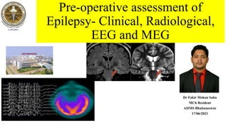
Pre-operative assessment of
Epilepsy- Clinical, Radiological,
EEG and MEG
Dr Fakir Mohan Sahu
MCh Resident
AIIMS Bhubaneswar
17/06/2021
Learning Objectives
• What is Refractory/ Drug resistance Epilepsy
• Goal of Pre-surgical evaluation
• Clinical Semiology
• Radiological Assessment
• Electroencephalography (EEG)- Scalp/Video
• Magnetoencephalography (MEG)
• Invasive Vs non-invasive assessment
• Recent Advances in Preoperative Epilepsy assessment
Introduction
• Epilepsy fail to respond to adequate antiepileptic treatment- 25% to 30%
• Sx success rate app. 75% (site & type of surgery and underlying etiology)
• Reduction in risk-
-a number of avoidable seizure related deaths,(Drowning, motor vehicle accident)
-fatal status epilepticus, & sudden unexpected death in epilepsy (SUDEP)
• In children, appropriate timing for surgical procedures is critical,
(lack of seizure control may interfere with consolidation of cognitive and motor functions in the developing brain)
Medically intractable (refractory) epilepsy
• When satisfactory seizure control cannot be achieved (ILAE)
-with any of the potentially available effective antiepileptic drugs (AEDs)
-alone or in combination, at doses or levels not a/w unacceptable side effects.
• First step of evaluation -to confirm the proper diagnosis and appropriate medical treatment has been utilized
• Poor response to medical treatment-
Noncompliance
Presence of pseudo-epileptic seizures alone or combined with epileptic ones
Incorrect classification of seizures & incorrect pharmacologic treatment.
Patient Perceptions
• Self-assessment of quality of life is an important step in evaluating the
patient’s perception of their disease.
• Some patients seek surgical treatment- for reasons other than medical
intractability
-intolerable side effects of AEDs,
-women who desire to get pregnant (concerned potential teratogenic)
-avoidance of the social stigmata
• Questionnaires QOLIE-31 for adults, and QOLIE-48-AD for adolescents
Goal of Pre-surgical evaluation
• Accurate and comprehensive mapping of the Anatomo-electro-clinical(AEC)
network defining the EZ
• Its possible overlap with clinically testable functional/ eloquent cortex
• To generate a “surgical diagram” depicting
 Epileptogenic region (ER)---Location, Extent
 Critical cortex ( CC)
• Surgical resection = ER - CC
• The MC location of seizure onset in adults -Temporal lobe,
Medial temporal lobe (hippocampus).
most amenable to surgical cure.
Classification of Presurgical evaluation
• Mandatory noninvasive – “universal
access”
 Clinical semiology and Exam
 Scalp EEG
 Imaging MRI
• Optional Noninvasive – “Limited Access “
Video EEG
Magnetoencephalography (MEG),
EEG-fMRI,
Nuclear imaging (PET,SPECT )
Neuropsychological testing
No single test adequately defines the epileptogenic region
Invasive test
invasive EEG
Depth, strip, grid electrodes
Wada (intracarotid amobarbital) test
Definition of abnormal brain areas
Clinical Semiology
• Temporal Lobe Epilepsy- an epigastric aura, followed by a quiet period of unresponsiveness
with staring, lipsmacking (oral automatisms).
-Picking at sheets or clothes (manual automatisms),
- contralateral dystonic posturing
-Postictal confusion and lethargy.
Dominant hemisphere- delayed recovery of language, often with transient aphasia on testing.
• Frontal lobe seizure-will occur from sleep with no warning,
-restlessness and/or prominent bilateral limb movements (bicycling/ asymmetric tonic posturing),
-end quickly with immediate recovery; this may recur several times in one night
• Occipital lobe seizures -visual aura, and may progress (due to electrical spread) into a temporal
lobe or frontal lobe type of seizure.
• Parietal lobe seizures are the least common, may have a sensory aura,
and tend to mimic frontal lobe seizures
Semiology Frontal Vs Temporal lobe
LANGUAGE ABNORMALITIES
AUTONOMIC FEATURES
MOTOR ABNORMALITIES
Ictal examination for Semiology
Ictal and postictal clinical feature with localization
Imaging CT
• Sturge weber syndrome – Tram track calcification.
• Calcification associated with neurocysticercosis.
• Tuberous sclerosis sub ependymal tubers.
• Abandoned in most epilepsy centers.
• 95 % Vs 32 % sensitivity (MRI Vs CT).
• Probability of positive CT in negative MR 0 %
while 80 % positive MR in CT negative cases.
Imaging MRI
• The primary role- to locate and define structural lesions.
• In TLE sensitivity is 75 % to 100 % for hippocampal sclerosis, Specificity 70- 85 %
• MRI also can depict relation of epileptogenic foci and functional tissue.
• Classic findings (MTS)-as a result of gliosis and the subsequent increase in free
water content.
• Associated findings -atrophy of the ipsilateral mammillary body, fornix, and other
parts of the limbic system
Imaging MRI
• Thin-section volumetric T1W imaging-used to calculate hippocampal volume
(Does not depict abnormal signal intensity, less useful than FLAIR and T2W for visual detection of MTS)
• T2W imaging is better than FLAIR for demonstrating the internal architecture of the hippocampus
• Coronal oblique MRI through the temporal lobes perpendicular to the plane of the hippocampus is the
(preferred modality)
• A FLAIR- nulls the CSF signal, Degree of signal abnormality is more obvious in hippocampus
• A pitfall of coronal FLAIR image- slight hyperintensity of all limbic structures relative to the neocortex.
T1 coronal images. Left hippocampal sclerosis is associated with widening of temporal horn and ipsilateral atrophy of fornix (A) and mammillary body (B) (arrows).
T1W Coronal
A and B, Axial and coronal images, showing migration and sulcation disorders, with gray matter nodular heterotopy. Note
the abnormal localization of gray matter in deep white matter lateral to right ventricle with ependymal extension (arrows).
T2W Axial & Coronal
A, T2 coronal image with region of interest measurement of the hippocampus. There is atrophy and hyperintensity on the left due to hippocampal sclerosis (arrow).
B, Flair sequence of coronal image showing left hippocampal hyperintensity (arrow), corresponding to sclerosis and atrophy.
T2W FLAIR
• When structural MR imaging does not demonstrate a lesion
• Volumetric neocortical measurements may provide an objective way
to evaluate the extent of resection and its relation to surgical results
Quantitative MR imaging
Diffusion Tensor Imaging
• In the white matter, diffusion
greatest parallel to the white matter tracts
minimal perpendicular to them.
• Malformations of cortical development and acquired lesions.
Increased mean diffusivity.
Increased perpendicular diffusivities.
Tractography of horizontal temporal-frontal and temporal-occipital (Meyer’s) tracts,
there is interruption of fibers of Meyer’s loop (arrow).
Magnetic Resonance Spectroscopy(MRS)
• MRS is a powerful adjunct to MR imaging.
• Consistent with the HPE characteristics of
reduced neuron cell counts or neuronal
dysfunction and increased glial cell numbers
• Reduced NAA and increased Cho in TLE.
• Extratemporal lobe epilepsy, the ability of MR
spectroscopy to lateralize the epilepsy is less
(50%).
Hippocampal univoxel spectroscopy. There is bilateral reduction of N-acetyl-aspartate
compatible with hippocampal sclerosis
Functional MR Imaging
• Used for mapping language, memory, and
sensorimotor location for presurgical planning.
• This imaging modality is based on
- the observation that increased neuronal activity
-a/w an increase in CBF
-therefore an increase in the
oxyhemoglobin/ deoxyhemoglobin ratio.
fMRI study showing left cerebral hemispheric dominance for language in a healthy subject using a phonologic
category
EEG (Electroencephalography)
• The inventor of EEG in 1924 Hans Berger German psychiatrist.
(alpha wave /"Berger wave.")
Used for recording the electrical activity of the brain,
• Awake & Sleep
• Ictal & Interictal
• Eye opening & Closure, Hyperventilation, Photic stimulation.
• Surface/ video/ Invasive / Combinations
• Lateralization (left vs. right hemisphere) and localization
• The presence of interictal (between seizures) epileptiform discharges. EDs:
sharp waves or spikes.
• Lack of EDs does NOT rule out epilepsy (up to 10%).
• Spike waves : negative transients with steep ascending and descending
limbs and a duration of 20 to 70 ms.
Scalp EEG
• Interictal s/o increased epileptogenic potential.
• Ictal Eds s/o classification of seizure, brain
region of seizure activity.
• Sensitivity and specificity depends upon,
Localization of epileptic brain tissue.
Duration of recording.
Use of supplementary electrodes.
Frequency of seizures
Timing in relation to last seizure.
(A) EEG showing left temporal interictal epileptiform discharges;
(B) 7-Hz-rhythmic theta activity over the left temporal region;
(C) left hippocampal atrophy and increased signal on T2W MRI sequence
(D) postoperative MRI showing complete resection of lt ant temporal
lobectomy with AHC.
Human EEG with prominent resting state activity – alpha-rhythm. Left: EEG traces (horizontal – time in seconds; vertical – amplitudes, scale 100 μV). Right: power spectra of shown signals (vertical lines – 10 and 20 Hz,
scale is linear). Alpha-rhythm consists of sinusoidal-like waves with frequencies in 8–12 Hz range (11 Hz in this case) more prominent in posterior sites. Alpha range is red at power spectrum graph.
Human EEG with prominent resting state activity
Common artifacts in human EEG. 1: Electrooculographic artifact caused by the excitation of eyeball's muscles (related to blinking, for example). Big-amplitude, slow, positive wave prominent in frontal electrodes. 2: Electrode's artifact caused by
bad contact (and thus bigger impedance) between P3 electrode and skin. 3: Swallowing artifact. 4: Common reference electrode's artifact caused by bad contact between reference electrode and skin. Huge wave similar in all channels.
Common artifacts in human EEG
EEG recording for temporal lobe area
Medial temporal lobe epilepsy usually have EDs from the anterior-mid temporal lobe
(electrodes F7/F8, T3/T4, as well as anterior temporal, sphenoidal, and inferior temporal electrodes if used).
Background EEG that showed mild independent bilateral temporal and generalized slowing;
(b) right anterior temporal spikes (∗); (c) left anterior temporal spikes (∗∗)
(right temporal IEDs to left temporal IEDs 70 : 30) in sleep; (d) left temporal type 2 ictal rhythm as rhythmic delta activity in the left temporal
regions (arrows) during one of her typical complex partial seizures (seizure onset zone).
Video EEG
• The combined use of video and EEG recording improves the
sensitivity and specificity over EEG recording alone.
• In many cases -as a first study in the proper classification of
the seizure disorder.
• A typical duration of inpatient monitoring is 3 to 5 days.
Video EEG
• Video-EEG monitoring has been demonstrated to-
 to accurately differentiate between epileptic and non-epileptic seizures
 to distinguish between primarily generalized and partial onset seizures,
 to determine seizure onset localization and lateralization
Stereo-EEG (SEEG)- is the surgical implantation of
electrodes into the brain in order to better localize the
seizure focus.
Magnetoencephalography (MEG)
• First MEG recording(1968) performed by Dr. Cohen using a single
channel, (more than 300 sensors)
• The MEG modality measures extracranial magnetic fields perpendicular
to the direction of intracellular currents in apical dendrites.
• Able to measure the epilepsy specific information (i.e., the brain activity
reflecting seizures and/or interictal epileptiform discharges) directly
• Non-invasively and with a very high temporal resolution (millisecond-
range).
• Advantages of MEG as opposed to EEG studies
-magnetic fields are minimally affected by conductivities of
intervening structures
-tissues between brain and scalp.
-superior than EEG in localizing the irritative zone
(Frontal, intrasylvian or insular focus )
Figure 1: MEG acquisition: (a) MEG (306 channel) dewar inside
a magnetically shielded room (MSR), (b) Five-head positioning
indicator (HPI) coils for head movement monitoring during
acquisition, (c) 3D-digitization carried out using a wooden chair,
goggles with transmitter, and a stylus with receiver, (d) patient
MEG data acquisition with simultaneous EEG in erect posture
MEG
Invasive EEG monitoring
INDICATIONS:
• Seizures are lateralized but not localized.
• Seizures are localized but not lateralized.
• Seizures are neither localized nor lateralized (eg, stereotyped complex partial seizures with diffuse ictal changes).
• Seizure localization is discordant with other data.
• Relationship of seizure onset to lesion must be determined (eg, dual pathology or multiple intracranial lesions).
Invasive EEG monitoring
• Temporal lobe seizures
• -Doubtful side
• -Normal MRI
• -Bilateral pathology
• - Discordant non-invasive testing
• Extratemporal seizures
• -Definition of extent of epileptogenic area
• -Extra-operative cortical mapping
 Depth,
 strip
 grid electrodes
• Implantable intracranial devices used to record the
electrocorticogram (ECoG).
• to stimulate the cortex to determine function.
• Depth electrodes are multicontact, thin, tubular, rigid or
semirigid electrodes.
• Penetrate the brain substances for the purpose of recording
from deep structures.
TYPES OF ELECTRODES
• Intracranial strip electrodes are a linear array of 2-16 disk electrodes
embedded in a strip of silastic or in tubular in structure
• Grid electrodes are parallel rows of similar numbers of electrodes that
can be configured in standard or custom designs.
• Grid and strip electrodes are designed to be in direct contact with
brain neocortex.
• Electrodes are placed in the subdural space/ the epidural space.
TYPES OF ELECTRODES
• Constructed from biologically inert materials (ie silastic, stainless
steel, platinum).
• Platinum electrodes are more easily seen on fluoroscopic images
and are compatible with MRI.
• The morbidity of surgery depends on the type of electrode
 intracranial strip electrodes-lowest morbidity.
Intracranial grid placement-highest morbidity.
• Transient neurological deficit, hematoma, cerebral edema with
increased intracranial pressure, and infarction.
Wada test
• Developed by Dr Jun Wada. AKA intracarotid sodium amobarbital procedure (ISAP)
• Neuroradiologist, Epileptologist, Neuropsychologist
• To preoperatively determine which hemisphere contains language function
• To test memory function within each hemisphere
Procedure:
• Accomplished by individually cannulating each internal carotid artery.
• After contrast arteriography verifies that blood flows to the corresponding hemisphere
and not to the brainstem or contralateral side,
• A dose of sodium amobarbital (sufficient to impede hemispheric function) is injected.
• Both sides of the brain are tested in one day. Usually, there is a wait of 30 to 60 minutes
before the second side is tested, Test is done as an outpatient test
Wada test
• Different word, object, and picture cards are shown, and the awake side tries to recognize and remember what it sees.
• If speech persists in the face of this hemiparesis, language function is assumed to not be represented within that
hemisphere.
• The deficiencies of this evaluation for memory function directly relate to the multiple problems.
Injection of a drug into the ICA does not assure drug effect in the basal
temporal area in general or the hippocampal region specifically.
(due to variations in the direct blood supply to the hippocampus)
• Complication like pain haemorrhage, infection
• Alternate possible test- fMRI
Recent Advances- Nuclear imaging
Positron emission tomography (PET)
• An injection of radio-labeled glucose (18FDG) to measure brain metabolism.
• Interictal PET -shows hypometabolism in the seizure focus, especially in TLE.
• Ictal PET is not practical due to the extremely short half life of the radiotracers used.
• PET is most useful in MRI-negative TLE, (non-lesional, extra-temporal epilepsy)
• FDG PET identifies sites of interictal metabolism in 70 % with TLE and 60 % frontal
lobe onset seizure.
• C -11 flumazenil PET effective in hippocampal sclerosis & shows pathological foci in a
more circumscribed fashion.
In 80% of patients there is an increase in blood
flow and glucose metabolism during a seizure in
the cerebral cortex. However, between seizure
there tends to be a lower than normal glucose
uptake and blood flow.
Single photon emission computed tomography(SPECT)
• Ictal SPECT ideal method for localization and lateralization of
seizure onset, Shows hypermetabolism
• SPECT also performed better in the setting of negative MRI.
 Inter ictal scan – Hypoperfusion large area in the hemisphere of
onset.
 Ictal /Interictal quantitative difference analysis provides for the
best and most reliable seizure focus localization
Subtraction ictal- interictal SPECT co-registered to MRI (SISCOM):
• A newer methodology that has greater accuracy than either ictal or
interictal SPECT scanning.
• This requires obtaining scans separated by at least 48 hrs to
accommodate radionucleotide wash out during an interictal period and
within seconds of seizure onset.
• These scans are then subtracted from one another with the use of
specialized computer software.
• This leaves a better indication of the cortical area of ictal onset.
• This subtracted scan can then be co-registered onto the patient's MRI to
provide support for the location of the focus.
Conclusion
• Epilepsy semiology-an improved understanding of epilepsy related issues and localizations
• MRI is most sensitive for identification of a structural lesion.
• A routine EEG- correlate cerebral dysfunction and a potential epileptogenic zone.(EZ)
• Ictal video-EEG -mirror foci, false localization & lateralization, secondary epileptogenesis
• Invasive video-EEG with extra-operative epidural, subdural, or depth electrodes-needed to define complex EZ
• The Wada test -determine lateralization of language and memory; fMRI is possible alternative
• SPECT and PET scans- confirm a functional relationship between radiologic lesion and EZ
Role of Patient management conference
• Epilepsy Patient Management Conference (EMC) - multidisciplinary weekly meeting
• Single most important discussion forum for the management of complex patients
• Epilepsy team – neurologist, surgeons, advanced practice nurses, administrators and
coordinators, neuropsychologists and neuroradiologists
• To discuss and synthesize the data gathered for particular patients who have undergone
Phase I monitoring.
• Discussion concludes with a formulation of a consensus management plan for the pt.
whether the patient would benefit from epilepsy surgery.
If so, further tests are usually necessary to confirm surgical candidacy.
and on the completion of those ancillary tests, the data are re-discussed
• In this iterative fashion a final management plan is made which is then communicated
to the patient by the team
Thank you

More Related Content

What's hot

Myoclonus
MyoclonusMyoclonus
MyoclonusPS Deb
 
Intra operative neurophysiological monitoring
Intra operative neurophysiological monitoringIntra operative neurophysiological monitoring
Intra operative neurophysiological monitoringKode Sashanka
 
Disconnection syndromes sharath
Disconnection syndromes sharathDisconnection syndromes sharath
Disconnection syndromes sharathPasham sharath
 
presurgical evaluation of epilepsy
presurgical evaluation of epilepsypresurgical evaluation of epilepsy
presurgical evaluation of epilepsySrirama Anjaneyulu
 
Frontal lobe epilepsy
Frontal lobe epilepsyFrontal lobe epilepsy
Frontal lobe epilepsyNeha Sharma
 
Neuroimaging in dementia
Neuroimaging in dementiaNeuroimaging in dementia
Neuroimaging in dementiaNeurologyKota
 
Intraoperative Neurophysiological Monitoring Brain
Intraoperative Neurophysiological Monitoring BrainIntraoperative Neurophysiological Monitoring Brain
Intraoperative Neurophysiological Monitoring BrainFarrukh Javeed
 
Mesial temporal lobe epilepsy
Mesial temporal lobe epilepsyMesial temporal lobe epilepsy
Mesial temporal lobe epilepsydr archana verma
 
Responsive Neurostimulation (RNS) for Intractable Epilepsy
Responsive Neurostimulation (RNS) for Intractable EpilepsyResponsive Neurostimulation (RNS) for Intractable Epilepsy
Responsive Neurostimulation (RNS) for Intractable EpilepsyAllina Health
 
Pet in parkinsonism
Pet  in parkinsonismPet  in parkinsonism
Pet in parkinsonismgulabsoni
 
Psp - Progressive Supranuclear Palsy
Psp - Progressive Supranuclear PalsyPsp - Progressive Supranuclear Palsy
Psp - Progressive Supranuclear PalsyJaber Samer
 

What's hot (20)

Epilepsy surgery
Epilepsy surgeryEpilepsy surgery
Epilepsy surgery
 
Epilepsy surgery
Epilepsy surgeryEpilepsy surgery
Epilepsy surgery
 
Myoclonus
MyoclonusMyoclonus
Myoclonus
 
Intra operative neurophysiological monitoring
Intra operative neurophysiological monitoringIntra operative neurophysiological monitoring
Intra operative neurophysiological monitoring
 
CP angle.pptx
CP angle.pptxCP angle.pptx
CP angle.pptx
 
Drug resistant epilepsy
Drug resistant epilepsyDrug resistant epilepsy
Drug resistant epilepsy
 
Myoclonus
MyoclonusMyoclonus
Myoclonus
 
Disconnection syndromes sharath
Disconnection syndromes sharathDisconnection syndromes sharath
Disconnection syndromes sharath
 
presurgical evaluation of epilepsy
presurgical evaluation of epilepsypresurgical evaluation of epilepsy
presurgical evaluation of epilepsy
 
Frontal lobe epilepsy
Frontal lobe epilepsyFrontal lobe epilepsy
Frontal lobe epilepsy
 
Neuroimaging in dementia
Neuroimaging in dementiaNeuroimaging in dementia
Neuroimaging in dementia
 
INSULAR GLIOMA SURGERY.pptx
INSULAR GLIOMA SURGERY.pptxINSULAR GLIOMA SURGERY.pptx
INSULAR GLIOMA SURGERY.pptx
 
Intraoperative Neurophysiological Monitoring Brain
Intraoperative Neurophysiological Monitoring BrainIntraoperative Neurophysiological Monitoring Brain
Intraoperative Neurophysiological Monitoring Brain
 
Mesial temporal lobe epilepsy
Mesial temporal lobe epilepsyMesial temporal lobe epilepsy
Mesial temporal lobe epilepsy
 
Responsive Neurostimulation (RNS) for Intractable Epilepsy
Responsive Neurostimulation (RNS) for Intractable EpilepsyResponsive Neurostimulation (RNS) for Intractable Epilepsy
Responsive Neurostimulation (RNS) for Intractable Epilepsy
 
Pet in parkinsonism
Pet  in parkinsonismPet  in parkinsonism
Pet in parkinsonism
 
Frontal lobe epilepsy
Frontal lobe epilepsyFrontal lobe epilepsy
Frontal lobe epilepsy
 
What's New in Multiple Sclerosis
What's New in Multiple SclerosisWhat's New in Multiple Sclerosis
What's New in Multiple Sclerosis
 
Epilepsy surgery
Epilepsy surgeryEpilepsy surgery
Epilepsy surgery
 
Psp - Progressive Supranuclear Palsy
Psp - Progressive Supranuclear PalsyPsp - Progressive Supranuclear Palsy
Psp - Progressive Supranuclear Palsy
 

Similar to Preoperative assessment of epilepsy clinical & radiological, eeg, meg

evaluation for epilepsy surgery.pptx
evaluation for epilepsy surgery.pptxevaluation for epilepsy surgery.pptx
evaluation for epilepsy surgery.pptxDr. Shahnawaz Alam
 
recent investigation in epilepsy
recent investigation in epilepsyrecent investigation in epilepsy
recent investigation in epilepsyOsama Ragab
 
Neurology advanced mr imaging in epilepsy v lai
Neurology advanced mr imaging in epilepsy v laiNeurology advanced mr imaging in epilepsy v lai
Neurology advanced mr imaging in epilepsy v laiJFIM
 
Somatosensory and motor evoked potentials by neelothpala
Somatosensory and motor evoked potentials by neelothpalaSomatosensory and motor evoked potentials by neelothpala
Somatosensory and motor evoked potentials by neelothpalavrkv2007
 
2 neuro. ixs 1
2 neuro. ixs 1 2 neuro. ixs 1
2 neuro. ixs 1 eliasmawla
 
Neuroimaging of alzheimer's
Neuroimaging of alzheimer'sNeuroimaging of alzheimer's
Neuroimaging of alzheimer'sDr Wasim
 
Somatosensory evoked potentials
Somatosensory evoked potentialsSomatosensory evoked potentials
Somatosensory evoked potentialsManideep Malaka
 
Advances in neuro anesthesia monitoring
Advances in neuro anesthesia monitoringAdvances in neuro anesthesia monitoring
Advances in neuro anesthesia monitoringWesam Mousa
 
Seminar on Brain Mapping techniques.pptx
Seminar on Brain Mapping techniques.pptxSeminar on Brain Mapping techniques.pptx
Seminar on Brain Mapping techniques.pptxSoumen Kanjilal
 
Evoked potential - An overview
Evoked potential - An overviewEvoked potential - An overview
Evoked potential - An overviewAnbarasi rajkumar
 
neuromonitoringinanesthesia-150217064704-conversion-gate02.pdf
neuromonitoringinanesthesia-150217064704-conversion-gate02.pdfneuromonitoringinanesthesia-150217064704-conversion-gate02.pdf
neuromonitoringinanesthesia-150217064704-conversion-gate02.pdfSaumya Singh
 
Neuromonitoring in anesthesia
Neuromonitoring in anesthesiaNeuromonitoring in anesthesia
Neuromonitoring in anesthesiaAntara Banerji
 
Vep and its practical importance
Vep and its practical importanceVep and its practical importance
Vep and its practical importancenikhilzandu
 

Similar to Preoperative assessment of epilepsy clinical & radiological, eeg, meg (20)

evaluation for epilepsy surgery.pptx
evaluation for epilepsy surgery.pptxevaluation for epilepsy surgery.pptx
evaluation for epilepsy surgery.pptx
 
Imaging in epilepsy1
Imaging in epilepsy1Imaging in epilepsy1
Imaging in epilepsy1
 
recent investigation in epilepsy
recent investigation in epilepsyrecent investigation in epilepsy
recent investigation in epilepsy
 
Epilepsy and anaesthesia
Epilepsy and anaesthesiaEpilepsy and anaesthesia
Epilepsy and anaesthesia
 
Neurology advanced mr imaging in epilepsy v lai
Neurology advanced mr imaging in epilepsy v laiNeurology advanced mr imaging in epilepsy v lai
Neurology advanced mr imaging in epilepsy v lai
 
Somatosensory and motor evoked potentials by neelothpala
Somatosensory and motor evoked potentials by neelothpalaSomatosensory and motor evoked potentials by neelothpala
Somatosensory and motor evoked potentials by neelothpala
 
Facial nerve palsy
Facial nerve palsyFacial nerve palsy
Facial nerve palsy
 
2 neuro. ixs 1
2 neuro. ixs 1 2 neuro. ixs 1
2 neuro. ixs 1
 
EEG & Evoked potentials
EEG & Evoked potentialsEEG & Evoked potentials
EEG & Evoked potentials
 
Neuroimaging of alzheimer's
Neuroimaging of alzheimer'sNeuroimaging of alzheimer's
Neuroimaging of alzheimer's
 
Somatosensory evoked potentials
Somatosensory evoked potentialsSomatosensory evoked potentials
Somatosensory evoked potentials
 
Advances in neuro anesthesia monitoring
Advances in neuro anesthesia monitoringAdvances in neuro anesthesia monitoring
Advances in neuro anesthesia monitoring
 
Medicine 5th year, 1st 2 lectures (Dr. Rasool)
Medicine 5th year, 1st 2 lectures (Dr. Rasool)Medicine 5th year, 1st 2 lectures (Dr. Rasool)
Medicine 5th year, 1st 2 lectures (Dr. Rasool)
 
NCS DDH.pptx
NCS DDH.pptxNCS DDH.pptx
NCS DDH.pptx
 
Seminar on Brain Mapping techniques.pptx
Seminar on Brain Mapping techniques.pptxSeminar on Brain Mapping techniques.pptx
Seminar on Brain Mapping techniques.pptx
 
Evoked potential - An overview
Evoked potential - An overviewEvoked potential - An overview
Evoked potential - An overview
 
neuromonitoringinanesthesia-150217064704-conversion-gate02.pdf
neuromonitoringinanesthesia-150217064704-conversion-gate02.pdfneuromonitoringinanesthesia-150217064704-conversion-gate02.pdf
neuromonitoringinanesthesia-150217064704-conversion-gate02.pdf
 
Neuromonitoring in anesthesia
Neuromonitoring in anesthesiaNeuromonitoring in anesthesia
Neuromonitoring in anesthesia
 
Chapter 7
Chapter 7Chapter 7
Chapter 7
 
Vep and its practical importance
Vep and its practical importanceVep and its practical importance
Vep and its practical importance
 

More from Dr Fakir Mohan Sahu

Facial and Hearing Preservation in Acoustic Neuroma Surgery
Facial and Hearing Preservation in Acoustic Neuroma SurgeryFacial and Hearing Preservation in Acoustic Neuroma Surgery
Facial and Hearing Preservation in Acoustic Neuroma SurgeryDr Fakir Mohan Sahu
 
CSF Production, Dynamics and Physiology
CSF Production, Dynamics and PhysiologyCSF Production, Dynamics and Physiology
CSF Production, Dynamics and PhysiologyDr Fakir Mohan Sahu
 
Neurological assessment of child
Neurological assessment of childNeurological assessment of child
Neurological assessment of childDr Fakir Mohan Sahu
 
Application of Neuronavigation in Brain Surgery
Application of Neuronavigation in Brain SurgeryApplication of Neuronavigation in Brain Surgery
Application of Neuronavigation in Brain SurgeryDr Fakir Mohan Sahu
 
Ophthalmological evalution in sellar suprasellar tumours
Ophthalmological evalution in sellar suprasellar tumoursOphthalmological evalution in sellar suprasellar tumours
Ophthalmological evalution in sellar suprasellar tumoursDr Fakir Mohan Sahu
 
Abdominal wall anatomy and its clinical importance
Abdominal wall anatomy and its clinical importanceAbdominal wall anatomy and its clinical importance
Abdominal wall anatomy and its clinical importanceDr Fakir Mohan Sahu
 
Parotid tumour asicon 2017 jaipur
Parotid tumour asicon 2017 jaipurParotid tumour asicon 2017 jaipur
Parotid tumour asicon 2017 jaipurDr Fakir Mohan Sahu
 
Vasospasm in subarachnoid haemorrhage pre finale
Vasospasm in subarachnoid haemorrhage pre finaleVasospasm in subarachnoid haemorrhage pre finale
Vasospasm in subarachnoid haemorrhage pre finaleDr Fakir Mohan Sahu
 
Neuro-otological aspects of Cerebellopontine angle SOL
Neuro-otological aspects of Cerebellopontine angle SOLNeuro-otological aspects of Cerebellopontine angle SOL
Neuro-otological aspects of Cerebellopontine angle SOLDr Fakir Mohan Sahu
 
Neurovascon 2019 Moya- Moya Disease case 3
Neurovascon 2019 Moya- Moya Disease case 3Neurovascon 2019 Moya- Moya Disease case 3
Neurovascon 2019 Moya- Moya Disease case 3Dr Fakir Mohan Sahu
 
Neurovascon 2019 Acom Aneurysm case 1
Neurovascon 2019 Acom Aneurysm case 1Neurovascon 2019 Acom Aneurysm case 1
Neurovascon 2019 Acom Aneurysm case 1Dr Fakir Mohan Sahu
 
Pulmonary Consideration and Complication in Neurosurgery
Pulmonary Consideration and Complication in NeurosurgeryPulmonary Consideration and Complication in Neurosurgery
Pulmonary Consideration and Complication in NeurosurgeryDr Fakir Mohan Sahu
 

More from Dr Fakir Mohan Sahu (20)

Surgical approach to thalamus
Surgical approach to thalamusSurgical approach to thalamus
Surgical approach to thalamus
 
Brain tumours
Brain tumoursBrain tumours
Brain tumours
 
Facial and Hearing Preservation in Acoustic Neuroma Surgery
Facial and Hearing Preservation in Acoustic Neuroma SurgeryFacial and Hearing Preservation in Acoustic Neuroma Surgery
Facial and Hearing Preservation in Acoustic Neuroma Surgery
 
CSF Production, Dynamics and Physiology
CSF Production, Dynamics and PhysiologyCSF Production, Dynamics and Physiology
CSF Production, Dynamics and Physiology
 
Neurological assessment of child
Neurological assessment of childNeurological assessment of child
Neurological assessment of child
 
Application of Neuronavigation in Brain Surgery
Application of Neuronavigation in Brain SurgeryApplication of Neuronavigation in Brain Surgery
Application of Neuronavigation in Brain Surgery
 
Pterional craniotomy
Pterional craniotomyPterional craniotomy
Pterional craniotomy
 
Ophthalmological evalution in sellar suprasellar tumours
Ophthalmological evalution in sellar suprasellar tumoursOphthalmological evalution in sellar suprasellar tumours
Ophthalmological evalution in sellar suprasellar tumours
 
Laparoscopic hernia anatomy
Laparoscopic hernia anatomyLaparoscopic hernia anatomy
Laparoscopic hernia anatomy
 
Abdominal wall anatomy and its clinical importance
Abdominal wall anatomy and its clinical importanceAbdominal wall anatomy and its clinical importance
Abdominal wall anatomy and its clinical importance
 
Meckels diverticulum
Meckels diverticulum Meckels diverticulum
Meckels diverticulum
 
Parotid tumour asicon 2017 jaipur
Parotid tumour asicon 2017 jaipurParotid tumour asicon 2017 jaipur
Parotid tumour asicon 2017 jaipur
 
Vasospasm in subarachnoid haemorrhage pre finale
Vasospasm in subarachnoid haemorrhage pre finaleVasospasm in subarachnoid haemorrhage pre finale
Vasospasm in subarachnoid haemorrhage pre finale
 
Neuro-otological aspects of Cerebellopontine angle SOL
Neuro-otological aspects of Cerebellopontine angle SOLNeuro-otological aspects of Cerebellopontine angle SOL
Neuro-otological aspects of Cerebellopontine angle SOL
 
Frontal and Parietal Lobe sign
Frontal and Parietal Lobe signFrontal and Parietal Lobe sign
Frontal and Parietal Lobe sign
 
Neurovascon 2019 Moya- Moya Disease case 3
Neurovascon 2019 Moya- Moya Disease case 3Neurovascon 2019 Moya- Moya Disease case 3
Neurovascon 2019 Moya- Moya Disease case 3
 
Neurovascon 2019 case 2
Neurovascon 2019 case 2Neurovascon 2019 case 2
Neurovascon 2019 case 2
 
Neurovascon 2019 Acom Aneurysm case 1
Neurovascon 2019 Acom Aneurysm case 1Neurovascon 2019 Acom Aneurysm case 1
Neurovascon 2019 Acom Aneurysm case 1
 
Pulmonary Consideration and Complication in Neurosurgery
Pulmonary Consideration and Complication in NeurosurgeryPulmonary Consideration and Complication in Neurosurgery
Pulmonary Consideration and Complication in Neurosurgery
 
Stem cell in Neurosurgery
Stem cell in NeurosurgeryStem cell in Neurosurgery
Stem cell in Neurosurgery
 

Recently uploaded

VIP Mumbai Call Girls Hiranandani Gardens Just Call 9920874524 with A/C Room ...
VIP Mumbai Call Girls Hiranandani Gardens Just Call 9920874524 with A/C Room ...VIP Mumbai Call Girls Hiranandani Gardens Just Call 9920874524 with A/C Room ...
VIP Mumbai Call Girls Hiranandani Gardens Just Call 9920874524 with A/C Room ...Garima Khatri
 
Call Girl Bangalore Nandini 7001305949 Independent Escort Service Bangalore
Call Girl Bangalore Nandini 7001305949 Independent Escort Service BangaloreCall Girl Bangalore Nandini 7001305949 Independent Escort Service Bangalore
Call Girl Bangalore Nandini 7001305949 Independent Escort Service Bangalorenarwatsonia7
 
Call Girls Service Chennai Jiya 7001305949 Independent Escort Service Chennai
Call Girls Service Chennai Jiya 7001305949 Independent Escort Service ChennaiCall Girls Service Chennai Jiya 7001305949 Independent Escort Service Chennai
Call Girls Service Chennai Jiya 7001305949 Independent Escort Service ChennaiNehru place Escorts
 
Russian Call Girls Chickpet - 7001305949 Booking and charges genuine rate for...
Russian Call Girls Chickpet - 7001305949 Booking and charges genuine rate for...Russian Call Girls Chickpet - 7001305949 Booking and charges genuine rate for...
Russian Call Girls Chickpet - 7001305949 Booking and charges genuine rate for...narwatsonia7
 
Call Girls Service Bellary Road Just Call 7001305949 Enjoy College Girls Service
Call Girls Service Bellary Road Just Call 7001305949 Enjoy College Girls ServiceCall Girls Service Bellary Road Just Call 7001305949 Enjoy College Girls Service
Call Girls Service Bellary Road Just Call 7001305949 Enjoy College Girls Servicenarwatsonia7
 
Call Girls Service Jaipur Grishma WhatsApp ❤8445551418 VIP Call Girls Jaipur
Call Girls Service Jaipur Grishma WhatsApp ❤8445551418 VIP Call Girls JaipurCall Girls Service Jaipur Grishma WhatsApp ❤8445551418 VIP Call Girls Jaipur
Call Girls Service Jaipur Grishma WhatsApp ❤8445551418 VIP Call Girls Jaipurparulsinha
 
Call Girls Doddaballapur Road Just Call 7001305949 Top Class Call Girl Servic...
Call Girls Doddaballapur Road Just Call 7001305949 Top Class Call Girl Servic...Call Girls Doddaballapur Road Just Call 7001305949 Top Class Call Girl Servic...
Call Girls Doddaballapur Road Just Call 7001305949 Top Class Call Girl Servic...narwatsonia7
 
Russian Call Girls Chennai Madhuri 9907093804 Independent Call Girls Service ...
Russian Call Girls Chennai Madhuri 9907093804 Independent Call Girls Service ...Russian Call Girls Chennai Madhuri 9907093804 Independent Call Girls Service ...
Russian Call Girls Chennai Madhuri 9907093804 Independent Call Girls Service ...Nehru place Escorts
 
Call Girls Whitefield Just Call 7001305949 Top Class Call Girl Service Available
Call Girls Whitefield Just Call 7001305949 Top Class Call Girl Service AvailableCall Girls Whitefield Just Call 7001305949 Top Class Call Girl Service Available
Call Girls Whitefield Just Call 7001305949 Top Class Call Girl Service Availablenarwatsonia7
 
Vip Call Girls Anna Salai Chennai 👉 8250192130 ❣️💯 Top Class Girls Available
Vip Call Girls Anna Salai Chennai 👉 8250192130 ❣️💯 Top Class Girls AvailableVip Call Girls Anna Salai Chennai 👉 8250192130 ❣️💯 Top Class Girls Available
Vip Call Girls Anna Salai Chennai 👉 8250192130 ❣️💯 Top Class Girls AvailableNehru place Escorts
 
Call Girls Service In Shyam Nagar Whatsapp 8445551418 Independent Escort Service
Call Girls Service In Shyam Nagar Whatsapp 8445551418 Independent Escort ServiceCall Girls Service In Shyam Nagar Whatsapp 8445551418 Independent Escort Service
Call Girls Service In Shyam Nagar Whatsapp 8445551418 Independent Escort Serviceparulsinha
 
College Call Girls Pune Mira 9907093804 Short 1500 Night 6000 Best call girls...
College Call Girls Pune Mira 9907093804 Short 1500 Night 6000 Best call girls...College Call Girls Pune Mira 9907093804 Short 1500 Night 6000 Best call girls...
College Call Girls Pune Mira 9907093804 Short 1500 Night 6000 Best call girls...Miss joya
 
Artifacts in Nuclear Medicine with Identifying and resolving artifacts.
Artifacts in Nuclear Medicine with Identifying and resolving artifacts.Artifacts in Nuclear Medicine with Identifying and resolving artifacts.
Artifacts in Nuclear Medicine with Identifying and resolving artifacts.MiadAlsulami
 
CALL ON ➥9907093804 🔝 Call Girls Hadapsar ( Pune) Girls Service
CALL ON ➥9907093804 🔝 Call Girls Hadapsar ( Pune)  Girls ServiceCALL ON ➥9907093804 🔝 Call Girls Hadapsar ( Pune)  Girls Service
CALL ON ➥9907093804 🔝 Call Girls Hadapsar ( Pune) Girls ServiceMiss joya
 
Call Girls Colaba Mumbai ❤️ 9920874524 👈 Cash on Delivery
Call Girls Colaba Mumbai ❤️ 9920874524 👈 Cash on DeliveryCall Girls Colaba Mumbai ❤️ 9920874524 👈 Cash on Delivery
Call Girls Colaba Mumbai ❤️ 9920874524 👈 Cash on Deliverynehamumbai
 
Call Girls Yelahanka Just Call 7001305949 Top Class Call Girl Service Available
Call Girls Yelahanka Just Call 7001305949 Top Class Call Girl Service AvailableCall Girls Yelahanka Just Call 7001305949 Top Class Call Girl Service Available
Call Girls Yelahanka Just Call 7001305949 Top Class Call Girl Service Availablenarwatsonia7
 
VIP Call Girls Indore Kirti 💚😋 9256729539 🚀 Indore Escorts
VIP Call Girls Indore Kirti 💚😋  9256729539 🚀 Indore EscortsVIP Call Girls Indore Kirti 💚😋  9256729539 🚀 Indore Escorts
VIP Call Girls Indore Kirti 💚😋 9256729539 🚀 Indore Escortsaditipandeya
 
Call Girls Chennai Megha 9907093804 Independent Call Girls Service Chennai
Call Girls Chennai Megha 9907093804 Independent Call Girls Service ChennaiCall Girls Chennai Megha 9907093804 Independent Call Girls Service Chennai
Call Girls Chennai Megha 9907093804 Independent Call Girls Service ChennaiNehru place Escorts
 

Recently uploaded (20)

VIP Mumbai Call Girls Hiranandani Gardens Just Call 9920874524 with A/C Room ...
VIP Mumbai Call Girls Hiranandani Gardens Just Call 9920874524 with A/C Room ...VIP Mumbai Call Girls Hiranandani Gardens Just Call 9920874524 with A/C Room ...
VIP Mumbai Call Girls Hiranandani Gardens Just Call 9920874524 with A/C Room ...
 
Escort Service Call Girls In Sarita Vihar,, 99530°56974 Delhi NCR
Escort Service Call Girls In Sarita Vihar,, 99530°56974 Delhi NCREscort Service Call Girls In Sarita Vihar,, 99530°56974 Delhi NCR
Escort Service Call Girls In Sarita Vihar,, 99530°56974 Delhi NCR
 
Call Girl Bangalore Nandini 7001305949 Independent Escort Service Bangalore
Call Girl Bangalore Nandini 7001305949 Independent Escort Service BangaloreCall Girl Bangalore Nandini 7001305949 Independent Escort Service Bangalore
Call Girl Bangalore Nandini 7001305949 Independent Escort Service Bangalore
 
Call Girls Service Chennai Jiya 7001305949 Independent Escort Service Chennai
Call Girls Service Chennai Jiya 7001305949 Independent Escort Service ChennaiCall Girls Service Chennai Jiya 7001305949 Independent Escort Service Chennai
Call Girls Service Chennai Jiya 7001305949 Independent Escort Service Chennai
 
Russian Call Girls Chickpet - 7001305949 Booking and charges genuine rate for...
Russian Call Girls Chickpet - 7001305949 Booking and charges genuine rate for...Russian Call Girls Chickpet - 7001305949 Booking and charges genuine rate for...
Russian Call Girls Chickpet - 7001305949 Booking and charges genuine rate for...
 
Call Girls Service Bellary Road Just Call 7001305949 Enjoy College Girls Service
Call Girls Service Bellary Road Just Call 7001305949 Enjoy College Girls ServiceCall Girls Service Bellary Road Just Call 7001305949 Enjoy College Girls Service
Call Girls Service Bellary Road Just Call 7001305949 Enjoy College Girls Service
 
Call Girls Service Jaipur Grishma WhatsApp ❤8445551418 VIP Call Girls Jaipur
Call Girls Service Jaipur Grishma WhatsApp ❤8445551418 VIP Call Girls JaipurCall Girls Service Jaipur Grishma WhatsApp ❤8445551418 VIP Call Girls Jaipur
Call Girls Service Jaipur Grishma WhatsApp ❤8445551418 VIP Call Girls Jaipur
 
Call Girls Doddaballapur Road Just Call 7001305949 Top Class Call Girl Servic...
Call Girls Doddaballapur Road Just Call 7001305949 Top Class Call Girl Servic...Call Girls Doddaballapur Road Just Call 7001305949 Top Class Call Girl Servic...
Call Girls Doddaballapur Road Just Call 7001305949 Top Class Call Girl Servic...
 
Russian Call Girls in Delhi Tanvi ➡️ 9711199012 💋📞 Independent Escort Service...
Russian Call Girls in Delhi Tanvi ➡️ 9711199012 💋📞 Independent Escort Service...Russian Call Girls in Delhi Tanvi ➡️ 9711199012 💋📞 Independent Escort Service...
Russian Call Girls in Delhi Tanvi ➡️ 9711199012 💋📞 Independent Escort Service...
 
Russian Call Girls Chennai Madhuri 9907093804 Independent Call Girls Service ...
Russian Call Girls Chennai Madhuri 9907093804 Independent Call Girls Service ...Russian Call Girls Chennai Madhuri 9907093804 Independent Call Girls Service ...
Russian Call Girls Chennai Madhuri 9907093804 Independent Call Girls Service ...
 
Call Girls Whitefield Just Call 7001305949 Top Class Call Girl Service Available
Call Girls Whitefield Just Call 7001305949 Top Class Call Girl Service AvailableCall Girls Whitefield Just Call 7001305949 Top Class Call Girl Service Available
Call Girls Whitefield Just Call 7001305949 Top Class Call Girl Service Available
 
Vip Call Girls Anna Salai Chennai 👉 8250192130 ❣️💯 Top Class Girls Available
Vip Call Girls Anna Salai Chennai 👉 8250192130 ❣️💯 Top Class Girls AvailableVip Call Girls Anna Salai Chennai 👉 8250192130 ❣️💯 Top Class Girls Available
Vip Call Girls Anna Salai Chennai 👉 8250192130 ❣️💯 Top Class Girls Available
 
Call Girls Service In Shyam Nagar Whatsapp 8445551418 Independent Escort Service
Call Girls Service In Shyam Nagar Whatsapp 8445551418 Independent Escort ServiceCall Girls Service In Shyam Nagar Whatsapp 8445551418 Independent Escort Service
Call Girls Service In Shyam Nagar Whatsapp 8445551418 Independent Escort Service
 
College Call Girls Pune Mira 9907093804 Short 1500 Night 6000 Best call girls...
College Call Girls Pune Mira 9907093804 Short 1500 Night 6000 Best call girls...College Call Girls Pune Mira 9907093804 Short 1500 Night 6000 Best call girls...
College Call Girls Pune Mira 9907093804 Short 1500 Night 6000 Best call girls...
 
Artifacts in Nuclear Medicine with Identifying and resolving artifacts.
Artifacts in Nuclear Medicine with Identifying and resolving artifacts.Artifacts in Nuclear Medicine with Identifying and resolving artifacts.
Artifacts in Nuclear Medicine with Identifying and resolving artifacts.
 
CALL ON ➥9907093804 🔝 Call Girls Hadapsar ( Pune) Girls Service
CALL ON ➥9907093804 🔝 Call Girls Hadapsar ( Pune)  Girls ServiceCALL ON ➥9907093804 🔝 Call Girls Hadapsar ( Pune)  Girls Service
CALL ON ➥9907093804 🔝 Call Girls Hadapsar ( Pune) Girls Service
 
Call Girls Colaba Mumbai ❤️ 9920874524 👈 Cash on Delivery
Call Girls Colaba Mumbai ❤️ 9920874524 👈 Cash on DeliveryCall Girls Colaba Mumbai ❤️ 9920874524 👈 Cash on Delivery
Call Girls Colaba Mumbai ❤️ 9920874524 👈 Cash on Delivery
 
Call Girls Yelahanka Just Call 7001305949 Top Class Call Girl Service Available
Call Girls Yelahanka Just Call 7001305949 Top Class Call Girl Service AvailableCall Girls Yelahanka Just Call 7001305949 Top Class Call Girl Service Available
Call Girls Yelahanka Just Call 7001305949 Top Class Call Girl Service Available
 
VIP Call Girls Indore Kirti 💚😋 9256729539 🚀 Indore Escorts
VIP Call Girls Indore Kirti 💚😋  9256729539 🚀 Indore EscortsVIP Call Girls Indore Kirti 💚😋  9256729539 🚀 Indore Escorts
VIP Call Girls Indore Kirti 💚😋 9256729539 🚀 Indore Escorts
 
Call Girls Chennai Megha 9907093804 Independent Call Girls Service Chennai
Call Girls Chennai Megha 9907093804 Independent Call Girls Service ChennaiCall Girls Chennai Megha 9907093804 Independent Call Girls Service Chennai
Call Girls Chennai Megha 9907093804 Independent Call Girls Service Chennai
 

Preoperative assessment of epilepsy clinical & radiological, eeg, meg

  • 1. Pre-operative assessment of Epilepsy- Clinical, Radiological, EEG and MEG Dr Fakir Mohan Sahu MCh Resident AIIMS Bhubaneswar 17/06/2021
  • 2. Learning Objectives • What is Refractory/ Drug resistance Epilepsy • Goal of Pre-surgical evaluation • Clinical Semiology • Radiological Assessment • Electroencephalography (EEG)- Scalp/Video • Magnetoencephalography (MEG) • Invasive Vs non-invasive assessment • Recent Advances in Preoperative Epilepsy assessment
  • 3. Introduction • Epilepsy fail to respond to adequate antiepileptic treatment- 25% to 30% • Sx success rate app. 75% (site & type of surgery and underlying etiology) • Reduction in risk- -a number of avoidable seizure related deaths,(Drowning, motor vehicle accident) -fatal status epilepticus, & sudden unexpected death in epilepsy (SUDEP) • In children, appropriate timing for surgical procedures is critical, (lack of seizure control may interfere with consolidation of cognitive and motor functions in the developing brain)
  • 4. Medically intractable (refractory) epilepsy • When satisfactory seizure control cannot be achieved (ILAE) -with any of the potentially available effective antiepileptic drugs (AEDs) -alone or in combination, at doses or levels not a/w unacceptable side effects. • First step of evaluation -to confirm the proper diagnosis and appropriate medical treatment has been utilized • Poor response to medical treatment- Noncompliance Presence of pseudo-epileptic seizures alone or combined with epileptic ones Incorrect classification of seizures & incorrect pharmacologic treatment.
  • 5. Patient Perceptions • Self-assessment of quality of life is an important step in evaluating the patient’s perception of their disease. • Some patients seek surgical treatment- for reasons other than medical intractability -intolerable side effects of AEDs, -women who desire to get pregnant (concerned potential teratogenic) -avoidance of the social stigmata • Questionnaires QOLIE-31 for adults, and QOLIE-48-AD for adolescents
  • 6. Goal of Pre-surgical evaluation • Accurate and comprehensive mapping of the Anatomo-electro-clinical(AEC) network defining the EZ • Its possible overlap with clinically testable functional/ eloquent cortex • To generate a “surgical diagram” depicting  Epileptogenic region (ER)---Location, Extent  Critical cortex ( CC) • Surgical resection = ER - CC • The MC location of seizure onset in adults -Temporal lobe, Medial temporal lobe (hippocampus). most amenable to surgical cure.
  • 7. Classification of Presurgical evaluation • Mandatory noninvasive – “universal access”  Clinical semiology and Exam  Scalp EEG  Imaging MRI • Optional Noninvasive – “Limited Access “ Video EEG Magnetoencephalography (MEG), EEG-fMRI, Nuclear imaging (PET,SPECT ) Neuropsychological testing No single test adequately defines the epileptogenic region Invasive test invasive EEG Depth, strip, grid electrodes Wada (intracarotid amobarbital) test
  • 9. Clinical Semiology • Temporal Lobe Epilepsy- an epigastric aura, followed by a quiet period of unresponsiveness with staring, lipsmacking (oral automatisms). -Picking at sheets or clothes (manual automatisms), - contralateral dystonic posturing -Postictal confusion and lethargy. Dominant hemisphere- delayed recovery of language, often with transient aphasia on testing. • Frontal lobe seizure-will occur from sleep with no warning, -restlessness and/or prominent bilateral limb movements (bicycling/ asymmetric tonic posturing), -end quickly with immediate recovery; this may recur several times in one night • Occipital lobe seizures -visual aura, and may progress (due to electrical spread) into a temporal lobe or frontal lobe type of seizure. • Parietal lobe seizures are the least common, may have a sensory aura, and tend to mimic frontal lobe seizures
  • 10. Semiology Frontal Vs Temporal lobe
  • 15. Ictal and postictal clinical feature with localization
  • 16. Imaging CT • Sturge weber syndrome – Tram track calcification. • Calcification associated with neurocysticercosis. • Tuberous sclerosis sub ependymal tubers. • Abandoned in most epilepsy centers. • 95 % Vs 32 % sensitivity (MRI Vs CT). • Probability of positive CT in negative MR 0 % while 80 % positive MR in CT negative cases.
  • 17. Imaging MRI • The primary role- to locate and define structural lesions. • In TLE sensitivity is 75 % to 100 % for hippocampal sclerosis, Specificity 70- 85 % • MRI also can depict relation of epileptogenic foci and functional tissue. • Classic findings (MTS)-as a result of gliosis and the subsequent increase in free water content. • Associated findings -atrophy of the ipsilateral mammillary body, fornix, and other parts of the limbic system
  • 18. Imaging MRI • Thin-section volumetric T1W imaging-used to calculate hippocampal volume (Does not depict abnormal signal intensity, less useful than FLAIR and T2W for visual detection of MTS) • T2W imaging is better than FLAIR for demonstrating the internal architecture of the hippocampus • Coronal oblique MRI through the temporal lobes perpendicular to the plane of the hippocampus is the (preferred modality) • A FLAIR- nulls the CSF signal, Degree of signal abnormality is more obvious in hippocampus • A pitfall of coronal FLAIR image- slight hyperintensity of all limbic structures relative to the neocortex.
  • 19. T1 coronal images. Left hippocampal sclerosis is associated with widening of temporal horn and ipsilateral atrophy of fornix (A) and mammillary body (B) (arrows). T1W Coronal
  • 20. A and B, Axial and coronal images, showing migration and sulcation disorders, with gray matter nodular heterotopy. Note the abnormal localization of gray matter in deep white matter lateral to right ventricle with ependymal extension (arrows). T2W Axial & Coronal
  • 21. A, T2 coronal image with region of interest measurement of the hippocampus. There is atrophy and hyperintensity on the left due to hippocampal sclerosis (arrow). B, Flair sequence of coronal image showing left hippocampal hyperintensity (arrow), corresponding to sclerosis and atrophy. T2W FLAIR • When structural MR imaging does not demonstrate a lesion • Volumetric neocortical measurements may provide an objective way to evaluate the extent of resection and its relation to surgical results Quantitative MR imaging
  • 22. Diffusion Tensor Imaging • In the white matter, diffusion greatest parallel to the white matter tracts minimal perpendicular to them. • Malformations of cortical development and acquired lesions. Increased mean diffusivity. Increased perpendicular diffusivities. Tractography of horizontal temporal-frontal and temporal-occipital (Meyer’s) tracts, there is interruption of fibers of Meyer’s loop (arrow).
  • 23. Magnetic Resonance Spectroscopy(MRS) • MRS is a powerful adjunct to MR imaging. • Consistent with the HPE characteristics of reduced neuron cell counts or neuronal dysfunction and increased glial cell numbers • Reduced NAA and increased Cho in TLE. • Extratemporal lobe epilepsy, the ability of MR spectroscopy to lateralize the epilepsy is less (50%). Hippocampal univoxel spectroscopy. There is bilateral reduction of N-acetyl-aspartate compatible with hippocampal sclerosis
  • 24. Functional MR Imaging • Used for mapping language, memory, and sensorimotor location for presurgical planning. • This imaging modality is based on - the observation that increased neuronal activity -a/w an increase in CBF -therefore an increase in the oxyhemoglobin/ deoxyhemoglobin ratio. fMRI study showing left cerebral hemispheric dominance for language in a healthy subject using a phonologic category
  • 25. EEG (Electroencephalography) • The inventor of EEG in 1924 Hans Berger German psychiatrist. (alpha wave /"Berger wave.") Used for recording the electrical activity of the brain, • Awake & Sleep • Ictal & Interictal • Eye opening & Closure, Hyperventilation, Photic stimulation. • Surface/ video/ Invasive / Combinations • Lateralization (left vs. right hemisphere) and localization • The presence of interictal (between seizures) epileptiform discharges. EDs: sharp waves or spikes. • Lack of EDs does NOT rule out epilepsy (up to 10%). • Spike waves : negative transients with steep ascending and descending limbs and a duration of 20 to 70 ms.
  • 26. Scalp EEG • Interictal s/o increased epileptogenic potential. • Ictal Eds s/o classification of seizure, brain region of seizure activity. • Sensitivity and specificity depends upon, Localization of epileptic brain tissue. Duration of recording. Use of supplementary electrodes. Frequency of seizures Timing in relation to last seizure. (A) EEG showing left temporal interictal epileptiform discharges; (B) 7-Hz-rhythmic theta activity over the left temporal region; (C) left hippocampal atrophy and increased signal on T2W MRI sequence (D) postoperative MRI showing complete resection of lt ant temporal lobectomy with AHC.
  • 27. Human EEG with prominent resting state activity – alpha-rhythm. Left: EEG traces (horizontal – time in seconds; vertical – amplitudes, scale 100 μV). Right: power spectra of shown signals (vertical lines – 10 and 20 Hz, scale is linear). Alpha-rhythm consists of sinusoidal-like waves with frequencies in 8–12 Hz range (11 Hz in this case) more prominent in posterior sites. Alpha range is red at power spectrum graph. Human EEG with prominent resting state activity Common artifacts in human EEG. 1: Electrooculographic artifact caused by the excitation of eyeball's muscles (related to blinking, for example). Big-amplitude, slow, positive wave prominent in frontal electrodes. 2: Electrode's artifact caused by bad contact (and thus bigger impedance) between P3 electrode and skin. 3: Swallowing artifact. 4: Common reference electrode's artifact caused by bad contact between reference electrode and skin. Huge wave similar in all channels. Common artifacts in human EEG
  • 28. EEG recording for temporal lobe area Medial temporal lobe epilepsy usually have EDs from the anterior-mid temporal lobe (electrodes F7/F8, T3/T4, as well as anterior temporal, sphenoidal, and inferior temporal electrodes if used).
  • 29. Background EEG that showed mild independent bilateral temporal and generalized slowing; (b) right anterior temporal spikes (∗); (c) left anterior temporal spikes (∗∗) (right temporal IEDs to left temporal IEDs 70 : 30) in sleep; (d) left temporal type 2 ictal rhythm as rhythmic delta activity in the left temporal regions (arrows) during one of her typical complex partial seizures (seizure onset zone).
  • 30. Video EEG • The combined use of video and EEG recording improves the sensitivity and specificity over EEG recording alone. • In many cases -as a first study in the proper classification of the seizure disorder. • A typical duration of inpatient monitoring is 3 to 5 days.
  • 31. Video EEG • Video-EEG monitoring has been demonstrated to-  to accurately differentiate between epileptic and non-epileptic seizures  to distinguish between primarily generalized and partial onset seizures,  to determine seizure onset localization and lateralization Stereo-EEG (SEEG)- is the surgical implantation of electrodes into the brain in order to better localize the seizure focus.
  • 32. Magnetoencephalography (MEG) • First MEG recording(1968) performed by Dr. Cohen using a single channel, (more than 300 sensors) • The MEG modality measures extracranial magnetic fields perpendicular to the direction of intracellular currents in apical dendrites. • Able to measure the epilepsy specific information (i.e., the brain activity reflecting seizures and/or interictal epileptiform discharges) directly • Non-invasively and with a very high temporal resolution (millisecond- range). • Advantages of MEG as opposed to EEG studies -magnetic fields are minimally affected by conductivities of intervening structures -tissues between brain and scalp. -superior than EEG in localizing the irritative zone (Frontal, intrasylvian or insular focus ) Figure 1: MEG acquisition: (a) MEG (306 channel) dewar inside a magnetically shielded room (MSR), (b) Five-head positioning indicator (HPI) coils for head movement monitoring during acquisition, (c) 3D-digitization carried out using a wooden chair, goggles with transmitter, and a stylus with receiver, (d) patient MEG data acquisition with simultaneous EEG in erect posture
  • 33. MEG
  • 34. Invasive EEG monitoring INDICATIONS: • Seizures are lateralized but not localized. • Seizures are localized but not lateralized. • Seizures are neither localized nor lateralized (eg, stereotyped complex partial seizures with diffuse ictal changes). • Seizure localization is discordant with other data. • Relationship of seizure onset to lesion must be determined (eg, dual pathology or multiple intracranial lesions).
  • 35. Invasive EEG monitoring • Temporal lobe seizures • -Doubtful side • -Normal MRI • -Bilateral pathology • - Discordant non-invasive testing • Extratemporal seizures • -Definition of extent of epileptogenic area • -Extra-operative cortical mapping  Depth,  strip  grid electrodes • Implantable intracranial devices used to record the electrocorticogram (ECoG). • to stimulate the cortex to determine function. • Depth electrodes are multicontact, thin, tubular, rigid or semirigid electrodes. • Penetrate the brain substances for the purpose of recording from deep structures.
  • 36. TYPES OF ELECTRODES • Intracranial strip electrodes are a linear array of 2-16 disk electrodes embedded in a strip of silastic or in tubular in structure • Grid electrodes are parallel rows of similar numbers of electrodes that can be configured in standard or custom designs. • Grid and strip electrodes are designed to be in direct contact with brain neocortex. • Electrodes are placed in the subdural space/ the epidural space.
  • 37. TYPES OF ELECTRODES • Constructed from biologically inert materials (ie silastic, stainless steel, platinum). • Platinum electrodes are more easily seen on fluoroscopic images and are compatible with MRI. • The morbidity of surgery depends on the type of electrode  intracranial strip electrodes-lowest morbidity. Intracranial grid placement-highest morbidity. • Transient neurological deficit, hematoma, cerebral edema with increased intracranial pressure, and infarction.
  • 38. Wada test • Developed by Dr Jun Wada. AKA intracarotid sodium amobarbital procedure (ISAP) • Neuroradiologist, Epileptologist, Neuropsychologist • To preoperatively determine which hemisphere contains language function • To test memory function within each hemisphere Procedure: • Accomplished by individually cannulating each internal carotid artery. • After contrast arteriography verifies that blood flows to the corresponding hemisphere and not to the brainstem or contralateral side, • A dose of sodium amobarbital (sufficient to impede hemispheric function) is injected. • Both sides of the brain are tested in one day. Usually, there is a wait of 30 to 60 minutes before the second side is tested, Test is done as an outpatient test
  • 39. Wada test • Different word, object, and picture cards are shown, and the awake side tries to recognize and remember what it sees. • If speech persists in the face of this hemiparesis, language function is assumed to not be represented within that hemisphere. • The deficiencies of this evaluation for memory function directly relate to the multiple problems. Injection of a drug into the ICA does not assure drug effect in the basal temporal area in general or the hippocampal region specifically. (due to variations in the direct blood supply to the hippocampus) • Complication like pain haemorrhage, infection • Alternate possible test- fMRI
  • 40. Recent Advances- Nuclear imaging Positron emission tomography (PET) • An injection of radio-labeled glucose (18FDG) to measure brain metabolism. • Interictal PET -shows hypometabolism in the seizure focus, especially in TLE. • Ictal PET is not practical due to the extremely short half life of the radiotracers used. • PET is most useful in MRI-negative TLE, (non-lesional, extra-temporal epilepsy) • FDG PET identifies sites of interictal metabolism in 70 % with TLE and 60 % frontal lobe onset seizure. • C -11 flumazenil PET effective in hippocampal sclerosis & shows pathological foci in a more circumscribed fashion. In 80% of patients there is an increase in blood flow and glucose metabolism during a seizure in the cerebral cortex. However, between seizure there tends to be a lower than normal glucose uptake and blood flow.
  • 41. Single photon emission computed tomography(SPECT) • Ictal SPECT ideal method for localization and lateralization of seizure onset, Shows hypermetabolism • SPECT also performed better in the setting of negative MRI.  Inter ictal scan – Hypoperfusion large area in the hemisphere of onset.  Ictal /Interictal quantitative difference analysis provides for the best and most reliable seizure focus localization
  • 42. Subtraction ictal- interictal SPECT co-registered to MRI (SISCOM): • A newer methodology that has greater accuracy than either ictal or interictal SPECT scanning. • This requires obtaining scans separated by at least 48 hrs to accommodate radionucleotide wash out during an interictal period and within seconds of seizure onset. • These scans are then subtracted from one another with the use of specialized computer software. • This leaves a better indication of the cortical area of ictal onset. • This subtracted scan can then be co-registered onto the patient's MRI to provide support for the location of the focus.
  • 43.
  • 44. Conclusion • Epilepsy semiology-an improved understanding of epilepsy related issues and localizations • MRI is most sensitive for identification of a structural lesion. • A routine EEG- correlate cerebral dysfunction and a potential epileptogenic zone.(EZ) • Ictal video-EEG -mirror foci, false localization & lateralization, secondary epileptogenesis • Invasive video-EEG with extra-operative epidural, subdural, or depth electrodes-needed to define complex EZ • The Wada test -determine lateralization of language and memory; fMRI is possible alternative • SPECT and PET scans- confirm a functional relationship between radiologic lesion and EZ
  • 45. Role of Patient management conference • Epilepsy Patient Management Conference (EMC) - multidisciplinary weekly meeting • Single most important discussion forum for the management of complex patients • Epilepsy team – neurologist, surgeons, advanced practice nurses, administrators and coordinators, neuropsychologists and neuroradiologists • To discuss and synthesize the data gathered for particular patients who have undergone Phase I monitoring. • Discussion concludes with a formulation of a consensus management plan for the pt. whether the patient would benefit from epilepsy surgery. If so, further tests are usually necessary to confirm surgical candidacy. and on the completion of those ancillary tests, the data are re-discussed • In this iterative fashion a final management plan is made which is then communicated to the patient by the team