SlideShare a Scribd company logo
1 of 715
• Name the type of neuron based on the
pictures below?
Sensory Neuron
Interneuron
Motor Neuron
Copyright © 2010 Ryan P. Murphy
Human Body Unit
Part X/XIII
Human Body Unit
Part X/XIII
Human Body Unit
Part X/XIII
• RED SLIDE: These are notes that are very
important and should be recorded in your
science journal.
Copyright © 2010 Ryan P. Murphy
-Nice neat notes that are legible and use
indentations when appropriate.
-Example of indent.
-Skip a line between topics
-Don’t skip pages
-Make visuals clear and well drawn. Please label.
Kidneys
Ureters
Urinary Bladder
Copyright © 2010 Ryan P. Murphy
• RED SLIDE: These are notes that are very
important and should be recorded in your
science journal.
• BLACK SLIDE: Pay attention, follow
directions, complete projects as described
and answer required questions neatly.
Copyright © 2010 Ryan P. Murphy
• Keep an eye out for “The-Owl” and raise
your hand as soon as you see him.
– He will be hiding somewhere in the slideshow
Copyright © 2010 Ryan P. Murphy
“Hoot, Hoot”
“Good Luck!”
Copyright © 2010 Ryan P. Murphy
 New Area of Focus: The Nervous System
Copyright © 2010 Ryan P. Murphy
• Everything we have learned so far, and
everything you will ever learn takes place
in the nervous system.
Copyright © 2010 Ryan P. Murphy
 The nervous system receives and then
sends out information about your body.
 It also monitors and responds to changes in
your environment.
Copyright © 2010 Ryan P. Murphy
• Your brain receives vast amounts of
information all of the time.
Copyright © 2010 Ryan P. Murphy
• Your brain receives vast amounts of
information all of the time.
– We will close our eyes for a second and rely
on other messages your brain receives.
Copyright © 2010 Ryan P. Murphy
• Your brain receives vast amounts of
information all of the time.
– We will close our eyes for a second and rely
on other messages your brain receives.
– For the next thirty seconds be absolutely
silent and be ready to report what you…
Copyright © 2010 Ryan P. Murphy
• Your brain receives vast amounts of
information all of the time.
– We will close our eyes for a second and rely
on other messages your brain receives.
– For the next thirty seconds be absolutely
silent and be ready to report what you…
Copyright © 2010 Ryan P. Murphy
Feel
Hear
Smell
Taste
Dream
Think
• Who thought about keeping their heartbeat
going?
• Who thought about blinking?
• Who thought about regulating hormones?
• Who thought about breathing normal?
Copyright © 2010 Ryan P. Murphy
• Who thought about keeping their heartbeat
going?
• Who thought about blinking?
• Who thought about their blood pressure?
• Who thought about regulating their body
temperature?
• Who thought about regulating hormones?
• Who thought about breathing normal?
Copyright © 2010 Ryan P. Murphy
• While you are using your nervous system
for all of your senses, it’s working double
controlling all of the things in your body to
keep you living?
Copyright © 2010 Ryan P. Murphy
• Changes that are happening all of the time
in your body and out are called stimuli.
Copyright © 2010 Ryan P. Murphy
• Activity! Ice Cube in your hand again.
– Feel the immediate stimulus sent to your brain.
• Activity Stimulus!
Copyright © 2010 Ryan P. Murphy
• Activity Stimulus!
– Very slowly move your finger until it touches
your eyelash.
• Activity Stimulus!
– Very slowly move your finger until it touches
your eyelash.
– A reflex action will cause your eye to blink.
• Activity Stimulus!
– Very slowly move your finger until it touches
your eyelash.
– A reflex action will cause your eye to blink.
• Activity Stimulus!
– You can now decide to flick the back of your
neck with your with your thumb and middle
finger causing pain.
• This is a conscious voluntary action that you have
control over.
Copyright © 2010 Ryan P. Murphy
• Activity Stimulus!
– You can now decide to flick the back of your
neck with your with your thumb and middle
finger causing pain.
• This is a conscious voluntary action that you have
control over.
Copyright © 2010 Ryan P. Murphy
• Activity Stimulus!
– You can now decide to flick the back of your
neck with your with your thumb and middle
finger causing pain.
• This is a conscious voluntary action that you have
control over.
Copyright © 2010 Ryan P. Murphy
• Activity Stimulus!
– You can now decide to flick the back of your
neck with your with your thumb and middle
finger causing pain.
• This is a conscious voluntary action that you have
control over.
Copyright © 2010 Ryan P. Murphy
• The messages that are constantly
traveling through your body are carried by
the neuron or nerve cells.
 Neuron: A specialized cell transmitting
nerve impulses.
 Electrical and chemical signaling.
.
Copyright © 2010 Ryan P. Murphy
 Neuron: A specialized cell transmitting
nerve impulses.
 Electrical and chemical signaling.
.
Copyright © 2010 Ryan P. Murphy
• Electrical signal: Changes + and – charges
from one end of a neuron to another.
Copyright © 2010 Ryan P. Murphy
• Electrical signal: Changes + and – charges
from one end of a neuron to another.
Copyright © 2010 Ryan P. Murphy
• Chemical signal: Chemicals allow signals to
go from one neuron to another by “jumping
the gap (synapse)”.
Copyright © 2010 Ryan P. Murphy
• Nervous System (A) Available Sheet.
• Drawing a nerve cell / neuron step by step
drawing in journal.
Copyright © 2010 Ryan P. Murphy
Cell Body
Copyright © 2010 Ryan P. Murphy
Cell Body
Copyright © 2010 Ryan P. Murphy
Cell Body
Copyright © 2010 Ryan P. Murphy
Cell Body
Dendrites
Copyright © 2010 Ryan P. Murphy
Cell Body
Dendrites
Muscle
Copyright © 2010 Ryan P. Murphy
Cell Body
Dendrites
Copyright © 2010 Ryan P. Murphy
Cell Body
Dendrites
Copyright © 2010 Ryan P. Murphy
Cell Body
Dendrites
Copyright © 2010 Ryan P. Murphy
Cell Body
Dendrites
Copyright © 2010 Ryan P. Murphy
Cell Body
Dendrites
Myelin
sheaths
Copyright © 2010 Ryan P. Murphy
Cell Body
Dendrites
Myelin
sheaths
Copyright © 2010 Ryan P. Murphy
Cell Body
Dendrites
Myelin
sheaths
Axon
Copyright © 2010 Ryan P. Murphy
Cell Body
Dendrites
Myelin
sheaths
Axon terminals
Axon
Copyright © 2010 Ryan P. Murphy
Cell Body
Dendrites
Myelin
sheaths
Axon terminals
Axon
Copyright © 2010 Ryan P. Murphy
Cell Body
Dendrites
Myelin
sheaths
Axon terminals
Axon
Copyright © 2010 Ryan P. Murphy
Cell Body
Dendrites
Myelin
sheaths
Axon terminals
Axon
Copyright © 2010 Ryan P. Murphy
Cell Body
Dendrites
Myelin
sheaths
Axon terminals
Axon
Copyright © 2010 Ryan P. Murphy
Cell Body
Dendrites
Myelin
sheaths
Axon terminals
Axon
Copyright © 2010 Ryan P. Murphy
Cell Body
Dendrites
Myelin
sheaths
Axon terminals
Axon
Copyright © 2010 Ryan P. Murphy
Cell Body
Dendrites
Myelin
sheaths
Axon terminals
Axon
1 mm to over 1 meter
in length
Copyright © 2010 Ryan P. Murphy
Cell Body
Dendrites
Myelin
sheaths
Axon terminals
Axon
1 mm to over 1 meter
in length
Copyright © 2010 Ryan P. Murphy
Cell Body
Dendrites
Myelin
sheaths
Axon terminals
Axon
1 mm to over 1 meter
in length
Copyright © 2010 Ryan P. Murphy
Cell Body
Dendrites
Myelin
sheaths
Axon terminals
Axon
1 mm to over 1 meter
in length
Copyright © 2010 Ryan P. Murphy
Cell Body
Dendrites
Myelin
sheaths
Axon terminals
Axon
1 mm to over 1 meter
in length
Copyright © 2010 Ryan P. Murphy
Cell Body
Dendrites
Myelin
sheaths
Axon terminals
Axon
1 mm to over 1 meter
in length
Copyright © 2010 Ryan P. Murphy
Cell Body
Dendrites
Myelin
sheaths
Axon terminals
Axon
1 mm to over 1 meter
in length
Copyright © 2010 Ryan P. Murphy
Cell Body
Dendrites
Myelin
sheaths
Axon terminals
Axon
1 mm to over 1 meter
in length
Copyright © 2010 Ryan P. Murphy
Cell Body
Dendrites
Myelin
sheaths
Axon terminals
Axon
1 mm to over 1 meter
in length
Another
Axon
Neurotransmitters
sent to receptors
Cell Body
Dendrites
Myelin
sheaths
Axon terminals
Axon
1 mm to over 1 meter
in length
Another
Axon
Neurotransmitters
sent to receptors
Cell Body
Dendrites
Myelin
sheaths
Axon terminals
Axon
1 mm to over 1 meter
in length
Another
Axon
Neurotransmitters
sent to receptors
Cell Body
Dendrites
Myelin
sheaths
Axon terminals
Axon
1 mm to over 1 meter
in length
Another
Axon
Neurotransmitters
sent to receptors
Copyright © 2010 Ryan P. Murphy
Fingers are dendrites
Hand is cell body
Fingers are dendrites
Hand is cell body
Arm is axon
Fingers are dendrites
Hand is cell body
Arm is axon
“He’ll need a finely tuned
nervous system to hit
that shot.”
Fingers are dendrites
Hand is cell body
Arm is axon
• Activity! Build a Neuron and label the
following.
– Cell Body
– Dendrites
– Axon
– Axon terminals.
Copyright © 2010 Ryan P. Murphy
• Some construction ideas.
Copyright © 2010 Ryan P. Murphy
• Some construction ideas.
– Out of clay
Copyright © 2010 Ryan P. Murphy
• Some construction ideas.
– Out of clay
– Out of beads
Copyright © 2010 Ryan P. Murphy
• Some construction ideas.
– Out of clay
– Out of beads
– Pipe cleaner
Copyright © 2010 Ryan P. Murphy
• Some construction ideas.
– Out of clay
– Out of beads
– Pipe cleaner
– Rope Neuron
Copyright © 2010 Ryan P. Murphy
• Some construction ideas.
– Out of clay
– Out of beads
– Pipe cleaner
– Rope Neuron
– Compact disc and string
• Holes in cd to attach dendrites
Copyright © 2010 Ryan P. Murphy
• Some construction ideas. (Others?)
– Out of clay
– Out of beads
– Pipe cleaner
– Rope Neuron
– Compact disc and string
• Holes in cd to attach dendrites
Copyright © 2010 Ryan P. Murphy
• You can complete this question.
• Activity! Simulation of a neuron / Saltatory
conduction.
– Whole class is one neuron (myelinated axons)
– Students stand at arms length from each other
and form a winding line through classroom.
– Teacher says “go” to start, and first student
gently slaps hand on person next to them.
– That person gently slaps the hand on the person
next to them and so on down the axon.
– Last person in line should toss an object into the
air representing the signal going to another
neuron.
– Teacher will time you.
Copyright © 2010 Ryan P. Murphy
Down line until last person
• Activity! Neurotransmitter.
– Each student is a neuron / nerve cell.
– Teacher passes out small object to each student that
easily fits into hand.
– Students should stand in a line at arms length from each
other. (Line can curve around room).
– Put object in left hand, have right hand open to accept
object.
– When teacher says go, students at the beginning of the
line place their object (Chemical signal) into the dendrites
of the student next to them.
• That student then passes their object, and so on down the line.
– We will time how fast it takes us. Our nervous system
can do it in less than seconds. Visual on next slide.
Copyright © 2010 Ryan P. Murphy
• Nervous System (A) Available Sheet.
• There are three types of neurons.
Copyright © 2010 Ryan P. Murphy
• There are three types of neurons.
– Sensory neurons
Copyright © 2010 Ryan P. Murphy
• There are three types of neurons.
– Sensory neurons
Copyright © 2010 Ryan P. Murphy
• There are three types of neurons.
– Sensory neurons
– Interneurons
Copyright © 2010 Ryan P. Murphy
• There are three types of neurons.
– Sensory neurons
– Interneurons
Copyright © 2010 Ryan P. Murphy
• There are three types of neurons.
– Sensory neurons
– Interneurons
– Motor neurons
Copyright © 2010 Ryan P. Murphy
• There are three types of neurons.
– Sensory neurons
– Interneurons
– Motor neurons
Copyright © 2010 Ryan P. Murphy
• Interneuron: Transmits impulses between
other neurons. (Brain and Spinal Column)
Copyright © 2010 Ryan P. Murphy
• Sensory neuron: Conducts impulses
inwards to the brain or spinal cord.
Copyright © 2010 Ryan P. Murphy
• Sensory neuron: Conducts impulses
inwards to the brain or spinal cord.
• touch
• odor
• taste
• sound
• vision
Copyright © 2010 Ryan P. Murphy
• Motor Neurons: Pathway along which
impulses pass from the brain or spinal
cord to a muscle or gland.
Copyright © 2010 Ryan P. Murphy
• Name the type of neuron based on the
pictures below?
Copyright © 2010 Ryan P. Murphy
• Name the type of neuron based on the
pictures below?
Copyright © 2010 Ryan P. Murphy
• Name the type of neuron based on the
pictures below?
Sensory Neuron
Copyright © 2010 Ryan P. Murphy
• Name the type of neuron based on the
pictures below?
Sensory Neuron
Copyright © 2010 Ryan P. Murphy
• Name the type of neuron based on the
pictures below?
Sensory Neuron
Interneuron
Copyright © 2010 Ryan P. Murphy
• Name the type of neuron based on the
pictures below?
Sensory Neuron
Interneuron
Copyright © 2010 Ryan P. Murphy
• Name the type of neuron based on the
pictures below?
Sensory Neuron
Interneuron
Motor Neuron
Copyright © 2010 Ryan P. Murphy
• Which one directs signals inward toward
the spinal column?
Sensory Neuron
Interneuron
Motor Neuron
Copyright © 2010 Ryan P. Murphy
• Which one directs signals inward toward
the spinal column?
Sensory Neuron
Interneuron
Motor Neuron
Copyright © 2010 Ryan P. Murphy
• Which one transmits impulses from
neurons to neurons?
Sensory Neuron
Interneuron
Motor Neuron
Copyright © 2010 Ryan P. Murphy
• Which one transmits impulses from
neurons to neurons?
Sensory Neuron
Interneuron
Motor Neuron
Copyright © 2010 Ryan P. Murphy
• Which one is a Pathway along which impulses
pass from the brain or spinal cord to a muscle or
gland?
Sensory Neuron
Interneuron
Motor Neuron
Copyright © 2010 Ryan P. Murphy
• Which one is a Pathway along which impulses
pass from the brain or spinal cord to a muscle or
gland?
Sensory Neuron
Interneuron
Motor Neuron
Copyright © 2010 Ryan P. Murphy
“Oh-no!” “My
neurons are telling
me we are trying it
one more time.”
• Name the type of neuron based on the
pictures below?
Copyright © 2010 Ryan P. Murphy
• Name the type of neuron based on the
pictures below?
Copyright © 2010 Ryan P. Murphy
• Name the type of neuron based on the
pictures below?
Sensory Neuron
Copyright © 2010 Ryan P. Murphy
• Name the type of neuron based on the
pictures below?
Sensory Neuron
Copyright © 2010 Ryan P. Murphy
• Name the type of neuron based on the
pictures below?
Sensory Neuron
Interneuron
Copyright © 2010 Ryan P. Murphy
• Name the type of neuron based on the
pictures below?
Sensory Neuron
Interneuron
Copyright © 2010 Ryan P. Murphy
• Name the type of neuron based on the
pictures below?
Sensory Neuron
Interneuron
Motor Neuron
Copyright © 2010 Ryan P. Murphy
• Receptors: Cells that receive messages
from your surroundings.
Copyright © 2010 Ryan P. Murphy
• Receptors: Cells that receive messages
from your surroundings. Receptor Cell 
Copyright © 2010 Ryan P. Murphy
• Receptors: Cells that receive messages
from your surroundings. Receptor Cell 
Interneurons  Brain  Neurons 
Effector Cell.
Copyright © 2010 Ryan P. Murphy
• Receptors: Cells that receive messages
from your surroundings. Receptor Cell 
Interneurons  Brain  Interneurons 
Effector Cell.
Copyright © 2010 Ryan P. Murphy
• Receptors: Cells that receive messages
from your surroundings. Receptor Cell 
Interneurons  Brain  Interneurons 
Copyright © 2010 Ryan P. Murphy
• Receptors: Cells that receive messages
from your surroundings. Receptor Cell 
Interneurons  Brain  Interneurons 
Effector Cell.
Copyright © 2010 Ryan P. Murphy
• Effectors: Cell that gets stimulated by a
neuron (Muscle cell)
Copyright © 2010 Ryan P. Murphy
• You can complete this question.
Learn more about neurons at…
http://faculty.washington.edu/chudler/cel
ls.html
 The Central Nervous System: Brain and
Spinal Cord  Control center of the body.
Copyright © 2010 Ryan P. Murphy
 The Central Nervous System: Brain and
Spinal Cord  Control center of the body.
Copyright © 2010 Ryan P. Murphy
 The Central Nervous System: Brain and
Spinal Cord  Control center of the body.
 Peripheral Nervous System: Network of
nerves throughout body.
Copyright © 2010 Ryan P. Murphy
 The Central Nervous System: Brain and
Spinal Cord  Control center of the body.
 Peripheral Nervous System: Network of
nerves throughout body.
Copyright © 2010 Ryan P. Murphy
• You can now complete this question.
• Activity! The connectivity of the brain
(Interneurons).
– The brain is an amazing organ that makes
many connections with other cells.
– Let’s understand this power with a little
exercise with twenty brain cells.
– An average brain may have 80-90 billion cells.
– Make ten dots on each side of your page
– (Please be organized and space them out so
they match)
– Draw line from the cell (dot on the right) to all
of the dots (cells) on the left.
Copyright © 2010 Ryan P. Murphy
• Activity! The connectivity of the brain
(Interneurons). (Optional)
– The brain is an amazing organ that makes
many connections with other cells.
– Let’s understand this power with a little
exercise with twenty brain cells.
– An average brain may have 80-90 billion cells.
– Make ten dots on each side of your page
– (Please be organized and space them out so
they match)
– Draw line from the cell (dot on the right) to all
of the dots (cells) on the left.
Copyright © 2010 Ryan P. Murphy
• Activity! The connectivity of the brain
(Interneurons).
– The brain is an amazing organ that makes
many connections with other cells.
– Let’s understand this power with a little
exercise with twenty brain cells.
– An average brain may have 80-90 billion cells.
– Make ten dots on each side of your page
– (Please be organized and space them out so
they match)
– Draw line from the cell (dot on the right) to all
of the dots (cells) on the left.
Copyright © 2010 Ryan P. Murphy
• Activity! The connectivity of the brain
(Interneurons).
– The brain is an amazing organ that makes
many connections with other cells.
– Let’s understand this power with a little
exercise with twenty brain cells.
– An average brain may have 80-90 billion cells.
– Make ten dots on each side of your page
– (Please be organized and space them out so
they match)
– Draw line from the cell (dot on the right) to all
of the dots (cells) on the left.
Copyright © 2010 Ryan P. Murphy
Neurons, Synapsis, Memories, and learn more at… http://www.human-
memory.net/brain_neurons.html
Copyright © 2010 Ryan P. Murphy
• Central Nervous System is very complex.
Your body is adjusting to constant change.
Copyright © 2010 Ryan P. Murphy
• Central Nervous System is very complex.
Your body is adjusting to constant change.
– On the next slide your central nervous system
will adjust to the amount of light that enters
the retina.
Copyright © 2010 Ryan P. Murphy
• All of the messages that are constantly
being sent in your body are interpreted in
the central nervous system.
Copyright © 2010 Ryan P. Murphy
• The Brain: An organ of soft nervous tissue
contained in the skull of vertebrates,
functioning as the coordinating center of
sensation and intellectual and nervous
activity.
Copyright © 2010 Ryan P. Murphy
• The brain is well protected by the skull.
– The brain is also covered in three layers of
connective tissue which nourish and protect.
Copyright © 2010 Ryan P. Murphy
• The brain is well protected by the skull.
– The brain is also covered in three layers of
connective tissue which nourish and protect.
Copyright © 2010 Ryan P. Murphy
• The brain is well protected by the skull.
– The brain is also covered in three layers of
connective tissue which nourish and protect.
Copyright © 2010 Ryan P. Murphy
• The brain is well protected by the skull.
– The brain is also covered in three layers of
connective tissue which nourish and protect.
Copyright © 2010 Ryan P. Murphy
• The brain is well protected by the skull.
– The brain is also covered in three layers of
connective tissue which nourish and protect.
Copyright © 2010 Ryan P. Murphy
• Thick outer layer that comes in contact with
the skull.
• Watery layer cushion brain
• Inner layer clings to the surface of the brain.
• Thick outer layer that comes in contact with
the skull.
• Watery layer cushions brain
• Inner layer clings to the surface of the brain.
• Thick outer layer that comes in contact with
the skull.
• Watery layer cushions brain
• Inner layer clings to the surface of the brain.
• Thick outer layer that comes in contact with
the skull.
• Watery layer cushions brain
• Inner layer clings to the surface of the brain.
Copyright © 2010 Ryan P. Murphy
• Nervous System (A) Available Sheet.
• Activity! How a watery layer (cerebrospinal
fluid (CSF) aids in cushioning the brain
from impacts.
Copyright © 2010 Ryan P. Murphy
• Activity! How a watery layer (cerebrospinal
fluid (CSF) aids in cushioning the brain
from impacts.
– Draw a face on two raw eggs.
Copyright © 2010 Ryan P. Murphy
• Activity! How a watery layer (cerebrospinal
fluid (CSF) aids in cushioning the brain
from impacts.
– Draw a face on two raw eggs.
– Place one in a clear container with sealing lid
slightly larger than the egg. (Shake five times
increasing in strength – Observe after each
shake)
– Place the other egg in the same container.
This time fill the container with water. Repeat
shaking process and make a conclusion
about (cerebrospinal fluid (CSF).
Copyright © 2010 Ryan P. Murphy
• You can now complete this question.
• Build a Brain Recipe Provided Sheet.
• Activity! Building a Brain.
• The brain should be about 3 lbs. (1.35 kg.)
and feel like a real brain.
– 1 gallon ZipLock Bag
– Add 1.5 cups (360 ml) instant potato flakes.
– Add 2.5 cup (600 ml) hot water
– Add 2 cups (480 ml) clean sand
Copyright © 2010 Ryan P. Murphy
• Build a Brain (More
difficult)
– 2 cups water
– 2 cups flour
– 4 teaspoons cream of
tartar
– 1 cup salt
– One quarter cup
vegetable oil
– Cook over low heat
until lumpy and then
let cool.
– Use hands to mold
into a brain.
Mix first
Add in after other
ingredients are well
mixed
Use toothpicks and
masking tape to create
signs for the lobes of the
brain on the next slide.
Copyright © 2010 Ryan P. Murphy
• Parts of the Brain
• Nervous System (A) Available Sheet.
• Step by step drawing of the brain.
– Do not make brain a whole page as you will
need to put text around it.
Copyright © 2010 Ryan P. Murphy
Cerebrum
Cerebrum
Cerebrum
Corpus Callosum
Cerebrum
Corpus Callosum
Thalmus
Cerebrum
Corpus Callosum
Thalmus
Cerebrum
Corpus Callosum
Thalmus
Cerebrum
Corpus Callosum
Thalmus
Cerebellum
Cerebrum
Corpus Callosum
Thalmus
Cerebellum
Medulla
Cerebrum
Corpus Callosum
Thalmus
Cerebellum
Medulla
Spinal Cord
Cerebrum
Corpus Callosum
Thalmus
Cerebellum
Medulla
Spinal Cord
Folds and wrinkles help
increase surface area
Cerebrum
Corpus Callosum
Thalmus
Cerebellum
Medulla
Spinal Cord
Folds and wrinkles help
increase surface area
Learning, Intelligence,
emotions, personality,
Judgment, and all
voluntary activities of
your body.
Cerebrum
Corpus Callosum
Thalmus
Cerebellum
Medulla
Spinal Cord
Folds and wrinkles help
increase surface area
Learning, Intelligence,
emotions, personality,
Judgment, and all
voluntary activities of
your body.
Cerebrum
Corpus Callosum
Thalmus
Cerebellum
Medulla
Spinal Cord
Folds and wrinkles help
increase surface area
Learning, Intelligence,
emotions, personality,
Judgment, and all
voluntary activities of
your body.
Copyright © 2010 Ryan P. Murphy
Big Picture-
Big Picture-
Record in your journal if
you think you are more left,
or more right brained.
• Nervous System (A) Available Sheet.
• Activity! Take a left or right brain test.
– (Keyword Search: left brain right brain quiz)
– http://www.web-
us.com/brain/braindominance.htm
– http://www.intelliscript.net/test_area/questionnair
e/questionnaire.cgi
Copyright © 2010 Ryan P. Murphy
• Do you see the dancer turning clockwise
or anti-clockwise on the next slide?
– If clockwise, then you use more of the right
side of the brain.
– If counterclockwise, then you use more of the
left side of your brain.
– I apologize that this image is risqué.
Copyright © 2010 Ryan P. Murphy
Right Brain
Right BrainLeft Brain
Cerebrum
Corpus Callosum
Thalmus
Cerebellum
Medulla
Spinal Cord
Folds and wrinkles help
increase surface area
Learning, Intelligence,
emotions, personality,
Judgment, and all
voluntary activities of
your body.
Cerebrum
Corpus Callosum
Thalmus
Cerebellum
Medulla
Spinal Cord
Folds and wrinkles help
increase surface area
Learning, Intelligence,
emotions, personality,
Judgment, and all
voluntary activities of
your body.
Medulla
connects brain
to spinal
column and
controls all
involuntary
activities.
Cerebrum
Corpus Callosum
Thalmus
Cerebellum
Medulla
Spinal Cord
Folds and wrinkles help
increase surface area
Learning, Intelligence,
emotions, personality,
Judgment, and all
voluntary activities of
your body.
Medulla
connects brain
to spinal
column and
controls all
involuntary
activities.
33 Vertebrae bones protect
the spinal cord that carries
impulses to and from body.
• Activity! Making a spinal column.
• Each table group uses empty spools of
thread or other cylinders to create a spinal
column.
– String is nerves.
– Columns are the vertebrae
– Info and visual on next slide.
Copyright © 2010 Ryan P. Murphy
• Spinal column.
• Note how final spinal column is flexible.
• 31 segments and 33 bones
– 7 cervical vertebrae.
– 12 thoracic.
– 5 lumbar.
– 5 sacral
– 4 coccygeal
Copyright © 2010 Ryan P. Murphy
The curves of
your spine are
important
because they
allow the spine
to support
more weight
than if it were
straight.
The curves of
your spine are
important
because they
allow the spine
to support
more weight
than if it were
straight.
Also allows for
compression.
• Your vertebrae protect your spinal cord but
are not indes t r u c t i b l e.
Copyright © 2010 Ryan P. Murphy
• Image of cracked spinal column and
severed spinal cord.
• Paralysis: Inability to move or function; total
stoppage or severe impairment of activity
• Again! Please wear your seatbelt.
Copyright © 2010 Ryan P. Murphy
• Again! Please wear your seatbelt.
– Besides possibly saving you from TBI
(Traumatic Brain Injury).
• Again! Please wear your seatbelt.
– Besides possibly saving you from TBI
(Traumatic Brain Injury).
Copyright © 2010 Ryan P. Murphy
• Again! Please wear your seatbelt.
– Besides possibly saving you from TBI
(Traumatic Brain Injury).
– It can also possibly save you from serious and
life altering spinal cord injury.
Copyright © 2010 Ryan P. Murphy
• Video Link! Spinal Cord and Spinal Cord
Injury.
– http://www.youtube.com/watch?v=zxpb1-
okVig&feature=relmfu
Learn more about spinal cord injuries at…
http://www.apparelyzed.com/spinal_cord_injury.html
Cerebrum
Corpus Callosum
Thalmus
Cerebellum
Medulla
Spinal Cord
Folds and wrinkles help
increase surface area
Learning, Intelligence,
emotions, personality,
Judgment, and all
voluntary activities of
your body.
Medulla
connects brain
to spinal
column and
controls all
involuntary
activities.
33 Vertebrae bones protect
the spinal cord that carries
impulses to and from body.
Cerebrum
Corpus Collosum
Thalmus
Cerebellum
Medulla
Spinal Cord
Folds and wrinkles help
increase surface area
Learning, Intelligence,
emotions, personality,
Judgment, and all
voluntary activities of
your body.
Medulla
connects brain
to spinal
column and
controls all
involuntary
activities.
33 Vertebrae bones protect
the spinal cord that carries
impulses to and from body.
• Thalmus: Lobed mass of grey matter buried
under the cerebral cortex. It is involved in
sensory perception and regulation of motor
functions.
• Thalmus: Lobed mass of grey matter buried
under the cerebral cortex. It is involved in
sensory perception and regulation of motor
functions.
– Also controls sleep and awake consciousness.
Cerebrum
Corpus Callosum
Thalmus
Cerebellum
Medulla
Spinal Cord
Folds and wrinkles help
increase surface area
Learning, Intelligence,
emotions, personality,
Judgment, and all
voluntary activities of
your body.
Medulla
connects brain
to spinal
column and
controls all
involuntary
activities.
33 Vertebrae bones protect
the spinal cord that carries
impulses to and from body.
• Corpus Callosum: Thick band of nerve
fibers that divides the cerebrum into left
and right hemispheres.
Copyright © 2010 Ryan P. Murphy
• Corpus Callosum: Thick band of nerve
fibers that divides the cerebrum into left
and right hemispheres.
– Allows communication between both
hemispheres.
Copyright © 2010 Ryan P. Murphy
• Visual information that we see on the left
gets processed by the right hemisphere.
Copyright © 2010 Ryan P. Murphy
• Visual information that we see on the left
gets processed by the right hemisphere.
• Information on the right gets processed by
the left hemisphere.
Copyright © 2010 Ryan P. Murphy
• Visual information that we see on the left
gets processed by the right hemisphere.
• Information on the right gets processed by
the left hemisphere.
– The neurons are "crossed"
Copyright © 2010 Ryan P. Murphy
• Activity! Seeing the mini hot dog finger.
• Touch your pointer fingers together in front
of your eye.
• Activity! Seeing the mini hot dog finger.
• Touch your pointer fingers together in front
of your eye.
• You should see this at some point.
Cerebrum
Corpus Callosum
Thalmus
Cerebellum
Medulla
Spinal Cord
Folds and wrinkles help
increase surface area
Learning, Intelligence,
emotions, personality,
Judgment, and all
voluntary activities of
your body.
Medulla
connects brain
to spinal
column and
controls all
involuntary
activities.
33 Vertebrae bones protect
the spinal cord that carries
impulses to and from body.
Controls motor
movement,
coordination, balance.
• You can now complete this question.
• Activity! Messing with the cerebellum.
– Teacher places a piece of tape on the floor for
several meters.
– Students try of walk the line looking through
the wrong end of binoculars.
Copyright © 2010 Ryan P. Murphy
• Sense Organs: They respond to changes
in light, sound, heat, pressure, and
chemicals.
Copyright © 2010 Ryan P. Murphy
• Sense Organs: They respond to changes
in light, sound, heat, pressure, and
chemicals.
• Some common sense organs
Copyright © 2010 Ryan P. Murphy
• Sense Organs: They respond to changes
in light, sound, heat, pressure, and
chemicals.
• Some common sense organs
• Sense Organs: They respond to changes
in light, sound, heat, pressure, and
chemicals.
• Some common sense organs
• Sense Organs: They respond to changes
in light, sound, heat, pressure, and
chemicals.
• Some common sense organs
• Sense Organs: They respond to changes
in light, sound, heat, pressure, and
chemicals.
• Some common sense organs
• Sense Organs: They respond to changes
in light, sound, heat, pressure, and
chemicals.
• Some common sense organs
• Can anyone name the mystery actor
below?
• Can anyone name the mystery actor
below?
Copyright © 2010 Ryan P. Murphy
• Can anyone name the mystery actor
below?
• Can anyone name the mystery actor
below?
• Can anyone name the mystery actor
below? Owen Wilson
• Ben Stiller and Owen Wilson have
appeared in 6 movies together.
Copyright © 2010 Ryan P. Murphy
• Ben Stiller and Owen Wilson have
appeared in 6 movies together.
Copyright © 2010 Ryan P. Murphy
• Ben Stiller and Owen Wilson have
appeared in 6 movies together.
Copyright © 2010 Ryan P. Murphy
• Ben Stiller and Owen Wilson have
appeared in 6 movies together.
Copyright © 2010 Ryan P. Murphy
• Ben Stiller and Owen Wilson have
appeared in 6 movies together.
Copyright © 2010 Ryan P. Murphy
• Ben Stiller and Owen Wilson have
appeared in 6 movies together.
Copyright © 2010 Ryan P. Murphy
• Ben Stiller and Owen Wilson have
appeared in 6 movies together.
• Ben Stiller and Owen Wilson have
appeared in 6 movies together.
• Ben Stiller and Owen Wilson have
appeared in 6 movies together.
• How are you doing on your bundle.
– You should be close to page 25.
• Nervous System (A) Available Sheet.
Copyright © 2010 Ryan P. Murphy
• Frontal Lobe-
associated with
reasoning, planning,
parts of speech,
movement, emotions,
and problem solving
Copyright © 2010 Ryan P. Murphy
• Frontal Lobe-
associated with
reasoning, planning,
parts of speech,
movement, emotions,
and problem solving
Copyright © 2010 Ryan P. Murphy
• Frontal Lobe- associated
with reasoning, planning,
parts of speech, movement,
emotions, and problem
solving
• Parietal Lobe- associated
with movement, orientation,
recognition, perception of
stimuli
• Occipital Lobe- associated
with visual processing
• Temporal Lobe- associated
with perception and
recognition of auditory
stimuli, memory, and speech
Copyright © 2010 Ryan P. Murphy
Learn more about the lobes of the brain
at…
http://faculty.washington.edu/chudler/lob
e.html
• You can now complete this question.
• Nervous System (A) Available Sheet.
• Functions of the Brain step by step
drawing.
Copyright © 2010 Ryan P. Murphy
Gently sketch folds, it
doesn’t need to be exact.
• When you bang the back of you head you
often see stars.
Copyright © 2010 Ryan P. Murphy
• When you bang the back of you head you
often see stars.
– These stars are the firing of neurons, which is
interpreted by the brain's visual cortex as
quick flashes of light.
Copyright © 2010 Ryan P. Murphy
• Eye: Organ that detects light and converts
it to electro-chemical impulses in neurons.
Copyright © 2010 Ryan P. Murphy
• The eye requires a brain to interpret these
electrochemical impulses.
Copyright © 2010 Ryan P. Murphy
• Nervous System (A) Available Sheet.
• A quick step by step drawing of an eye.
– Place eye on next page beneath your brain.
Copyright © 2010 Ryan P. Murphy
• A quick step by step drawing of an eye.
– Place eye on next page beneath your brain.
Copyright © 2010 Ryan P. Murphy
Use pencil
because you will
need to erase
parts of the
circle.
-or use very
light pen
• A quick step by step drawing of an eye.
– Place eye on next page beneath your brain.
• A quick step by step drawing of an eye.
– Place eye on next page beneath your brain.
• A quick step by step drawing of an eye.
– Place eye on next page beneath your brain.
• A quick step by step drawing of an eye.
– Place eye on next page beneath your brain.
• A quick step by step drawing of an eye.
– Place eye on next page beneath your brain.
• A quick step by step drawing of an eye.
– Place eye on next page beneath your brain.
• A quick step by step drawing of an eye.
– Place eye on next page beneath your brain.
• A quick step by step drawing of an eye.
– Place eye on next page beneath your brain.
• A quick step by step drawing of an eye.
– Place eye on next page beneath your brain.
• A quick step by step drawing of an eye.
– Place eye on next page beneath your brain.
• A quick step by step drawing of an eye.
– Place eye on next page beneath your brain.
• A quick step by step drawing of an eye.
– Place eye on next page beneath your brain.
• A quick step by step drawing of an eye.
– Place eye on next page beneath your brain.
• A quick step by step drawing of an eye.
– Place eye on next page beneath your brain.
Tear Duct
• A quick step by step drawing of an eye.
– Place eye on next page beneath your brain.
Tear Duct
The tear
glands protect
and water
your eyes by
washing away
unwanted
particles.
• Nervous System (A) Available Sheet.
• A quick step by step drawing of an eye.
– Place next to other eye drawing.
• A quick step by step drawing of an eye.
– Place next to other eye drawing.
Copyright © 2010 Ryan P. Murphy
• A quick step by step drawing of an eye.
– Place next to other eye drawing.
Copyright © 2010 Ryan P. Murphy
• A quick step by step drawing of an eye.
– Place next to other eye drawing.
Copyright © 2010 Ryan P. Murphy
• A quick step by step drawing of an eye.
– Place next to other eye drawing.
Copyright © 2010 Ryan P. Murphy
• A quick step by step drawing of an eye.
– Place next to other eye drawing.
Copyright © 2010 Ryan P. Murphy
• A quick step by step drawing of an eye.
– Place next to other eye drawing.
Copyright © 2010 Ryan P. Murphy
• A quick step by step drawing of an eye.
– Place next to other eye drawing.
Copyright © 2010 Ryan P. Murphy
• A quick step by step drawing of an eye.
– Place next to other eye drawing.
Copyright © 2010 Ryan P. Murphy
• A quick step by step drawing of an eye.
– Place next to other eye drawing.
Copyright © 2010 Ryan P. Murphy
• A quick step by step drawing of an eye.
– Place next to other eye drawing.
Copyright © 2010 Ryan P. Murphy
• A quick step by step drawing of an eye.
– Place next to other eye drawing.
Lens
Copyright © 2010 Ryan P. Murphy
• A quick step by step drawing of an eye.
– Place next to other eye drawing.
Lens
Iris
• A quick step by step drawing of an eye.
– Place next to other eye drawing.
Lens
Iris
Pupil
Copyright © 2010 Ryan P. Murphy
• A quick step by step drawing of an eye.
– Place next to other eye drawing.
Lens
Iris
Pupil
Acqueous
Humor
Copyright © 2010 Ryan P. Murphy
• A quick step by step drawing of an eye.
– Place next to other eye drawing.
Lens
Iris
Pupil
Acqueous
Humor
Cornea
Copyright © 2010 Ryan P. Murphy
• A quick step by step drawing of an eye.
– Place next to other eye drawing.
Lens
Iris
Pupil
Acqueous
Humor
Cornea
Sclera
Copyright © 2010 Ryan P. Murphy
• A quick step by step drawing of an eye.
– Place next to other eye drawing.
Lens
Iris
Pupil
Acqueous
Humor
Cornea
Sclera
Retina
Copyright © 2010 Ryan P. Murphy
• A quick step by step drawing of an eye.
– Place next to other eye drawing.
Lens
Iris
Pupil
Acqueous
Humor
Cornea
Sclera
Retina
Macula
Copyright © 2010 Ryan P. Murphy
• A quick step by step drawing of an eye.
– Place next to other eye drawing.
Lens
Iris
Pupil
Acqueous
Humor
Cornea
Sclera
Retina
Macula
Copyright © 2010 Ryan P. Murphy
• A quick step by step drawing of an eye.
– Place next to other eye drawing.
Lens
Iris
Pupil
Acqueous
Humor
Cornea
Sclera
Retina
Macula
Optic
Nerve
Vitreous Fluid
Copyright © 2010 Ryan P. Murphy
• A quick step by step drawing of an eye.
– Place next to other eye drawing.
Lens
Iris
Pupil
Acqueous
Humor
Cornea
Sclera
Retina
Macula
Optic
Nerve
Vitreous Humor
Copyright © 2010 Ryan P. Murphy
• A quick step by step drawing of an eye.
– Place next to other eye drawing.
Lens
Iris
Pupil
Acqueous
Humor
Cornea
Sclera (Clear Membrane)
Retina
Macula
Optic
Nerve
Vitreous Humor
Copyright © 2010 Ryan P. Murphy
• A quick step by step drawing of an eye.
– Place next to other eye drawing.
Lens
Iris
Pupil
Acqueous
Humor
Cornea
Sclera (Clear Membrane)
Retina
Macula
Optic
Nerve
Vitreous Humor
• A quick step by step drawing of an eye.
– Place next to other eye drawing.
Lens
Iris
Pupil
Acqueous
Humor
Cornea
Clear, Light
passes through,
Protects, fixed
focus.
Sclera (Clear Membrane)
Retina
Macula
Optic
Nerve
Vitreous Humor
Copyright © 2010 Ryan P. Murphy
• A quick step by step drawing of an eye.
– Place next to other eye drawing.
Lens
Iris
Pupil
Acqueous
Humor
Cornea
Clear, Light
passes through,
Protects, fixed
focus.
Sclera (Clear Membrane)
Retina
Macula
Optic
Nerve
Vitreous Humor
Copyright © 2010 Ryan P. Murphy
Most of the focusing
occurs here (Fixed)
• A quick step by step drawing of an eye.
– Place next to other eye drawing.
Lens
Iris
Pupil
Acqueous
Humor
Cornea
Clear, Light
passes through,
Protects, fixed
focus.
Sclera (Clear Membrane)
Retina
Macula
Optic
Nerve
Vitreous Fluid
Copyright © 2010 Ryan P. Murphy
• A quick step by step drawing of an eye.
– Place next to other eye drawing.
Lens
Pupil
Acqueous
Humor
Cornea
Clear, Light
passes through,
Protects, fixed
focus.
Sclera (Clear Membrane)
Retina
Macula
Optic
Nerve
Vitreous Humor
Gives our eyes color,
enlarging in dim light and
contracting in bright light.
known as the pupil.
Copyright © 2010 Ryan P. Murphy
• Activity!
Copyright © 2010 Ryan P. Murphy
• Activity!
– Dark Classroom: Observe the size of the
pupils of the person next to you.
• They should be large to let in light?
Copyright © 2010 Ryan P. Murphy
• Activity!
– Dark Classroom: Observe the size of the
pupils of the person next to you.
• They should be large to let in light?
Copyright © 2010 Ryan P. Murphy
• Activity!
– Dark Classroom: Observe the size of the
pupils of the person next to you.
• They should be large to let in light?
– Outside: Head outside into the light and let
your eyes adjust.
• Did the pupils become smaller to let in less light?
Copyright © 2010 Ryan P. Murphy
• Activity!
– Dark Classroom: Observe the size of the
pupils of the person next to you.
• They should be large to let in light?
– Outside: Head outside into the light and let
your eyes adjust.
• Did the pupils become smaller to let in less light?
Copyright © 2010 Ryan P. Murphy
• A quick step by step drawing of an eye.
– Place next to other eye drawing.
Lens
Pupil
Acqueous
Humor
Cornea
Clear, Light
passes through,
Protects
Sclera (Clear Membrane)
Retina
Macula
Optic
Nerve
Vitreous Fluid
Gives our eyes color,
enlarging in dim light and
contracting in bright light.
known as the pupil.
Copyright © 2010 Ryan P. Murphy
• A quick step by step drawing of an eye.
– Place next to other eye drawing.
Lens
Pupil
Acqueous
Humor
Cornea
Clear, Light
passes through,
Protects, fixed
focus.
Sclera (Clear Membrane)
Retina
Macula
Optic
Nerve
Vitreous Fluid
Gives our eyes color,
enlarging in dim light and
contracting in bright light.
known as the pupil.
Vitreous is transparent, colorless
mass of soft, gelatinous material
filling the eyeball
Copyright © 2010 Ryan P. Murphy
• A quick step by step drawing of an eye.
– Place next to other eye drawing.
Lens helps to
focus light on the
retina
Pupil
Acqueous
Humor
Cornea
Clear, Light
passes through,
Protects, fixed
focus.
Sclera (Clear Membrane)
Retina
Macula
Optic
Nerve
Vitreous Humor
Gives our eyes color,
enlarging in dim light and
contracting in bright light.
known as the pupil.
Vitreous is transparent, colorless
mass of soft, gelatinous material
filling the eyeball
Copyright © 2010 Ryan P. Murphy
• Nervous System (A) Available Sheet.
• Convex lens: A convex lens bends the
light that goes through it toward a focal
point. The light spreads out again past this
focal point. (Image reverses)
e
e
Copyright © 2010 Ryan P. Murphy
• Lens: A transparent optical device used to
converge or diverge transmitted light.
Copyright © 2010 Ryan P. Murphy
• Lens: A transparent optical device used to
converge or diverge transmitted light.
Copyright © 2010 Ryan P. Murphy
Which lens is diverging light?
• Lens: A transparent optical device used to
converge or diverge transmitted light.
Copyright © 2010 Ryan P. Murphy
Diverging Light
• Lens: A transparent optical device used to
converge or diverge transmitted light.
Copyright © 2010 Ryan P. Murphy
Diverging Light
• Lens: A transparent optical device used to
converge or diverge transmitted light.
Copyright © 2010 Ryan P. Murphy
Converging light
• Lens: A transparent optical device used to
converge or diverge transmitted light.
Copyright © 2010 Ryan P. Murphy
Converging light
Copyright © 2010 Ryan P. Murphy
Convex
Copyright © 2010 Ryan P. Murphy
Convex
Concave
Copyright © 2010 Ryan P. Murphy
Convex
Concave
Copyright © 2010 Ryan P. Murphy
Convex
Concave
Copyright © 2010 Ryan P. Murphy
“Get out of
my cave.”
• A quick step by step drawing of an eye.
– Place next to other eye drawing.
Lens helps to
focus light on the
retina
Pupil
Acqueous
Humor
Cornea
Clear, Light
passes through,
Protects, fixed
focus.
Sclera (Clear Membrane)
Retina
Macula
Optic
Nerve
Vitreous Humor
Gives our eyes color,
enlarging in dim light and
contracting in bright light.
known as the pupil.
Vitreous is transparent, colorless
mass of soft, gelatinous material
filling the eyeball
Copyright © 2010 Ryan P. Murphy
• A quick step by step drawing of an eye.
– Place next to other eye drawing.
Lens helps to
focus light on the
retina
Pupil
Acqueous
Humor
Sclera (Clear Membrane)
Retina
Macula
Optic
Nerve
Vitreous Humor
Gives our eyes color,
enlarging in dim light and
contracting in bright light.
known as the pupil.
Vitreous is transparent, colorless
mass of soft, gelatinous material
filling the eyeball
Copyright © 2010 Ryan P. Murphy
Which lens is converging light?
Copyright © 2010 Ryan P. Murphy
Which lens is converging light?
Convex
Copyright © 2010 Ryan P. Murphy
Convex
Concave
Copyright © 2010 Ryan P. Murphy
Convex
Concave
Copyright © 2010 Ryan P. Murphy
Convex
Concave
Copyright © 2010 Ryan P. Murphy
“Get out of
my cave.”
• Concavo-convex
• Concavo-convex
• Which a concave polygon?
Copyright © 2010 Ryan P. Murphy
• Which is double convex?
Copyright © 2010 Ryan P. Murphy
• Which is double convex?
Copyright © 2010 Ryan P. Murphy
• Which is double concave?
Copyright © 2010 Ryan P. Murphy
• For those who wear eyeglasses, the shape
of the lens in the eye glasses help to correct
the focus point.
Copyright © 2010 Ryan P. Murphy
• These eyeglasses are double_________?
Copyright © 2010 Ryan P. Murphy
• These eyeglasses are double_________?
Copyright © 2010 Ryan P. Murphy
Concave
• These eyeglasses are double_________?
Copyright © 2010 Ryan P. Murphy
Concave
• Nearsighted
Copyright © 2010 Ryan P. Murphy
• Nearsighted
Copyright © 2010 Ryan P. Murphy
• Farsighted
Copyright © 2010 Ryan P. Murphy
• Farsighted
Copyright © 2010 Ryan P. Murphy
• Which is nearsightedness, and which is far
sightedness?
Farsightedness
Farsightedness Nearsightedness
• Is this person nearsighted or far sighted?
Copyright © 2010 Ryan P. Murphy
• Answer! Farsighted
Copyright © 2010 Ryan P. Murphy
• Answer! Farsighted
Copyright © 2010 Ryan P. Murphy
• Activity! Vision test. Second from the
bottom row from the back of the room.
Copyright © 2010 Ryan P. Murphy
• Cataract: A clouding of the lens of the eye.
Copyright © 2010 Ryan P. Murphy
• Cataract: A clouding of the lens of the eye.
“C’mon Boy.”
• Glaucoma: Eye conditions that lead to
damage to the optic nerve, the nerve that
carries visual information from the eye to
the brain.
Copyright © 2010 Ryan P. Murphy
• Glaucoma: Eye conditions that lead to
damage to the optic nerve, the nerve that
carries visual information from the eye to
the brain.
– Increased pressure from aqueous humor.
Copyright © 2010 Ryan P. Murphy
• Glaucoma: Eye conditions that lead to
damage to the optic nerve, the nerve that
carries visual information from the eye to
the brain.
– Increased pressure from aqueous humor.
Copyright © 2010 Ryan P. Murphy
• A quick step by step drawing of an eye.
– Place next to other eye drawing.
Lens helps to
focus light on the
retina
Pupil
Acqueous
Humor
Cornea
Clear, Light
passes through,
Protects
Sclera (Clear Membrane)
Retina
Macula
Optic
Nerve
Vitreous Humor
Gives our eyes color,
enlarging in dim light and
contracting in bright light.
known as the pupil.
Vitreous is transparent, colorless
mass of soft, gelatinous material
filling the eyeball
• A quick step by step drawing of an eye.
– Place next to other eye drawing.
Lens helps to
focus light on the
retina
Pupil
Acqueous
Humor
Cornea
Clear, Light
passes through,
Protects. Fixed
focus.
Sclera (Clear Membrane)
Retina back of the
eye, formed of light-
sensitive nerve
endings that carry
the visual impulse
to the optic nerve..
Macula
Optic
Nerve
Vitreous Humor
Gives our eyes color,
enlarging in dim light and
contracting in bright light.
known as the pupil.
Vitreous is transparent, colorless
mass of soft, gelatinous material
filling the eyeball
• A quick step by step drawing of an eye.
– Place next to other eye drawing.
Lens helps to
focus light on the
retina
Pupil
Acqueous
Humor
Cornea
Clear, Light
passes through,
Protects, fixed
focus.
Sclera (Clear Membrane)
Retina back of the
eye, formed of light-
sensitive nerve
endings that carry
the visual impulse
to the optic nerve..
Macula
Optic
Nerve
Vitreous Humor
Gives our eyes color,
enlarging in dim light and
contracting in bright light.
known as the pupil.
Vitreous is transparent, colorless
mass of soft, gelatinous material
filling the eyeball
• Diabetes: Can cause Retinopathy which
damages the eye.
• Diabetes: Can cause Retinopathy which
damages the eye.
• A quick step by step drawing of an eye.
– Place next to other eye drawing.
Lens helps to
focus light on the
retina
Pupil
Acqueous
Humor
Cornea
Clear, Light
passes through,
Protects
Sclera (Clear Membrane)
Retina back of the
eye, formed of light-
sensitive nerve
endings that carry
the visual impulse
to the optic nerve..
Macula
Optic
Nerve
Vitreous Humor
Gives our eyes color,
enlarging in dim light and
contracting in bright light.
known as the pupil.
Vitreous is transparent, colorless
mass of soft, gelatinous material
filling the eyeball
• A quick step by step drawing of an eye.
– Place next to other eye drawing.
Lens helps to
focus light on the
retina
Pupil
Acqueous
Humor
Cornea
Clear, Light
passes through,
Protects, fixed
focus.
Sclera (Clear Membrane)
Retina back of the
eye, formed of light-
sensitive nerve
endings that carry
the visual impulse
to the optic nerve..
Macula
Optic
Nerve
Vitreous Humor
Gives our eyes color,
enlarging in dim light and
contracting in bright light.
known as the pupil.
Vitreous is transparent, colorless
mass of soft, gelatinous material
filling the eyeball
• A quick step by step drawing of an eye.
– Place next to other eye drawing.
Lens helps to
focus light on the
retina
Pupil
Acqueous
Humor
Cornea
Clear, Light
passes through,
Protects
Sclera (Clear Membrane)
Retina back of the
eye, formed of light-
sensitive nerve
endings that carry
the visual impulse
to the optic nerve..
Macula small area in
the retina that provides
our most central, acute
vision.
Optic
Nerve
Vitreous Humor
Gives our eyes color,
enlarging in dim light and
contracting in bright light.
known as the pupil.
Vitreous is transparent, colorless
mass of soft, gelatinous material
filling the eyeball
• A quick step by step drawing of an eye.
– Place next to other eye drawing.
Lens helps to
focus light on the
retina
Pupil
Acqueous
Humor
Cornea
Clear, Light
passes through,
Protects, fixed
focus.
Sclera (Clear Membrane)
Retina back of the
eye, formed of light-
sensitive nerve
endings that carry
the visual impulse
to the optic nerve..
Macula small area in
the retina that provides
our most central, acute
vision.
Optic
Nerve
Vitreous Humor
Gives our eyes color,
enlarging in dim light and
contracting in bright light.
known as the pupil.
Vitreous is transparent, colorless
mass of soft, gelatinous material
filling the eyeball
• Rod and Cones: The two types of
photoreceptors in the eye.
• Rod and Cones: The two types of
photoreceptors in the eye.
– Rods are more numerous (120 million) and
work well in dim light.
• That is why you don’t really see colors at night.
• Rod and Cones: The two types of
photoreceptors in the eye.
– Rods are more numerous (120 million) and
work well in dim light.
– Cones see color (6-7 million – macula) and
don’t work well in dim light.
• That is why you don’t really see colors at night.
• Which is a rod and which is a cone?
Cone
Cone
Cone
Rod
• Activity! Night Vision
– Our eyes have a large range. They can see in
very bright, and very dark conditions.
• Activity! Night Vision
– Our eyes have a large range. They can see in
very bright, and very dark conditions.
– Rhodopsin which is found in rods is the key to
night vision.
• Activity! Night Vision
– Our eyes have a large range. They can see in
very bright, and very dark conditions.
– Rhodopsin which is found in rods is the key to
night vision.
• It is the chemical that the rods use to absorb
photons and perceive light. 
• When you expose your eyes to bright light,
the rhodopsin breaks down into retinal and
opsin.
• If you then turn out the lights and try to see
in the dark you can’t because cones need
a lot of light and there is no rhodopsin so
the rods don’t work.
• Over the course of several minutes,
however, the retinal and opsin recombine
back into rhodopsin, and you can see
again.
• Activity! Night Vision
• You must go to a room in the building that is
complete darkness.
• Just pulling the blinds won’t work, bring a towel
to cover up the light that may shine under the
door.
• Everyone enters bright room and immediately
covers right eye with hand so that single photon
is not allowed to enter that eye. Left is allowed
to see the brightness.
• After 7+ minutes, teacher will shut off the lights.
• Take your right hand off your eye and cover your
left, then alternate back and fourth between
covering your right eye and then your left while
looking around.
• Summary.
• The Rhodopsin built-up in you right eye
that was covered to help you see in the
dark. At the same time the rhodopsin was
being broken down by light. When the
lights went out, you had one eye adjusted
to night vision, and one eye that was not.
• Summary.
• The Rhodopsin built-up in your right eye
that was covered to help you see in the
dark. At the same time the rhodopsin was
being broken down by light. When the
lights went out, you had one eye adjusted
to night vision, and one eye that was not.
• Summary.
• The Rhodopsin built-up in your right eye
that was covered to help you see in the
dark. At the same time the rhodopsin was
being broken down by light. When the
lights went out, you had one eye adjusted
to night vision, and one eye that was not.
• Summary.
• The Rhodopsin built-up in your right eye
that was covered to help you see in the
dark. At the same time the rhodopsin was
being broken down by light. When the
lights went out, you had one eye adjusted
to night vision, and one eye that was not.
• Summary.
• The Rhodopsin built-up in your right eye
that was covered to help you see in the
dark. At the same time the rhodopsin was
being broken down by light. When the
lights went out, you had one eye adjusted
to night vision, and one eye that was not.
• A quick step by step drawing of an eye.
– Place next to other eye drawing.
Lens helps to
focus light on the
retina
Pupil
Acqueous
Humor
Cornea
Clear, Light
passes through,
Protects
Sclera (Clear Membrane)
Retina back of the
eye, formed of light-
sensitive nerve
endings that carry
the visual impulse
to the optic nerve..
Macula small area in
the retina that provides
our most central, acute
vision.
Optic
Nerve
Vitreous Humor
Gives our eyes color,
enlarging in dim light and
contracting in bright light.
known as the pupil.
Vitreous is transparent, colorless
mass of soft, gelatinous material
filling the eyeball
Copyright © 2010 Ryan P. Murphy
• A quick step by step drawing of an eye.
– Place next to other eye drawing.
Lens helps to
focus light on the
retina
Pupil
Acqueous
Humor
Cornea
Clear, Light
passes through,
Protects, fixed
focus.
Sclera (Clear Membrane)
Retina back of the
eye, formed of light-
sensitive nerve
endings that carry
the visual impulse
to the optic nerve..
Macula small area in
the retina that provides
our most central, acute
vision.
Optic
Nerve
Vitreous Humor
Gives our eyes color,
enlarging in dim light and
contracting in bright light.
known as the pupil.
Vitreous is transparent, colorless
mass of soft, gelatinous material
filling the eyeball
Copyright © 2010 Ryan P. Murphy
• A quick step by step drawing of an eye.
– Place next to other eye drawing.
Lens helps to
focus light on the
retina
Pupil
Acqueous
Humor
Cornea
Clear, Light
passes through,
Protects
Sclera (Clear Membrane)
Retina back of the
eye, formed of light-
sensitive nerve
endings that carry
the visual impulse
to the optic nerve..
Macula small area in
the retina that provides
our most central, acute
vision.
Optic Nerve conducts
visual impulses to the
brain from the retina.
Vitreous Humor
Gives our eyes color,
enlarging in dim light and
contracting in bright light.
known as the pupil.
Vitreous is transparent, colorless
mass of soft, gelatinous material
filling the eyeball
Copyright © 2010 Ryan P. Murphy
• A quick step by step drawing of an eye.
– Place next to other eye drawing.
Lens helps to
focus light on the
retina
Pupil
Acqueous
Humor
Cornea
Clear, Light
passes through,
Protects, fixed
focus.
Sclera (Clear Membrane)
Retina back of the
eye, formed of light-
sensitive nerve
endings that carry
the visual impulse
to the optic nerve.
Macula small area in
the retina that provides
our most central, acute
vision.
Optic Nerve conducts
visual impulses to the
brain from the retina.
Vitreous Humor
Gives our eyes color,
enlarging in dim light and
contracting in bright light.
known as the pupil.
Vitreous is transparent, colorless
mass of soft, gelatinous material
filling the eyeball
Copyright © 2010 Ryan P. Murphy
• You can now complete this question.
• A quick step by step drawing of an eye.
– Place eye on next page beneath your brain.
Tear Duct
Learn more at… http://www.lensshopper.com/eye-anatomy.asp
or http://www.tedmontgomery.com/the_eye/
• Everything you see is actually upside
down.
Copyright © 2010 Ryan P. Murphy
• Everything you see is actually upside
down.
Copyright © 2010 Ryan P. Murphy
• Everything you see is actually upside
down.
– Your visual processing center in your brain
then processes this message quickly.
Copyright © 2010 Ryan P. Murphy
• Video Link! Anatomy of the Eye
– Kind of a Bausch and Lomb ad but a really nice
animation.
• http://www.youtube.com/watch?v=gvozcv8pS
3c
Copyright © 2010 Ryan P. Murphy
• Try and guess the mystery picture beneath
the boxes.
– Raise your hand when you think you know.
You only get one guess.
Copyright © 2010 Ryan P. Murphy
• Try and guess the mystery picture beneath
the boxes.
– Raise your hand when you think you know.
You only get one guess.
Copyright © 2010 Ryan P. Murphy
• Try and guess the mystery picture beneath
the boxes.
– Raise your hand when you think you know.
You only get one guess.
Copyright © 2010 Ryan P. Murphy
• You can now lightly color these pictures and
provide informative text in the white space.
Atom
Molecule
Cell
Organelle
Cell
Tissue
Organ
Organ
System
Individual
orm
ollows
function
Homeostasis: The
ability of an organism or
cell to maintain internal
equilibrium by adjusting
its physiological
processes.regardless of
outside conditions.
Cells are the
structural and
functional units
of all living
organisms.
–
Humans have
some 75-100
Trillion
Name the
major
bones
shown
below. Use
your
resource
sheets
•Long Bones
•Flat Bones
•Irregular Bones
•Short Bones
–Spongy Bone
–Compact Bone
Tendon
Ligament
Name
these
muscles Fast
Food 
Name them
Villi
Human Body Unit
Part X/XIII
Human Body Unit
Part X/XIII
Human Body Unit
Part X/XIII
Identify Neuron Types

More Related Content

Viewers also liked

neuron structure and function
neuron structure and functionneuron structure and function
neuron structure and functionHuma Shafique
 
Neuron Structure and Types
Neuron Structure and TypesNeuron Structure and Types
Neuron Structure and TypesFZ M Seven
 
10 Brain.ppt
10 Brain.ppt10 Brain.ppt
10 Brain.pptShama
 
Craft the Perfect Posts for the "Big 3" Social Networks
Craft the Perfect Posts for the "Big 3" Social NetworksCraft the Perfect Posts for the "Big 3" Social Networks
Craft the Perfect Posts for the "Big 3" Social NetworksHubSpot
 

Viewers also liked (6)

Neurons
NeuronsNeurons
Neurons
 
Structure of neuron
Structure of neuronStructure of neuron
Structure of neuron
 
neuron structure and function
neuron structure and functionneuron structure and function
neuron structure and function
 
Neuron Structure and Types
Neuron Structure and TypesNeuron Structure and Types
Neuron Structure and Types
 
10 Brain.ppt
10 Brain.ppt10 Brain.ppt
10 Brain.ppt
 
Craft the Perfect Posts for the "Big 3" Social Networks
Craft the Perfect Posts for the "Big 3" Social NetworksCraft the Perfect Posts for the "Big 3" Social Networks
Craft the Perfect Posts for the "Big 3" Social Networks
 

Similar to Identify Neuron Types

Skeletal System Unit PowerPoint, Bones, Human Body, Skeleton Lesson
Skeletal System Unit PowerPoint, Bones, Human Body, Skeleton LessonSkeletal System Unit PowerPoint, Bones, Human Body, Skeleton Lesson
Skeletal System Unit PowerPoint, Bones, Human Body, Skeleton Lessonwww.sciencepowerpoint.com
 
Muscular System Lesson PowerPoint, Muscles, Human Body Unit
Muscular System Lesson PowerPoint, Muscles, Human Body UnitMuscular System Lesson PowerPoint, Muscles, Human Body Unit
Muscular System Lesson PowerPoint, Muscles, Human Body Unitwww.sciencepowerpoint.com
 
Endocrine System Lesson PowerPoint, Hormones, Drugs, Additiction, Puberty, an...
Endocrine System Lesson PowerPoint, Hormones, Drugs, Additiction, Puberty, an...Endocrine System Lesson PowerPoint, Hormones, Drugs, Additiction, Puberty, an...
Endocrine System Lesson PowerPoint, Hormones, Drugs, Additiction, Puberty, an...www.sciencepowerpoint.com
 
Tendon or Ligament Quiz, Build a Arm with Popsicle Sticks Lesson
Tendon or Ligament Quiz, Build a Arm with Popsicle Sticks LessonTendon or Ligament Quiz, Build a Arm with Popsicle Sticks Lesson
Tendon or Ligament Quiz, Build a Arm with Popsicle Sticks Lessonwww.sciencepowerpoint.com
 
Predator Prey Relationships, Lesson PowerPoint
Predator Prey Relationships, Lesson PowerPointPredator Prey Relationships, Lesson PowerPoint
Predator Prey Relationships, Lesson PowerPointwww.sciencepowerpoint.com
 
Biomagnification and Bioaccumulation Lesson PowerPoint, Food Chain, Pollution...
Biomagnification and Bioaccumulation Lesson PowerPoint, Food Chain, Pollution...Biomagnification and Bioaccumulation Lesson PowerPoint, Food Chain, Pollution...
Biomagnification and Bioaccumulation Lesson PowerPoint, Food Chain, Pollution...www.sciencepowerpoint.com
 
The Excretory System Lesson PowerPoint, Kidneys, Bladder, Skinn and more
The Excretory System Lesson PowerPoint, Kidneys, Bladder, Skinn and moreThe Excretory System Lesson PowerPoint, Kidneys, Bladder, Skinn and more
The Excretory System Lesson PowerPoint, Kidneys, Bladder, Skinn and morewww.sciencepowerpoint.com
 
Nervous System (Part B), Senses, Hearing, Touch, Brain, Observation Skills an...
Nervous System (Part B), Senses, Hearing, Touch, Brain, Observation Skills an...Nervous System (Part B), Senses, Hearing, Touch, Brain, Observation Skills an...
Nervous System (Part B), Senses, Hearing, Touch, Brain, Observation Skills an...www.sciencepowerpoint.com
 
Ecology Abiotic Factors, Moisture, Animals and Plants, Isopod Lab Lesson Powe...
Ecology Abiotic Factors, Moisture, Animals and Plants, Isopod Lab Lesson Powe...Ecology Abiotic Factors, Moisture, Animals and Plants, Isopod Lab Lesson Powe...
Ecology Abiotic Factors, Moisture, Animals and Plants, Isopod Lab Lesson Powe...www.sciencepowerpoint.com
 
Animal Dentition Lesson PowerPoint, Teeth, Quiz, Types of Teeth
Animal Dentition Lesson PowerPoint, Teeth, Quiz, Types of TeethAnimal Dentition Lesson PowerPoint, Teeth, Quiz, Types of Teeth
Animal Dentition Lesson PowerPoint, Teeth, Quiz, Types of Teethwww.sciencepowerpoint.com
 
Mammals Lesson Biology PowerPoint, Class Mammalia, Taxonomy and Classification
Mammals Lesson Biology PowerPoint, Class Mammalia, Taxonomy and ClassificationMammals Lesson Biology PowerPoint, Class Mammalia, Taxonomy and Classification
Mammals Lesson Biology PowerPoint, Class Mammalia, Taxonomy and Classificationwww.sciencepowerpoint.com
 
Kingdom Animalia Biology Lesson PowerPoint, Taxonomy, Animal Phylums
Kingdom Animalia Biology Lesson PowerPoint, Taxonomy, Animal PhylumsKingdom Animalia Biology Lesson PowerPoint, Taxonomy, Animal Phylums
Kingdom Animalia Biology Lesson PowerPoint, Taxonomy, Animal Phylumswww.sciencepowerpoint.com
 
Digestive System Lesson PowerPoint, Digestion, Stomach, Intestine, Anatomy Lesso
Digestive System Lesson PowerPoint, Digestion, Stomach, Intestine, Anatomy LessoDigestive System Lesson PowerPoint, Digestion, Stomach, Intestine, Anatomy Lesso
Digestive System Lesson PowerPoint, Digestion, Stomach, Intestine, Anatomy Lessowww.sciencepowerpoint.com
 
The Circulatory System, Cardiovascular, Lesson PowerPoint, Heat, Blood and more
The Circulatory System, Cardiovascular, Lesson PowerPoint, Heat, Blood and moreThe Circulatory System, Cardiovascular, Lesson PowerPoint, Heat, Blood and more
The Circulatory System, Cardiovascular, Lesson PowerPoint, Heat, Blood and morewww.sciencepowerpoint.com
 
Human Population Growth PowerPoint, Sustainability
Human Population Growth PowerPoint, SustainabilityHuman Population Growth PowerPoint, Sustainability
Human Population Growth PowerPoint, Sustainabilitywww.sciencepowerpoint.com
 
Flowers and Fruits Biology Lesson PowerPoint, Parts of Flower, Dissection
Flowers and Fruits Biology Lesson PowerPoint, Parts of Flower, DissectionFlowers and Fruits Biology Lesson PowerPoint, Parts of Flower, Dissection
Flowers and Fruits Biology Lesson PowerPoint, Parts of Flower, Dissectionwww.sciencepowerpoint.com
 

Similar to Identify Neuron Types (20)

Skeletal System Unit PowerPoint, Bones, Human Body, Skeleton Lesson
Skeletal System Unit PowerPoint, Bones, Human Body, Skeleton LessonSkeletal System Unit PowerPoint, Bones, Human Body, Skeleton Lesson
Skeletal System Unit PowerPoint, Bones, Human Body, Skeleton Lesson
 
Muscular System Lesson PowerPoint, Muscles, Human Body Unit
Muscular System Lesson PowerPoint, Muscles, Human Body UnitMuscular System Lesson PowerPoint, Muscles, Human Body Unit
Muscular System Lesson PowerPoint, Muscles, Human Body Unit
 
Endocrine System Lesson PowerPoint, Hormones, Drugs, Additiction, Puberty, an...
Endocrine System Lesson PowerPoint, Hormones, Drugs, Additiction, Puberty, an...Endocrine System Lesson PowerPoint, Hormones, Drugs, Additiction, Puberty, an...
Endocrine System Lesson PowerPoint, Hormones, Drugs, Additiction, Puberty, an...
 
Tendon or Ligament Quiz, Build a Arm with Popsicle Sticks Lesson
Tendon or Ligament Quiz, Build a Arm with Popsicle Sticks LessonTendon or Ligament Quiz, Build a Arm with Popsicle Sticks Lesson
Tendon or Ligament Quiz, Build a Arm with Popsicle Sticks Lesson
 
Predator Prey Relationships, Lesson PowerPoint
Predator Prey Relationships, Lesson PowerPointPredator Prey Relationships, Lesson PowerPoint
Predator Prey Relationships, Lesson PowerPoint
 
Dna 130805152406-phpapp01
Dna 130805152406-phpapp01Dna 130805152406-phpapp01
Dna 130805152406-phpapp01
 
Biomagnification and Bioaccumulation Lesson PowerPoint, Food Chain, Pollution...
Biomagnification and Bioaccumulation Lesson PowerPoint, Food Chain, Pollution...Biomagnification and Bioaccumulation Lesson PowerPoint, Food Chain, Pollution...
Biomagnification and Bioaccumulation Lesson PowerPoint, Food Chain, Pollution...
 
Parasites Lesson PowerPoint
Parasites Lesson PowerPointParasites Lesson PowerPoint
Parasites Lesson PowerPoint
 
The Excretory System Lesson PowerPoint, Kidneys, Bladder, Skinn and more
The Excretory System Lesson PowerPoint, Kidneys, Bladder, Skinn and moreThe Excretory System Lesson PowerPoint, Kidneys, Bladder, Skinn and more
The Excretory System Lesson PowerPoint, Kidneys, Bladder, Skinn and more
 
Nervous System (Part B), Senses, Hearing, Touch, Brain, Observation Skills an...
Nervous System (Part B), Senses, Hearing, Touch, Brain, Observation Skills an...Nervous System (Part B), Senses, Hearing, Touch, Brain, Observation Skills an...
Nervous System (Part B), Senses, Hearing, Touch, Brain, Observation Skills an...
 
Lab Safety PowerPoint
Lab Safety PowerPointLab Safety PowerPoint
Lab Safety PowerPoint
 
Ecology Abiotic Factors, Moisture, Animals and Plants, Isopod Lab Lesson Powe...
Ecology Abiotic Factors, Moisture, Animals and Plants, Isopod Lab Lesson Powe...Ecology Abiotic Factors, Moisture, Animals and Plants, Isopod Lab Lesson Powe...
Ecology Abiotic Factors, Moisture, Animals and Plants, Isopod Lab Lesson Powe...
 
Animal Dentition Lesson PowerPoint, Teeth, Quiz, Types of Teeth
Animal Dentition Lesson PowerPoint, Teeth, Quiz, Types of TeethAnimal Dentition Lesson PowerPoint, Teeth, Quiz, Types of Teeth
Animal Dentition Lesson PowerPoint, Teeth, Quiz, Types of Teeth
 
Mammals Lesson Biology PowerPoint, Class Mammalia, Taxonomy and Classification
Mammals Lesson Biology PowerPoint, Class Mammalia, Taxonomy and ClassificationMammals Lesson Biology PowerPoint, Class Mammalia, Taxonomy and Classification
Mammals Lesson Biology PowerPoint, Class Mammalia, Taxonomy and Classification
 
Kingdom Animalia Biology Lesson PowerPoint, Taxonomy, Animal Phylums
Kingdom Animalia Biology Lesson PowerPoint, Taxonomy, Animal PhylumsKingdom Animalia Biology Lesson PowerPoint, Taxonomy, Animal Phylums
Kingdom Animalia Biology Lesson PowerPoint, Taxonomy, Animal Phylums
 
Digestive System Lesson PowerPoint, Digestion, Stomach, Intestine, Anatomy Lesso
Digestive System Lesson PowerPoint, Digestion, Stomach, Intestine, Anatomy LessoDigestive System Lesson PowerPoint, Digestion, Stomach, Intestine, Anatomy Lesso
Digestive System Lesson PowerPoint, Digestion, Stomach, Intestine, Anatomy Lesso
 
The Circulatory System, Cardiovascular, Lesson PowerPoint, Heat, Blood and more
The Circulatory System, Cardiovascular, Lesson PowerPoint, Heat, Blood and moreThe Circulatory System, Cardiovascular, Lesson PowerPoint, Heat, Blood and more
The Circulatory System, Cardiovascular, Lesson PowerPoint, Heat, Blood and more
 
Environmental Studies Lesson PowerPoint
Environmental Studies Lesson PowerPointEnvironmental Studies Lesson PowerPoint
Environmental Studies Lesson PowerPoint
 
Human Population Growth PowerPoint, Sustainability
Human Population Growth PowerPoint, SustainabilityHuman Population Growth PowerPoint, Sustainability
Human Population Growth PowerPoint, Sustainability
 
Flowers and Fruits Biology Lesson PowerPoint, Parts of Flower, Dissection
Flowers and Fruits Biology Lesson PowerPoint, Parts of Flower, DissectionFlowers and Fruits Biology Lesson PowerPoint, Parts of Flower, Dissection
Flowers and Fruits Biology Lesson PowerPoint, Parts of Flower, Dissection
 

More from www.sciencepowerpoint.com

Flooding, Dams, Levee's PowerPoint Quiz Game
Flooding, Dams, Levee's PowerPoint Quiz GameFlooding, Dams, Levee's PowerPoint Quiz Game
Flooding, Dams, Levee's PowerPoint Quiz Gamewww.sciencepowerpoint.com
 
Flooding PowerPoint, Flood, Hydroelectric Dams
Flooding PowerPoint, Flood, Hydroelectric DamsFlooding PowerPoint, Flood, Hydroelectric Dams
Flooding PowerPoint, Flood, Hydroelectric Damswww.sciencepowerpoint.com
 
Name that Benthic Macroinvertebrates Quiz, Aquatic Insects
Name that Benthic Macroinvertebrates Quiz, Aquatic InsectsName that Benthic Macroinvertebrates Quiz, Aquatic Insects
Name that Benthic Macroinvertebrates Quiz, Aquatic Insectswww.sciencepowerpoint.com
 
Benthic Macroinvertebrates Lesson PowerPoint, Water Quality, Insects
Benthic Macroinvertebrates Lesson PowerPoint, Water Quality, InsectsBenthic Macroinvertebrates Lesson PowerPoint, Water Quality, Insects
Benthic Macroinvertebrates Lesson PowerPoint, Water Quality, Insectswww.sciencepowerpoint.com
 
Stream Table Lesson PowerPoint, Parts of River, River Erosion
Stream Table Lesson PowerPoint, Parts of River, River ErosionStream Table Lesson PowerPoint, Parts of River, River Erosion
Stream Table Lesson PowerPoint, Parts of River, River Erosionwww.sciencepowerpoint.com
 
Stream Order Lesson PowerPoint, River and Water Unit
Stream Order Lesson PowerPoint, River and Water UnitStream Order Lesson PowerPoint, River and Water Unit
Stream Order Lesson PowerPoint, River and Water Unitwww.sciencepowerpoint.com
 
Features of a River Visual Quiz, Stream Table
Features of a River Visual Quiz, Stream TableFeatures of a River Visual Quiz, Stream Table
Features of a River Visual Quiz, Stream Tablewww.sciencepowerpoint.com
 
River Unit Lesson PowerPoint, Watersheds, Stream Table,
River Unit Lesson PowerPoint, Watersheds, Stream Table, River Unit Lesson PowerPoint, Watersheds, Stream Table,
River Unit Lesson PowerPoint, Watersheds, Stream Table, www.sciencepowerpoint.com
 
Lake Turnover PowerPoint, Summer Stagnation, Epilimnion, Thermocline, Hypolim...
Lake Turnover PowerPoint, Summer Stagnation, Epilimnion, Thermocline, Hypolim...Lake Turnover PowerPoint, Summer Stagnation, Epilimnion, Thermocline, Hypolim...
Lake Turnover PowerPoint, Summer Stagnation, Epilimnion, Thermocline, Hypolim...www.sciencepowerpoint.com
 
Properties of Water PowerPoint, Adhesion, Cohesion, Surface Tension
Properties of Water PowerPoint, Adhesion, Cohesion, Surface TensionProperties of Water PowerPoint, Adhesion, Cohesion, Surface Tension
Properties of Water PowerPoint, Adhesion, Cohesion, Surface Tensionwww.sciencepowerpoint.com
 
Science Skills Unit Quiz Game, Metric System
Science Skills Unit Quiz Game, Metric SystemScience Skills Unit Quiz Game, Metric System
Science Skills Unit Quiz Game, Metric Systemwww.sciencepowerpoint.com
 

More from www.sciencepowerpoint.com (20)

Bacteria Quiz Game Lesson
Bacteria Quiz Game LessonBacteria Quiz Game Lesson
Bacteria Quiz Game Lesson
 
Rivers and Water Quality Quiz Game
Rivers and Water Quality Quiz GameRivers and Water Quality Quiz Game
Rivers and Water Quality Quiz Game
 
Flooding, Dams, Levee's PowerPoint Quiz Game
Flooding, Dams, Levee's PowerPoint Quiz GameFlooding, Dams, Levee's PowerPoint Quiz Game
Flooding, Dams, Levee's PowerPoint Quiz Game
 
Salmon and Fish Unit PowerPoint
Salmon and Fish Unit PowerPointSalmon and Fish Unit PowerPoint
Salmon and Fish Unit PowerPoint
 
Flooding PowerPoint, Flood, Hydroelectric Dams
Flooding PowerPoint, Flood, Hydroelectric DamsFlooding PowerPoint, Flood, Hydroelectric Dams
Flooding PowerPoint, Flood, Hydroelectric Dams
 
Name that Benthic Macroinvertebrates Quiz, Aquatic Insects
Name that Benthic Macroinvertebrates Quiz, Aquatic InsectsName that Benthic Macroinvertebrates Quiz, Aquatic Insects
Name that Benthic Macroinvertebrates Quiz, Aquatic Insects
 
Benthic Macroinvertebrates Lesson PowerPoint, Water Quality, Insects
Benthic Macroinvertebrates Lesson PowerPoint, Water Quality, InsectsBenthic Macroinvertebrates Lesson PowerPoint, Water Quality, Insects
Benthic Macroinvertebrates Lesson PowerPoint, Water Quality, Insects
 
Water Quality Unit Lesson PowerPoint
Water Quality Unit Lesson PowerPointWater Quality Unit Lesson PowerPoint
Water Quality Unit Lesson PowerPoint
 
Stream Table Lesson PowerPoint, Parts of River, River Erosion
Stream Table Lesson PowerPoint, Parts of River, River ErosionStream Table Lesson PowerPoint, Parts of River, River Erosion
Stream Table Lesson PowerPoint, Parts of River, River Erosion
 
Stream Order Lesson PowerPoint, River and Water Unit
Stream Order Lesson PowerPoint, River and Water UnitStream Order Lesson PowerPoint, River and Water Unit
Stream Order Lesson PowerPoint, River and Water Unit
 
Features of a River Visual Quiz, Stream Table
Features of a River Visual Quiz, Stream TableFeatures of a River Visual Quiz, Stream Table
Features of a River Visual Quiz, Stream Table
 
Watersheds PowerPoint
Watersheds PowerPointWatersheds PowerPoint
Watersheds PowerPoint
 
River Unit Lesson PowerPoint, Watersheds, Stream Table,
River Unit Lesson PowerPoint, Watersheds, Stream Table, River Unit Lesson PowerPoint, Watersheds, Stream Table,
River Unit Lesson PowerPoint, Watersheds, Stream Table,
 
Water Unit PowerPoint Quiz Game
Water Unit PowerPoint Quiz GameWater Unit PowerPoint Quiz Game
Water Unit PowerPoint Quiz Game
 
Lake Turnover PowerPoint, Summer Stagnation, Epilimnion, Thermocline, Hypolim...
Lake Turnover PowerPoint, Summer Stagnation, Epilimnion, Thermocline, Hypolim...Lake Turnover PowerPoint, Summer Stagnation, Epilimnion, Thermocline, Hypolim...
Lake Turnover PowerPoint, Summer Stagnation, Epilimnion, Thermocline, Hypolim...
 
Properties of Water Visual Quiz Game
Properties of Water Visual Quiz GameProperties of Water Visual Quiz Game
Properties of Water Visual Quiz Game
 
pH Lesson PowerPoint, Acid, Base, Science
pH Lesson PowerPoint, Acid, Base, Science pH Lesson PowerPoint, Acid, Base, Science
pH Lesson PowerPoint, Acid, Base, Science
 
Properties of Water PowerPoint, Adhesion, Cohesion, Surface Tension
Properties of Water PowerPoint, Adhesion, Cohesion, Surface TensionProperties of Water PowerPoint, Adhesion, Cohesion, Surface Tension
Properties of Water PowerPoint, Adhesion, Cohesion, Surface Tension
 
Water on Earth, H2O Lesson PowerPoint
Water on Earth, H2O Lesson PowerPointWater on Earth, H2O Lesson PowerPoint
Water on Earth, H2O Lesson PowerPoint
 
Science Skills Unit Quiz Game, Metric System
Science Skills Unit Quiz Game, Metric SystemScience Skills Unit Quiz Game, Metric System
Science Skills Unit Quiz Game, Metric System
 

Recently uploaded

Introduction to ArtificiaI Intelligence in Higher Education
Introduction to ArtificiaI Intelligence in Higher EducationIntroduction to ArtificiaI Intelligence in Higher Education
Introduction to ArtificiaI Intelligence in Higher Educationpboyjonauth
 
Romantic Opera MUSIC FOR GRADE NINE pptx
Romantic Opera MUSIC FOR GRADE NINE pptxRomantic Opera MUSIC FOR GRADE NINE pptx
Romantic Opera MUSIC FOR GRADE NINE pptxsqpmdrvczh
 
ECONOMIC CONTEXT - LONG FORM TV DRAMA - PPT
ECONOMIC CONTEXT - LONG FORM TV DRAMA - PPTECONOMIC CONTEXT - LONG FORM TV DRAMA - PPT
ECONOMIC CONTEXT - LONG FORM TV DRAMA - PPTiammrhaywood
 
Framing an Appropriate Research Question 6b9b26d93da94caf993c038d9efcdedb.pdf
Framing an Appropriate Research Question 6b9b26d93da94caf993c038d9efcdedb.pdfFraming an Appropriate Research Question 6b9b26d93da94caf993c038d9efcdedb.pdf
Framing an Appropriate Research Question 6b9b26d93da94caf993c038d9efcdedb.pdfUjwalaBharambe
 
Gas measurement O2,Co2,& ph) 04/2024.pptx
Gas measurement O2,Co2,& ph) 04/2024.pptxGas measurement O2,Co2,& ph) 04/2024.pptx
Gas measurement O2,Co2,& ph) 04/2024.pptxDr.Ibrahim Hassaan
 
Types of Journalistic Writing Grade 8.pptx
Types of Journalistic Writing Grade 8.pptxTypes of Journalistic Writing Grade 8.pptx
Types of Journalistic Writing Grade 8.pptxEyham Joco
 
Keynote by Prof. Wurzer at Nordex about IP-design
Keynote by Prof. Wurzer at Nordex about IP-designKeynote by Prof. Wurzer at Nordex about IP-design
Keynote by Prof. Wurzer at Nordex about IP-designMIPLM
 
Difference Between Search & Browse Methods in Odoo 17
Difference Between Search & Browse Methods in Odoo 17Difference Between Search & Browse Methods in Odoo 17
Difference Between Search & Browse Methods in Odoo 17Celine George
 
Alper Gobel In Media Res Media Component
Alper Gobel In Media Res Media ComponentAlper Gobel In Media Res Media Component
Alper Gobel In Media Res Media ComponentInMediaRes1
 
DATA STRUCTURE AND ALGORITHM for beginners
DATA STRUCTURE AND ALGORITHM for beginnersDATA STRUCTURE AND ALGORITHM for beginners
DATA STRUCTURE AND ALGORITHM for beginnersSabitha Banu
 
AmericanHighSchoolsprezentacijaoskolama.
AmericanHighSchoolsprezentacijaoskolama.AmericanHighSchoolsprezentacijaoskolama.
AmericanHighSchoolsprezentacijaoskolama.arsicmarija21
 
Like-prefer-love -hate+verb+ing & silent letters & citizenship text.pdf
Like-prefer-love -hate+verb+ing & silent letters & citizenship text.pdfLike-prefer-love -hate+verb+ing & silent letters & citizenship text.pdf
Like-prefer-love -hate+verb+ing & silent letters & citizenship text.pdfMr Bounab Samir
 
ROOT CAUSE ANALYSIS PowerPoint Presentation
ROOT CAUSE ANALYSIS PowerPoint PresentationROOT CAUSE ANALYSIS PowerPoint Presentation
ROOT CAUSE ANALYSIS PowerPoint PresentationAadityaSharma884161
 
Field Attribute Index Feature in Odoo 17
Field Attribute Index Feature in Odoo 17Field Attribute Index Feature in Odoo 17
Field Attribute Index Feature in Odoo 17Celine George
 

Recently uploaded (20)

Introduction to ArtificiaI Intelligence in Higher Education
Introduction to ArtificiaI Intelligence in Higher EducationIntroduction to ArtificiaI Intelligence in Higher Education
Introduction to ArtificiaI Intelligence in Higher Education
 
Romantic Opera MUSIC FOR GRADE NINE pptx
Romantic Opera MUSIC FOR GRADE NINE pptxRomantic Opera MUSIC FOR GRADE NINE pptx
Romantic Opera MUSIC FOR GRADE NINE pptx
 
ECONOMIC CONTEXT - LONG FORM TV DRAMA - PPT
ECONOMIC CONTEXT - LONG FORM TV DRAMA - PPTECONOMIC CONTEXT - LONG FORM TV DRAMA - PPT
ECONOMIC CONTEXT - LONG FORM TV DRAMA - PPT
 
Framing an Appropriate Research Question 6b9b26d93da94caf993c038d9efcdedb.pdf
Framing an Appropriate Research Question 6b9b26d93da94caf993c038d9efcdedb.pdfFraming an Appropriate Research Question 6b9b26d93da94caf993c038d9efcdedb.pdf
Framing an Appropriate Research Question 6b9b26d93da94caf993c038d9efcdedb.pdf
 
Gas measurement O2,Co2,& ph) 04/2024.pptx
Gas measurement O2,Co2,& ph) 04/2024.pptxGas measurement O2,Co2,& ph) 04/2024.pptx
Gas measurement O2,Co2,& ph) 04/2024.pptx
 
Types of Journalistic Writing Grade 8.pptx
Types of Journalistic Writing Grade 8.pptxTypes of Journalistic Writing Grade 8.pptx
Types of Journalistic Writing Grade 8.pptx
 
Model Call Girl in Tilak Nagar Delhi reach out to us at 🔝9953056974🔝
Model Call Girl in Tilak Nagar Delhi reach out to us at 🔝9953056974🔝Model Call Girl in Tilak Nagar Delhi reach out to us at 🔝9953056974🔝
Model Call Girl in Tilak Nagar Delhi reach out to us at 🔝9953056974🔝
 
9953330565 Low Rate Call Girls In Rohini Delhi NCR
9953330565 Low Rate Call Girls In Rohini  Delhi NCR9953330565 Low Rate Call Girls In Rohini  Delhi NCR
9953330565 Low Rate Call Girls In Rohini Delhi NCR
 
Keynote by Prof. Wurzer at Nordex about IP-design
Keynote by Prof. Wurzer at Nordex about IP-designKeynote by Prof. Wurzer at Nordex about IP-design
Keynote by Prof. Wurzer at Nordex about IP-design
 
OS-operating systems- ch04 (Threads) ...
OS-operating systems- ch04 (Threads) ...OS-operating systems- ch04 (Threads) ...
OS-operating systems- ch04 (Threads) ...
 
Difference Between Search & Browse Methods in Odoo 17
Difference Between Search & Browse Methods in Odoo 17Difference Between Search & Browse Methods in Odoo 17
Difference Between Search & Browse Methods in Odoo 17
 
Alper Gobel In Media Res Media Component
Alper Gobel In Media Res Media ComponentAlper Gobel In Media Res Media Component
Alper Gobel In Media Res Media Component
 
DATA STRUCTURE AND ALGORITHM for beginners
DATA STRUCTURE AND ALGORITHM for beginnersDATA STRUCTURE AND ALGORITHM for beginners
DATA STRUCTURE AND ALGORITHM for beginners
 
Model Call Girl in Bikash Puri Delhi reach out to us at 🔝9953056974🔝
Model Call Girl in Bikash Puri  Delhi reach out to us at 🔝9953056974🔝Model Call Girl in Bikash Puri  Delhi reach out to us at 🔝9953056974🔝
Model Call Girl in Bikash Puri Delhi reach out to us at 🔝9953056974🔝
 
AmericanHighSchoolsprezentacijaoskolama.
AmericanHighSchoolsprezentacijaoskolama.AmericanHighSchoolsprezentacijaoskolama.
AmericanHighSchoolsprezentacijaoskolama.
 
Rapple "Scholarly Communications and the Sustainable Development Goals"
Rapple "Scholarly Communications and the Sustainable Development Goals"Rapple "Scholarly Communications and the Sustainable Development Goals"
Rapple "Scholarly Communications and the Sustainable Development Goals"
 
Like-prefer-love -hate+verb+ing & silent letters & citizenship text.pdf
Like-prefer-love -hate+verb+ing & silent letters & citizenship text.pdfLike-prefer-love -hate+verb+ing & silent letters & citizenship text.pdf
Like-prefer-love -hate+verb+ing & silent letters & citizenship text.pdf
 
ROOT CAUSE ANALYSIS PowerPoint Presentation
ROOT CAUSE ANALYSIS PowerPoint PresentationROOT CAUSE ANALYSIS PowerPoint Presentation
ROOT CAUSE ANALYSIS PowerPoint Presentation
 
TataKelola dan KamSiber Kecerdasan Buatan v022.pdf
TataKelola dan KamSiber Kecerdasan Buatan v022.pdfTataKelola dan KamSiber Kecerdasan Buatan v022.pdf
TataKelola dan KamSiber Kecerdasan Buatan v022.pdf
 
Field Attribute Index Feature in Odoo 17
Field Attribute Index Feature in Odoo 17Field Attribute Index Feature in Odoo 17
Field Attribute Index Feature in Odoo 17
 

Identify Neuron Types

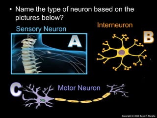
  • 1. • Name the type of neuron based on the pictures below? Sensory Neuron Interneuron Motor Neuron Copyright © 2010 Ryan P. Murphy
  • 2.
  • 6. • RED SLIDE: These are notes that are very important and should be recorded in your science journal. Copyright © 2010 Ryan P. Murphy
  • 7. -Nice neat notes that are legible and use indentations when appropriate. -Example of indent. -Skip a line between topics -Don’t skip pages -Make visuals clear and well drawn. Please label. Kidneys Ureters Urinary Bladder Copyright © 2010 Ryan P. Murphy
  • 8. • RED SLIDE: These are notes that are very important and should be recorded in your science journal. • BLACK SLIDE: Pay attention, follow directions, complete projects as described and answer required questions neatly. Copyright © 2010 Ryan P. Murphy
  • 9.
  • 10.
  • 11.
  • 12.
  • 13.
  • 14.
  • 15. • Keep an eye out for “The-Owl” and raise your hand as soon as you see him. – He will be hiding somewhere in the slideshow Copyright © 2010 Ryan P. Murphy
  • 17.  New Area of Focus: The Nervous System Copyright © 2010 Ryan P. Murphy
  • 18. • Everything we have learned so far, and everything you will ever learn takes place in the nervous system. Copyright © 2010 Ryan P. Murphy
  • 19.  The nervous system receives and then sends out information about your body.  It also monitors and responds to changes in your environment. Copyright © 2010 Ryan P. Murphy
  • 20. • Your brain receives vast amounts of information all of the time. Copyright © 2010 Ryan P. Murphy
  • 21. • Your brain receives vast amounts of information all of the time. – We will close our eyes for a second and rely on other messages your brain receives. Copyright © 2010 Ryan P. Murphy
  • 22. • Your brain receives vast amounts of information all of the time. – We will close our eyes for a second and rely on other messages your brain receives. – For the next thirty seconds be absolutely silent and be ready to report what you… Copyright © 2010 Ryan P. Murphy
  • 23. • Your brain receives vast amounts of information all of the time. – We will close our eyes for a second and rely on other messages your brain receives. – For the next thirty seconds be absolutely silent and be ready to report what you… Copyright © 2010 Ryan P. Murphy Feel Hear Smell Taste Dream Think
  • 24.
  • 25. • Who thought about keeping their heartbeat going? • Who thought about blinking? • Who thought about regulating hormones? • Who thought about breathing normal? Copyright © 2010 Ryan P. Murphy
  • 26. • Who thought about keeping their heartbeat going? • Who thought about blinking? • Who thought about their blood pressure? • Who thought about regulating their body temperature? • Who thought about regulating hormones? • Who thought about breathing normal? Copyright © 2010 Ryan P. Murphy
  • 27. • While you are using your nervous system for all of your senses, it’s working double controlling all of the things in your body to keep you living? Copyright © 2010 Ryan P. Murphy
  • 28. • Changes that are happening all of the time in your body and out are called stimuli. Copyright © 2010 Ryan P. Murphy
  • 29.
  • 30. • Activity! Ice Cube in your hand again. – Feel the immediate stimulus sent to your brain.
  • 31. • Activity Stimulus! Copyright © 2010 Ryan P. Murphy
  • 32. • Activity Stimulus! – Very slowly move your finger until it touches your eyelash.
  • 33. • Activity Stimulus! – Very slowly move your finger until it touches your eyelash. – A reflex action will cause your eye to blink.
  • 34. • Activity Stimulus! – Very slowly move your finger until it touches your eyelash. – A reflex action will cause your eye to blink.
  • 35. • Activity Stimulus! – You can now decide to flick the back of your neck with your with your thumb and middle finger causing pain. • This is a conscious voluntary action that you have control over. Copyright © 2010 Ryan P. Murphy
  • 36. • Activity Stimulus! – You can now decide to flick the back of your neck with your with your thumb and middle finger causing pain. • This is a conscious voluntary action that you have control over. Copyright © 2010 Ryan P. Murphy
  • 37. • Activity Stimulus! – You can now decide to flick the back of your neck with your with your thumb and middle finger causing pain. • This is a conscious voluntary action that you have control over. Copyright © 2010 Ryan P. Murphy
  • 38. • Activity Stimulus! – You can now decide to flick the back of your neck with your with your thumb and middle finger causing pain. • This is a conscious voluntary action that you have control over. Copyright © 2010 Ryan P. Murphy
  • 39. • The messages that are constantly traveling through your body are carried by the neuron or nerve cells.
  • 40.  Neuron: A specialized cell transmitting nerve impulses.  Electrical and chemical signaling. . Copyright © 2010 Ryan P. Murphy
  • 41.  Neuron: A specialized cell transmitting nerve impulses.  Electrical and chemical signaling. . Copyright © 2010 Ryan P. Murphy
  • 42. • Electrical signal: Changes + and – charges from one end of a neuron to another. Copyright © 2010 Ryan P. Murphy
  • 43. • Electrical signal: Changes + and – charges from one end of a neuron to another. Copyright © 2010 Ryan P. Murphy
  • 44. • Chemical signal: Chemicals allow signals to go from one neuron to another by “jumping the gap (synapse)”. Copyright © 2010 Ryan P. Murphy
  • 45. Copyright © 2010 Ryan P. Murphy
  • 46. Copyright © 2010 Ryan P. Murphy
  • 47. Copyright © 2010 Ryan P. Murphy
  • 48. Copyright © 2010 Ryan P. Murphy
  • 49. Copyright © 2010 Ryan P. Murphy
  • 50. Copyright © 2010 Ryan P. Murphy
  • 51. Copyright © 2010 Ryan P. Murphy
  • 52. Copyright © 2010 Ryan P. Murphy
  • 53. Copyright © 2010 Ryan P. Murphy
  • 54. Copyright © 2010 Ryan P. Murphy
  • 55. Copyright © 2010 Ryan P. Murphy
  • 56. Copyright © 2010 Ryan P. Murphy
  • 57. • Nervous System (A) Available Sheet.
  • 58. • Nervous System (A) Available Sheet.
  • 59. • Drawing a nerve cell / neuron step by step drawing in journal. Copyright © 2010 Ryan P. Murphy
  • 60. Copyright © 2010 Ryan P. Murphy Copyright © 2010 Ryan P. Murphy
  • 61. Copyright © 2010 Ryan P. Murphy Copyright © 2010 Ryan P. Murphy
  • 62. Cell Body Copyright © 2010 Ryan P. Murphy Copyright © 2010 Ryan P. Murphy
  • 63. Cell Body Copyright © 2010 Ryan P. Murphy Copyright © 2010 Ryan P. Murphy
  • 64. Cell Body Copyright © 2010 Ryan P. Murphy
  • 65. Cell Body Dendrites Copyright © 2010 Ryan P. Murphy
  • 67. Cell Body Dendrites Copyright © 2010 Ryan P. Murphy
  • 68. Cell Body Dendrites Copyright © 2010 Ryan P. Murphy
  • 69. Cell Body Dendrites Copyright © 2010 Ryan P. Murphy
  • 70. Cell Body Dendrites Copyright © 2010 Ryan P. Murphy
  • 73. Copyright © 2010 Ryan P. Murphy
  • 82. Cell Body Dendrites Myelin sheaths Axon terminals Axon 1 mm to over 1 meter in length Copyright © 2010 Ryan P. Murphy
  • 83. Cell Body Dendrites Myelin sheaths Axon terminals Axon 1 mm to over 1 meter in length Copyright © 2010 Ryan P. Murphy
  • 84. Cell Body Dendrites Myelin sheaths Axon terminals Axon 1 mm to over 1 meter in length Copyright © 2010 Ryan P. Murphy
  • 85. Cell Body Dendrites Myelin sheaths Axon terminals Axon 1 mm to over 1 meter in length Copyright © 2010 Ryan P. Murphy
  • 86. Cell Body Dendrites Myelin sheaths Axon terminals Axon 1 mm to over 1 meter in length Copyright © 2010 Ryan P. Murphy
  • 87. Cell Body Dendrites Myelin sheaths Axon terminals Axon 1 mm to over 1 meter in length Copyright © 2010 Ryan P. Murphy
  • 88. Cell Body Dendrites Myelin sheaths Axon terminals Axon 1 mm to over 1 meter in length Copyright © 2010 Ryan P. Murphy
  • 89. Cell Body Dendrites Myelin sheaths Axon terminals Axon 1 mm to over 1 meter in length Copyright © 2010 Ryan P. Murphy
  • 90. Cell Body Dendrites Myelin sheaths Axon terminals Axon 1 mm to over 1 meter in length Another Axon Neurotransmitters sent to receptors
  • 91. Cell Body Dendrites Myelin sheaths Axon terminals Axon 1 mm to over 1 meter in length Another Axon Neurotransmitters sent to receptors
  • 92. Cell Body Dendrites Myelin sheaths Axon terminals Axon 1 mm to over 1 meter in length Another Axon Neurotransmitters sent to receptors
  • 93. Cell Body Dendrites Myelin sheaths Axon terminals Axon 1 mm to over 1 meter in length Another Axon Neurotransmitters sent to receptors Copyright © 2010 Ryan P. Murphy
  • 94.
  • 97. Fingers are dendrites Hand is cell body Arm is axon
  • 98. Fingers are dendrites Hand is cell body Arm is axon “He’ll need a finely tuned nervous system to hit that shot.”
  • 99. Fingers are dendrites Hand is cell body Arm is axon
  • 100. • Activity! Build a Neuron and label the following. – Cell Body – Dendrites – Axon – Axon terminals. Copyright © 2010 Ryan P. Murphy
  • 101. • Some construction ideas. Copyright © 2010 Ryan P. Murphy
  • 102. • Some construction ideas. – Out of clay Copyright © 2010 Ryan P. Murphy
  • 103. • Some construction ideas. – Out of clay – Out of beads Copyright © 2010 Ryan P. Murphy
  • 104. • Some construction ideas. – Out of clay – Out of beads – Pipe cleaner Copyright © 2010 Ryan P. Murphy
  • 105. • Some construction ideas. – Out of clay – Out of beads – Pipe cleaner – Rope Neuron Copyright © 2010 Ryan P. Murphy
  • 106. • Some construction ideas. – Out of clay – Out of beads – Pipe cleaner – Rope Neuron – Compact disc and string • Holes in cd to attach dendrites Copyright © 2010 Ryan P. Murphy
  • 107. • Some construction ideas. (Others?) – Out of clay – Out of beads – Pipe cleaner – Rope Neuron – Compact disc and string • Holes in cd to attach dendrites Copyright © 2010 Ryan P. Murphy
  • 108. • You can complete this question.
  • 109.
  • 110.
  • 111. • Activity! Simulation of a neuron / Saltatory conduction. – Whole class is one neuron (myelinated axons) – Students stand at arms length from each other and form a winding line through classroom. – Teacher says “go” to start, and first student gently slaps hand on person next to them. – That person gently slaps the hand on the person next to them and so on down the axon. – Last person in line should toss an object into the air representing the signal going to another neuron. – Teacher will time you. Copyright © 2010 Ryan P. Murphy
  • 112. • Activity! Simulation of a neuron / Saltatory conduction. – Whole class is one neuron (myelinated axons) – Students stand at arms length from each other and form a winding line through classroom. – Teacher says “go” to start, and first student gently slaps hand on person next to them. – That person gently slaps the hand on the person next to them and so on down the axon. – Last person in line should toss an object into the air representing the signal going to another neuron. – Teacher will time you. Copyright © 2010 Ryan P. Murphy
  • 113. • Activity! Simulation of a neuron / Saltatory conduction. – Whole class is one neuron (myelinated axons) – Students stand at arms length from each other and form a winding line through classroom. – Teacher says “go” to start, and first student gently slaps hand on person next to them. – That person gently slaps the hand on the person next to them and so on down the axon. – Last person in line should toss an object into the air representing the signal going to another neuron. – Teacher will time you. Copyright © 2010 Ryan P. Murphy
  • 114. • Activity! Simulation of a neuron / Saltatory conduction. – Whole class is one neuron (myelinated axons) – Students stand at arms length from each other and form a winding line through classroom. – Teacher says “go” to start, and first student gently slaps hand on person next to them. – That person gently slaps the hand on the person next to them and so on down the axon. – Last person in line should toss an object into the air representing the signal going to another neuron. – Teacher will time you. Copyright © 2010 Ryan P. Murphy
  • 115. • Activity! Simulation of a neuron / Saltatory conduction. – Whole class is one neuron (myelinated axons) – Students stand at arms length from each other and form a winding line through classroom. – Teacher says “go” to start, and first student gently slaps hand on person next to them. – That person gently slaps the hand on the person next to them and so on down the axon. – Last person in line should toss an object into the air representing the signal going to another neuron. – Teacher will time you. Copyright © 2010 Ryan P. Murphy
  • 116. • Activity! Simulation of a neuron / Saltatory conduction. – Whole class is one neuron (myelinated axons) – Students stand at arms length from each other and form a winding line through classroom. – Teacher says “go” to start, and first student gently slaps hand on person next to them. – That person gently slaps the hand on the person next to them and so on down the axon. – Last person in line should toss an object into the air representing the signal going to another neuron. – Teacher will time you. Copyright © 2010 Ryan P. Murphy
  • 117. • Activity! Simulation of a neuron / Saltatory conduction. – Whole class is one neuron (myelinated axons) – Students stand at arms length from each other and form a winding line through classroom. – Teacher says “go” to start, and first student gently slaps hand on person next to them. – That person gently slaps the hand on the person next to them and so on down the axon. – Last person in line should toss an object into the air representing the signal going to another neuron. – Teacher will time you. Copyright © 2010 Ryan P. Murphy
  • 118.
  • 119.
  • 120.
  • 121.
  • 122. Down line until last person
  • 123. • Activity! Neurotransmitter. – Each student is a neuron / nerve cell. – Teacher passes out small object to each student that easily fits into hand. – Students should stand in a line at arms length from each other. (Line can curve around room). – Put object in left hand, have right hand open to accept object. – When teacher says go, students at the beginning of the line place their object (Chemical signal) into the dendrites of the student next to them. • That student then passes their object, and so on down the line. – We will time how fast it takes us. Our nervous system can do it in less than seconds. Visual on next slide. Copyright © 2010 Ryan P. Murphy
  • 124. • Activity! Neurotransmitter. – Each student is a neuron / nerve cell. – Teacher passes out small object to each student that easily fits into hand. – Students should stand in a line at arms length from each other. (Line can curve around room). – Put object in left hand, have right hand open to accept object. – When teacher says go, students at the beginning of the line place their object (Chemical signal) into the dendrites of the student next to them. • That student then passes their object, and so on down the line. – We will time how fast it takes us. Our nervous system can do it in less than seconds. Visual on next slide. Copyright © 2010 Ryan P. Murphy
  • 125. • Activity! Neurotransmitter. – Each student is a neuron / nerve cell. – Teacher passes out small object to each student that easily fits into hand. – Students should stand in a line at arms length from each other. (Line can curve around room). – Put object in left hand, have right hand open to accept object. – When teacher says go, students at the beginning of the line place their object (Chemical signal) into the dendrites of the student next to them. • That student then passes their object, and so on down the line. – We will time how fast it takes us. Our nervous system can do it in less than seconds. Visual on next slide. Copyright © 2010 Ryan P. Murphy
  • 126. • Activity! Neurotransmitter. – Each student is a neuron / nerve cell. – Teacher passes out small object to each student that easily fits into hand. – Students should stand in a line at arms length from each other. (Line can curve around room). – Put object in left hand, have right hand open to accept object. – When teacher says go, students at the beginning of the line place their object (Chemical signal) into the dendrites of the student next to them. • That student then passes their object, and so on down the line. – We will time how fast it takes us. Our nervous system can do it in less than seconds. Visual on next slide. Copyright © 2010 Ryan P. Murphy
  • 127. • Activity! Neurotransmitter. – Each student is a neuron / nerve cell. – Teacher passes out small object to each student that easily fits into hand. – Students should stand in a line at arms length from each other. (Line can curve around room). – Put object in left hand, have right hand open to accept object. – When teacher says go, students at the beginning of the line place their object (Chemical signal) into the dendrites of the student next to them. • That student then passes their object, and so on down the line. – We will time how fast it takes us. Our nervous system can do it in less than seconds. Visual on next slide. Copyright © 2010 Ryan P. Murphy
  • 128. • Activity! Neurotransmitter. – Each student is a neuron / nerve cell. – Teacher passes out small object to each student that easily fits into hand. – Students should stand in a line at arms length from each other. (Line can curve around room). – Put object in left hand, have right hand open to accept object. – When teacher says go, students at the beginning of the line place their object (Chemical signal) into the dendrites of the student next to them. • That student then passes their object, and so on down the line. – We will time how fast it takes us. Our nervous system can do it in less than seconds. Visual on next slide. Copyright © 2010 Ryan P. Murphy
  • 129. • Activity! Neurotransmitter. – Each student is a neuron / nerve cell. – Teacher passes out small object to each student that easily fits into hand. – Students should stand in a line at arms length from each other. (Line can curve around room). – Put object in left hand, have right hand open to accept object. – When teacher says go, students at the beginning of the line place their object (Chemical signal) into the dendrites of the student next to them. • That student then passes their object, and so on down the line. – We will time how fast it takes us. Our nervous system can do it in less than seconds. Visual on next slide. Copyright © 2010 Ryan P. Murphy
  • 130. Copyright © 2010 Ryan P. Murphy
  • 131. Copyright © 2010 Ryan P. Murphy
  • 132. Copyright © 2010 Ryan P. Murphy Copyright © 2010 Ryan P. Murphy
  • 133.
  • 134. Copyright © 2010 Ryan P. Murphy
  • 135. Copyright © 2010 Ryan P. Murphy
  • 136. Copyright © 2010 Ryan P. Murphy
  • 137. Copyright © 2010 Ryan P. Murphy
  • 138. Copyright © 2010 Ryan P. Murphy
  • 139. Copyright © 2010 Ryan P. Murphy
  • 140. Copyright © 2010 Ryan P. Murphy
  • 141. Copyright © 2010 Ryan P. Murphy
  • 142. Copyright © 2010 Ryan P. Murphy
  • 143. Copyright © 2010 Ryan P. Murphy
  • 144. Copyright © 2010 Ryan P. Murphy
  • 145. Copyright © 2010 Ryan P. Murphy
  • 146. Copyright © 2010 Ryan P. Murphy
  • 147. Copyright © 2010 Ryan P. Murphy
  • 148. Copyright © 2010 Ryan P. Murphy
  • 149. Copyright © 2010 Ryan P. Murphy
  • 150. Copyright © 2010 Ryan P. Murphy
  • 151. • Nervous System (A) Available Sheet.
  • 152. • There are three types of neurons. Copyright © 2010 Ryan P. Murphy
  • 153. • There are three types of neurons. – Sensory neurons Copyright © 2010 Ryan P. Murphy
  • 154. • There are three types of neurons. – Sensory neurons Copyright © 2010 Ryan P. Murphy
  • 155. • There are three types of neurons. – Sensory neurons – Interneurons Copyright © 2010 Ryan P. Murphy
  • 156. • There are three types of neurons. – Sensory neurons – Interneurons Copyright © 2010 Ryan P. Murphy
  • 157. • There are three types of neurons. – Sensory neurons – Interneurons – Motor neurons Copyright © 2010 Ryan P. Murphy
  • 158. • There are three types of neurons. – Sensory neurons – Interneurons – Motor neurons Copyright © 2010 Ryan P. Murphy
  • 159. • Interneuron: Transmits impulses between other neurons. (Brain and Spinal Column) Copyright © 2010 Ryan P. Murphy
  • 160. • Sensory neuron: Conducts impulses inwards to the brain or spinal cord. Copyright © 2010 Ryan P. Murphy
  • 161. • Sensory neuron: Conducts impulses inwards to the brain or spinal cord. • touch • odor • taste • sound • vision Copyright © 2010 Ryan P. Murphy
  • 162. • Motor Neurons: Pathway along which impulses pass from the brain or spinal cord to a muscle or gland. Copyright © 2010 Ryan P. Murphy
  • 163.
  • 164. • Name the type of neuron based on the pictures below? Copyright © 2010 Ryan P. Murphy
  • 165. • Name the type of neuron based on the pictures below? Copyright © 2010 Ryan P. Murphy
  • 166. • Name the type of neuron based on the pictures below? Sensory Neuron Copyright © 2010 Ryan P. Murphy
  • 167. • Name the type of neuron based on the pictures below? Sensory Neuron Copyright © 2010 Ryan P. Murphy
  • 168. • Name the type of neuron based on the pictures below? Sensory Neuron Interneuron Copyright © 2010 Ryan P. Murphy
  • 169. • Name the type of neuron based on the pictures below? Sensory Neuron Interneuron Copyright © 2010 Ryan P. Murphy
  • 170. • Name the type of neuron based on the pictures below? Sensory Neuron Interneuron Motor Neuron Copyright © 2010 Ryan P. Murphy
  • 171.
  • 172. • Which one directs signals inward toward the spinal column? Sensory Neuron Interneuron Motor Neuron Copyright © 2010 Ryan P. Murphy
  • 173. • Which one directs signals inward toward the spinal column? Sensory Neuron Interneuron Motor Neuron Copyright © 2010 Ryan P. Murphy
  • 174.
  • 175. • Which one transmits impulses from neurons to neurons? Sensory Neuron Interneuron Motor Neuron Copyright © 2010 Ryan P. Murphy
  • 176. • Which one transmits impulses from neurons to neurons? Sensory Neuron Interneuron Motor Neuron Copyright © 2010 Ryan P. Murphy
  • 177.
  • 178. • Which one is a Pathway along which impulses pass from the brain or spinal cord to a muscle or gland? Sensory Neuron Interneuron Motor Neuron Copyright © 2010 Ryan P. Murphy
  • 179. • Which one is a Pathway along which impulses pass from the brain or spinal cord to a muscle or gland? Sensory Neuron Interneuron Motor Neuron Copyright © 2010 Ryan P. Murphy
  • 180. “Oh-no!” “My neurons are telling me we are trying it one more time.”
  • 181. • Name the type of neuron based on the pictures below? Copyright © 2010 Ryan P. Murphy
  • 182. • Name the type of neuron based on the pictures below? Copyright © 2010 Ryan P. Murphy
  • 183. • Name the type of neuron based on the pictures below? Sensory Neuron Copyright © 2010 Ryan P. Murphy
  • 184. • Name the type of neuron based on the pictures below? Sensory Neuron Copyright © 2010 Ryan P. Murphy
  • 185. • Name the type of neuron based on the pictures below? Sensory Neuron Interneuron Copyright © 2010 Ryan P. Murphy
  • 186. • Name the type of neuron based on the pictures below? Sensory Neuron Interneuron Copyright © 2010 Ryan P. Murphy
  • 187. • Name the type of neuron based on the pictures below? Sensory Neuron Interneuron Motor Neuron Copyright © 2010 Ryan P. Murphy
  • 188. • Receptors: Cells that receive messages from your surroundings. Copyright © 2010 Ryan P. Murphy
  • 189. • Receptors: Cells that receive messages from your surroundings. Receptor Cell  Copyright © 2010 Ryan P. Murphy
  • 190. • Receptors: Cells that receive messages from your surroundings. Receptor Cell  Interneurons  Brain  Neurons  Effector Cell. Copyright © 2010 Ryan P. Murphy
  • 191. • Receptors: Cells that receive messages from your surroundings. Receptor Cell  Interneurons  Brain  Interneurons  Effector Cell. Copyright © 2010 Ryan P. Murphy
  • 192. • Receptors: Cells that receive messages from your surroundings. Receptor Cell  Interneurons  Brain  Interneurons  Copyright © 2010 Ryan P. Murphy
  • 193. • Receptors: Cells that receive messages from your surroundings. Receptor Cell  Interneurons  Brain  Interneurons  Effector Cell. Copyright © 2010 Ryan P. Murphy
  • 194. • Effectors: Cell that gets stimulated by a neuron (Muscle cell) Copyright © 2010 Ryan P. Murphy
  • 195. • You can complete this question.
  • 196. • You can complete this question. Learn more about neurons at… http://faculty.washington.edu/chudler/cel ls.html
  • 197.
  • 198.  The Central Nervous System: Brain and Spinal Cord  Control center of the body. Copyright © 2010 Ryan P. Murphy
  • 199.  The Central Nervous System: Brain and Spinal Cord  Control center of the body. Copyright © 2010 Ryan P. Murphy
  • 200.  The Central Nervous System: Brain and Spinal Cord  Control center of the body.  Peripheral Nervous System: Network of nerves throughout body. Copyright © 2010 Ryan P. Murphy
  • 201.  The Central Nervous System: Brain and Spinal Cord  Control center of the body.  Peripheral Nervous System: Network of nerves throughout body. Copyright © 2010 Ryan P. Murphy
  • 202. • You can now complete this question.
  • 203.
  • 204. • Activity! The connectivity of the brain (Interneurons). – The brain is an amazing organ that makes many connections with other cells. – Let’s understand this power with a little exercise with twenty brain cells. – An average brain may have 80-90 billion cells. – Make ten dots on each side of your page – (Please be organized and space them out so they match) – Draw line from the cell (dot on the right) to all of the dots (cells) on the left. Copyright © 2010 Ryan P. Murphy
  • 205. • Activity! The connectivity of the brain (Interneurons). (Optional) – The brain is an amazing organ that makes many connections with other cells. – Let’s understand this power with a little exercise with twenty brain cells. – An average brain may have 80-90 billion cells. – Make ten dots on each side of your page – (Please be organized and space them out so they match) – Draw line from the cell (dot on the right) to all of the dots (cells) on the left. Copyright © 2010 Ryan P. Murphy
  • 206. • Activity! The connectivity of the brain (Interneurons). – The brain is an amazing organ that makes many connections with other cells. – Let’s understand this power with a little exercise with twenty brain cells. – An average brain may have 80-90 billion cells. – Make ten dots on each side of your page – (Please be organized and space them out so they match) – Draw line from the cell (dot on the right) to all of the dots (cells) on the left. Copyright © 2010 Ryan P. Murphy
  • 207. • Activity! The connectivity of the brain (Interneurons). – The brain is an amazing organ that makes many connections with other cells. – Let’s understand this power with a little exercise with twenty brain cells. – An average brain may have 80-90 billion cells. – Make ten dots on each side of your page – (Please be organized and space them out so they match) – Draw line from the cell (dot on the right) to all of the dots (cells) on the left. Copyright © 2010 Ryan P. Murphy
  • 208. • Activity! The connectivity of the brain (Interneurons). – The brain is an amazing organ that makes many connections with other cells. – Let’s understand this power with a little exercise with twenty brain cells. – An average brain may have 80-90 billion cells. – Make ten dots on each side of your page – (Please be organized and space them out so they match) – Draw line from the cell (dot on the right) to all of the dots (cells) on the left. Copyright © 2010 Ryan P. Murphy
  • 209. • Activity! The connectivity of the brain (Interneurons). – The brain is an amazing organ that makes many connections with other cells. – Let’s understand this power with a little exercise with twenty brain cells. – An average brain may have 80-90 billion cells. – Make ten dots on each side of your page – (Please be organized and space them out so they match) – Draw line from the cell (dot on the right) to all of the dots (cells) on the left. Copyright © 2010 Ryan P. Murphy
  • 210. • Activity! The connectivity of the brain (Interneurons). – The brain is an amazing organ that makes many connections with other cells. – Let’s understand this power with a little exercise with twenty brain cells. – An average brain may have 80-90 billion cells. – Make ten dots on each side of your page – (Please be organized and space them out so they match) – Draw line from the cell (dot on the right) to all of the dots (cells) on the left. Copyright © 2010 Ryan P. Murphy
  • 211. • Activity! The connectivity of the brain (Interneurons). – The brain is an amazing organ that makes many connections with other cells. – Let’s understand this power with a little exercise with twenty brain cells. – An average brain may have 80-90 billion cells. – Make ten dots on each side of your page – (Please be organized and space them out so they match) – Draw line from the cell (dot on the right) to all of the dots (cells) on the left. Copyright © 2010 Ryan P. Murphy
  • 212.
  • 213.
  • 214.
  • 215.
  • 216.
  • 217.
  • 218.
  • 219.
  • 220.
  • 221.
  • 222.
  • 223.
  • 224.
  • 225.
  • 226.
  • 227. Copyright © 2010 Ryan P. Murphy Neurons, Synapsis, Memories, and learn more at… http://www.human- memory.net/brain_neurons.html
  • 228.
  • 229.
  • 230.
  • 231. Copyright © 2010 Ryan P. Murphy
  • 232. • Central Nervous System is very complex. Your body is adjusting to constant change. Copyright © 2010 Ryan P. Murphy
  • 233. • Central Nervous System is very complex. Your body is adjusting to constant change. – On the next slide your central nervous system will adjust to the amount of light that enters the retina. Copyright © 2010 Ryan P. Murphy
  • 234.
  • 235.
  • 236.
  • 237. • All of the messages that are constantly being sent in your body are interpreted in the central nervous system. Copyright © 2010 Ryan P. Murphy
  • 238. • The Brain: An organ of soft nervous tissue contained in the skull of vertebrates, functioning as the coordinating center of sensation and intellectual and nervous activity. Copyright © 2010 Ryan P. Murphy
  • 239. • The brain is well protected by the skull. – The brain is also covered in three layers of connective tissue which nourish and protect. Copyright © 2010 Ryan P. Murphy
  • 240. • The brain is well protected by the skull. – The brain is also covered in three layers of connective tissue which nourish and protect. Copyright © 2010 Ryan P. Murphy
  • 241. • The brain is well protected by the skull. – The brain is also covered in three layers of connective tissue which nourish and protect. Copyright © 2010 Ryan P. Murphy
  • 242. • The brain is well protected by the skull. – The brain is also covered in three layers of connective tissue which nourish and protect. Copyright © 2010 Ryan P. Murphy
  • 243. • The brain is well protected by the skull. – The brain is also covered in three layers of connective tissue which nourish and protect. Copyright © 2010 Ryan P. Murphy
  • 244.
  • 245. • Thick outer layer that comes in contact with the skull. • Watery layer cushion brain • Inner layer clings to the surface of the brain.
  • 246. • Thick outer layer that comes in contact with the skull. • Watery layer cushions brain • Inner layer clings to the surface of the brain.
  • 247. • Thick outer layer that comes in contact with the skull. • Watery layer cushions brain • Inner layer clings to the surface of the brain.
  • 248. • Thick outer layer that comes in contact with the skull. • Watery layer cushions brain • Inner layer clings to the surface of the brain. Copyright © 2010 Ryan P. Murphy
  • 249. • Nervous System (A) Available Sheet.
  • 250. • Activity! How a watery layer (cerebrospinal fluid (CSF) aids in cushioning the brain from impacts. Copyright © 2010 Ryan P. Murphy
  • 251. • Activity! How a watery layer (cerebrospinal fluid (CSF) aids in cushioning the brain from impacts. – Draw a face on two raw eggs. Copyright © 2010 Ryan P. Murphy
  • 252. • Activity! How a watery layer (cerebrospinal fluid (CSF) aids in cushioning the brain from impacts. – Draw a face on two raw eggs. – Place one in a clear container with sealing lid slightly larger than the egg. (Shake five times increasing in strength – Observe after each shake) Copyright © 2010 Ryan P. Murphy
  • 253. • Activity! How a watery layer (cerebrospinal fluid (CSF) aids in cushioning the brain from impacts. – Draw a face on two raw eggs. – Place one in a clear container with sealing lid slightly larger than the egg. (Shake five times increasing in strength – Observe after each shake) – Place the other egg in the same container. This time fill the container with water. Repeat shaking process and make a conclusion about (cerebrospinal fluid (CSF). Copyright © 2010 Ryan P. Murphy
  • 254. Copyright © 2010 Ryan P. Murphy
  • 255. Copyright © 2010 Ryan P. Murphy
  • 256. Copyright © 2010 Ryan P. Murphy
  • 257. • You can now complete this question.
  • 258.
  • 259. • Build a Brain Recipe Provided Sheet.
  • 260. • Activity! Building a Brain. • The brain should be about 3 lbs. (1.35 kg.) and feel like a real brain. – 1 gallon ZipLock Bag – Add 1.5 cups (360 ml) instant potato flakes. – Add 2.5 cup (600 ml) hot water – Add 2 cups (480 ml) clean sand Copyright © 2010 Ryan P. Murphy
  • 261. • Build a Brain (More difficult) – 2 cups water – 2 cups flour – 4 teaspoons cream of tartar – 1 cup salt – One quarter cup vegetable oil – Cook over low heat until lumpy and then let cool. – Use hands to mold into a brain. Mix first Add in after other ingredients are well mixed Copyright © 2010 Ryan P. Murphy
  • 262. • Build a Brain (More difficult) – 2 cups water – 2 cups flour – 4 teaspoons cream of tartar – 1 cup salt – One quarter cup vegetable oil – Cook over low heat until lumpy and then let cool. – Use hands to mold into a brain. Mix first Add in after other ingredients are well mixed Use toothpicks and masking tape to create signs for the lobes of the brain on the next slide. Copyright © 2010 Ryan P. Murphy
  • 263.
  • 264. • Parts of the Brain
  • 265. • Nervous System (A) Available Sheet.
  • 266. • Step by step drawing of the brain. – Do not make brain a whole page as you will need to put text around it. Copyright © 2010 Ryan P. Murphy
  • 267.
  • 268.
  • 269.
  • 270.
  • 281. Cerebrum Corpus Callosum Thalmus Cerebellum Medulla Spinal Cord Folds and wrinkles help increase surface area Learning, Intelligence, emotions, personality, Judgment, and all voluntary activities of your body.
  • 282. Cerebrum Corpus Callosum Thalmus Cerebellum Medulla Spinal Cord Folds and wrinkles help increase surface area Learning, Intelligence, emotions, personality, Judgment, and all voluntary activities of your body.
  • 283. Cerebrum Corpus Callosum Thalmus Cerebellum Medulla Spinal Cord Folds and wrinkles help increase surface area Learning, Intelligence, emotions, personality, Judgment, and all voluntary activities of your body. Copyright © 2010 Ryan P. Murphy
  • 284.
  • 285.
  • 286.
  • 287.
  • 289. Big Picture- Record in your journal if you think you are more left, or more right brained.
  • 290. • Nervous System (A) Available Sheet.
  • 291. • Activity! Take a left or right brain test. – (Keyword Search: left brain right brain quiz) – http://www.web- us.com/brain/braindominance.htm – http://www.intelliscript.net/test_area/questionnair e/questionnaire.cgi Copyright © 2010 Ryan P. Murphy
  • 292. • Do you see the dancer turning clockwise or anti-clockwise on the next slide? – If clockwise, then you use more of the right side of the brain. – If counterclockwise, then you use more of the left side of your brain. – I apologize that this image is risqué. Copyright © 2010 Ryan P. Murphy
  • 293.
  • 294.
  • 295.
  • 298. Cerebrum Corpus Callosum Thalmus Cerebellum Medulla Spinal Cord Folds and wrinkles help increase surface area Learning, Intelligence, emotions, personality, Judgment, and all voluntary activities of your body.
  • 299. Cerebrum Corpus Callosum Thalmus Cerebellum Medulla Spinal Cord Folds and wrinkles help increase surface area Learning, Intelligence, emotions, personality, Judgment, and all voluntary activities of your body. Medulla connects brain to spinal column and controls all involuntary activities.
  • 300. Cerebrum Corpus Callosum Thalmus Cerebellum Medulla Spinal Cord Folds and wrinkles help increase surface area Learning, Intelligence, emotions, personality, Judgment, and all voluntary activities of your body. Medulla connects brain to spinal column and controls all involuntary activities. 33 Vertebrae bones protect the spinal cord that carries impulses to and from body.
  • 301.
  • 302.
  • 303.
  • 304.
  • 305.
  • 306.
  • 307.
  • 308. • Activity! Making a spinal column. • Each table group uses empty spools of thread or other cylinders to create a spinal column. – String is nerves. – Columns are the vertebrae – Info and visual on next slide. Copyright © 2010 Ryan P. Murphy
  • 309. • Spinal column. • Note how final spinal column is flexible. • 31 segments and 33 bones – 7 cervical vertebrae. – 12 thoracic. – 5 lumbar. – 5 sacral – 4 coccygeal Copyright © 2010 Ryan P. Murphy
  • 310.
  • 311.
  • 312.
  • 313.
  • 314.
  • 315. The curves of your spine are important because they allow the spine to support more weight than if it were straight.
  • 316. The curves of your spine are important because they allow the spine to support more weight than if it were straight. Also allows for compression.
  • 317.
  • 318. • Your vertebrae protect your spinal cord but are not indes t r u c t i b l e. Copyright © 2010 Ryan P. Murphy
  • 319. Copyright © 2010 Ryan P. Murphy
  • 320. • Image of cracked spinal column and severed spinal cord.
  • 321. • Paralysis: Inability to move or function; total stoppage or severe impairment of activity
  • 322.
  • 323.
  • 324.
  • 325.
  • 326.
  • 327.
  • 328.
  • 329.
  • 330. • Again! Please wear your seatbelt. Copyright © 2010 Ryan P. Murphy
  • 331. • Again! Please wear your seatbelt. – Besides possibly saving you from TBI (Traumatic Brain Injury).
  • 332. • Again! Please wear your seatbelt. – Besides possibly saving you from TBI (Traumatic Brain Injury). Copyright © 2010 Ryan P. Murphy
  • 333. • Again! Please wear your seatbelt. – Besides possibly saving you from TBI (Traumatic Brain Injury). – It can also possibly save you from serious and life altering spinal cord injury. Copyright © 2010 Ryan P. Murphy
  • 334. • Video Link! Spinal Cord and Spinal Cord Injury. – http://www.youtube.com/watch?v=zxpb1- okVig&feature=relmfu Learn more about spinal cord injuries at… http://www.apparelyzed.com/spinal_cord_injury.html
  • 335. Cerebrum Corpus Callosum Thalmus Cerebellum Medulla Spinal Cord Folds and wrinkles help increase surface area Learning, Intelligence, emotions, personality, Judgment, and all voluntary activities of your body. Medulla connects brain to spinal column and controls all involuntary activities. 33 Vertebrae bones protect the spinal cord that carries impulses to and from body.
  • 336. Cerebrum Corpus Collosum Thalmus Cerebellum Medulla Spinal Cord Folds and wrinkles help increase surface area Learning, Intelligence, emotions, personality, Judgment, and all voluntary activities of your body. Medulla connects brain to spinal column and controls all involuntary activities. 33 Vertebrae bones protect the spinal cord that carries impulses to and from body.
  • 337. • Thalmus: Lobed mass of grey matter buried under the cerebral cortex. It is involved in sensory perception and regulation of motor functions.
  • 338. • Thalmus: Lobed mass of grey matter buried under the cerebral cortex. It is involved in sensory perception and regulation of motor functions. – Also controls sleep and awake consciousness.
  • 339. Cerebrum Corpus Callosum Thalmus Cerebellum Medulla Spinal Cord Folds and wrinkles help increase surface area Learning, Intelligence, emotions, personality, Judgment, and all voluntary activities of your body. Medulla connects brain to spinal column and controls all involuntary activities. 33 Vertebrae bones protect the spinal cord that carries impulses to and from body.
  • 340. • Corpus Callosum: Thick band of nerve fibers that divides the cerebrum into left and right hemispheres. Copyright © 2010 Ryan P. Murphy
  • 341. • Corpus Callosum: Thick band of nerve fibers that divides the cerebrum into left and right hemispheres. – Allows communication between both hemispheres. Copyright © 2010 Ryan P. Murphy
  • 342. • Visual information that we see on the left gets processed by the right hemisphere. Copyright © 2010 Ryan P. Murphy
  • 343. • Visual information that we see on the left gets processed by the right hemisphere. • Information on the right gets processed by the left hemisphere. Copyright © 2010 Ryan P. Murphy
  • 344. • Visual information that we see on the left gets processed by the right hemisphere. • Information on the right gets processed by the left hemisphere. – The neurons are "crossed" Copyright © 2010 Ryan P. Murphy
  • 345. • Activity! Seeing the mini hot dog finger. • Touch your pointer fingers together in front of your eye.
  • 346. • Activity! Seeing the mini hot dog finger. • Touch your pointer fingers together in front of your eye. • You should see this at some point.
  • 347. Cerebrum Corpus Callosum Thalmus Cerebellum Medulla Spinal Cord Folds and wrinkles help increase surface area Learning, Intelligence, emotions, personality, Judgment, and all voluntary activities of your body. Medulla connects brain to spinal column and controls all involuntary activities. 33 Vertebrae bones protect the spinal cord that carries impulses to and from body.
  • 348. Cerebrum Corpus Callosum Thalmus Cerebellum Medulla Spinal Cord Folds and wrinkles help increase surface area Learning, Intelligence, emotions, personality, Judgment, and all voluntary activities of your body. Medulla connects brain to spinal column and controls all involuntary activities. 33 Vertebrae bones protect the spinal cord that carries impulses to and from body. Controls motor movement, coordination, balance.
  • 349. • You can now complete this question.
  • 350.
  • 351. • Activity! Messing with the cerebellum. – Teacher places a piece of tape on the floor for several meters. – Students try of walk the line looking through the wrong end of binoculars. Copyright © 2010 Ryan P. Murphy
  • 352. • Sense Organs: They respond to changes in light, sound, heat, pressure, and chemicals. Copyright © 2010 Ryan P. Murphy
  • 353. • Sense Organs: They respond to changes in light, sound, heat, pressure, and chemicals. • Some common sense organs Copyright © 2010 Ryan P. Murphy
  • 354. • Sense Organs: They respond to changes in light, sound, heat, pressure, and chemicals. • Some common sense organs
  • 355. • Sense Organs: They respond to changes in light, sound, heat, pressure, and chemicals. • Some common sense organs
  • 356. • Sense Organs: They respond to changes in light, sound, heat, pressure, and chemicals. • Some common sense organs
  • 357. • Sense Organs: They respond to changes in light, sound, heat, pressure, and chemicals. • Some common sense organs
  • 358. • Sense Organs: They respond to changes in light, sound, heat, pressure, and chemicals. • Some common sense organs
  • 359. • Can anyone name the mystery actor below?
  • 360. • Can anyone name the mystery actor below? Copyright © 2010 Ryan P. Murphy
  • 361. • Can anyone name the mystery actor below?
  • 362. • Can anyone name the mystery actor below?
  • 363. • Can anyone name the mystery actor below? Owen Wilson
  • 364.
  • 365.
  • 366.
  • 367.
  • 368.
  • 369.
  • 370. • Ben Stiller and Owen Wilson have appeared in 6 movies together. Copyright © 2010 Ryan P. Murphy
  • 371. • Ben Stiller and Owen Wilson have appeared in 6 movies together. Copyright © 2010 Ryan P. Murphy
  • 372. • Ben Stiller and Owen Wilson have appeared in 6 movies together. Copyright © 2010 Ryan P. Murphy
  • 373. • Ben Stiller and Owen Wilson have appeared in 6 movies together. Copyright © 2010 Ryan P. Murphy
  • 374. • Ben Stiller and Owen Wilson have appeared in 6 movies together. Copyright © 2010 Ryan P. Murphy
  • 375. • Ben Stiller and Owen Wilson have appeared in 6 movies together. Copyright © 2010 Ryan P. Murphy
  • 376. • Ben Stiller and Owen Wilson have appeared in 6 movies together.
  • 377. • Ben Stiller and Owen Wilson have appeared in 6 movies together.
  • 378. • Ben Stiller and Owen Wilson have appeared in 6 movies together.
  • 379.
  • 380.
  • 381. • How are you doing on your bundle. – You should be close to page 25.
  • 382. • Nervous System (A) Available Sheet.
  • 383. Copyright © 2010 Ryan P. Murphy
  • 384. Copyright © 2010 Ryan P. Murphy
  • 385. Copyright © 2010 Ryan P. Murphy
  • 386. Copyright © 2010 Ryan P. Murphy
  • 387. • Frontal Lobe- associated with reasoning, planning, parts of speech, movement, emotions, and problem solving Copyright © 2010 Ryan P. Murphy
  • 388. • Frontal Lobe- associated with reasoning, planning, parts of speech, movement, emotions, and problem solving Copyright © 2010 Ryan P. Murphy
  • 389. • Frontal Lobe- associated with reasoning, planning, parts of speech, movement, emotions, and problem solving • Parietal Lobe- associated with movement, orientation, recognition, perception of stimuli • Occipital Lobe- associated with visual processing • Temporal Lobe- associated with perception and recognition of auditory stimuli, memory, and speech Copyright © 2010 Ryan P. Murphy
  • 390. • Frontal Lobe- associated with reasoning, planning, parts of speech, movement, emotions, and problem solving • Parietal Lobe- associated with movement, orientation, recognition, perception of stimuli • Occipital Lobe- associated with visual processing • Temporal Lobe- associated with perception and recognition of auditory stimuli, memory, and speech Copyright © 2010 Ryan P. Murphy
  • 391. • Frontal Lobe- associated with reasoning, planning, parts of speech, movement, emotions, and problem solving • Parietal Lobe- associated with movement, orientation, recognition, perception of stimuli • Occipital Lobe- associated with visual processing • Temporal Lobe- associated with perception and recognition of auditory stimuli, memory, and speech Copyright © 2010 Ryan P. Murphy
  • 392. • Frontal Lobe- associated with reasoning, planning, parts of speech, movement, emotions, and problem solving • Parietal Lobe- associated with movement, orientation, recognition, perception of stimuli • Occipital Lobe- associated with visual processing • Temporal Lobe- associated with perception and recognition of auditory stimuli, memory, and speech Copyright © 2010 Ryan P. Murphy
  • 393. • Frontal Lobe- associated with reasoning, planning, parts of speech, movement, emotions, and problem solving • Parietal Lobe- associated with movement, orientation, recognition, perception of stimuli • Occipital Lobe- associated with visual processing • Temporal Lobe- associated with perception and recognition of auditory stimuli, memory, and speech Copyright © 2010 Ryan P. Murphy
  • 394. • Frontal Lobe- associated with reasoning, planning, parts of speech, movement, emotions, and problem solving • Parietal Lobe- associated with movement, orientation, recognition, perception of stimuli • Occipital Lobe- associated with visual processing • Temporal Lobe- associated with perception and recognition of auditory stimuli, memory, and speech Copyright © 2010 Ryan P. Murphy Learn more about the lobes of the brain at… http://faculty.washington.edu/chudler/lob e.html
  • 395. • You can now complete this question.
  • 396.
  • 397. • Nervous System (A) Available Sheet.
  • 398. • Functions of the Brain step by step drawing. Copyright © 2010 Ryan P. Murphy
  • 399.
  • 400. Gently sketch folds, it doesn’t need to be exact.
  • 401.
  • 402.
  • 403.
  • 404. • When you bang the back of you head you often see stars. Copyright © 2010 Ryan P. Murphy
  • 405. • When you bang the back of you head you often see stars. – These stars are the firing of neurons, which is interpreted by the brain's visual cortex as quick flashes of light. Copyright © 2010 Ryan P. Murphy
  • 406.
  • 407.
  • 408.
  • 409. • Eye: Organ that detects light and converts it to electro-chemical impulses in neurons. Copyright © 2010 Ryan P. Murphy
  • 410. • The eye requires a brain to interpret these electrochemical impulses. Copyright © 2010 Ryan P. Murphy
  • 411. • Nervous System (A) Available Sheet.
  • 412. • A quick step by step drawing of an eye. – Place eye on next page beneath your brain. Copyright © 2010 Ryan P. Murphy
  • 413. • A quick step by step drawing of an eye. – Place eye on next page beneath your brain. Copyright © 2010 Ryan P. Murphy Use pencil because you will need to erase parts of the circle. -or use very light pen
  • 414. • A quick step by step drawing of an eye. – Place eye on next page beneath your brain.
  • 415. • A quick step by step drawing of an eye. – Place eye on next page beneath your brain.
  • 416. • A quick step by step drawing of an eye. – Place eye on next page beneath your brain.
  • 417. • A quick step by step drawing of an eye. – Place eye on next page beneath your brain.
  • 418. • A quick step by step drawing of an eye. – Place eye on next page beneath your brain.
  • 419. • A quick step by step drawing of an eye. – Place eye on next page beneath your brain.
  • 420. • A quick step by step drawing of an eye. – Place eye on next page beneath your brain.
  • 421. • A quick step by step drawing of an eye. – Place eye on next page beneath your brain.
  • 422. • A quick step by step drawing of an eye. – Place eye on next page beneath your brain.
  • 423. • A quick step by step drawing of an eye. – Place eye on next page beneath your brain.
  • 424. • A quick step by step drawing of an eye. – Place eye on next page beneath your brain.
  • 425. • A quick step by step drawing of an eye. – Place eye on next page beneath your brain.
  • 426. • A quick step by step drawing of an eye. – Place eye on next page beneath your brain.
  • 427. • A quick step by step drawing of an eye. – Place eye on next page beneath your brain. Tear Duct
  • 428. • A quick step by step drawing of an eye. – Place eye on next page beneath your brain. Tear Duct The tear glands protect and water your eyes by washing away unwanted particles.
  • 429. • Nervous System (A) Available Sheet.
  • 430. • A quick step by step drawing of an eye. – Place next to other eye drawing.
  • 431. • A quick step by step drawing of an eye. – Place next to other eye drawing. Copyright © 2010 Ryan P. Murphy
  • 432. • A quick step by step drawing of an eye. – Place next to other eye drawing. Copyright © 2010 Ryan P. Murphy
  • 433. • A quick step by step drawing of an eye. – Place next to other eye drawing. Copyright © 2010 Ryan P. Murphy
  • 434. • A quick step by step drawing of an eye. – Place next to other eye drawing. Copyright © 2010 Ryan P. Murphy
  • 435. • A quick step by step drawing of an eye. – Place next to other eye drawing. Copyright © 2010 Ryan P. Murphy
  • 436. • A quick step by step drawing of an eye. – Place next to other eye drawing. Copyright © 2010 Ryan P. Murphy
  • 437. • A quick step by step drawing of an eye. – Place next to other eye drawing. Copyright © 2010 Ryan P. Murphy
  • 438. • A quick step by step drawing of an eye. – Place next to other eye drawing. Copyright © 2010 Ryan P. Murphy
  • 439. • A quick step by step drawing of an eye. – Place next to other eye drawing. Copyright © 2010 Ryan P. Murphy
  • 440. • A quick step by step drawing of an eye. – Place next to other eye drawing. Copyright © 2010 Ryan P. Murphy
  • 441. • A quick step by step drawing of an eye. – Place next to other eye drawing. Lens Copyright © 2010 Ryan P. Murphy
  • 442. • A quick step by step drawing of an eye. – Place next to other eye drawing. Lens Iris
  • 443. • A quick step by step drawing of an eye. – Place next to other eye drawing. Lens Iris Pupil Copyright © 2010 Ryan P. Murphy
  • 444. • A quick step by step drawing of an eye. – Place next to other eye drawing. Lens Iris Pupil Acqueous Humor Copyright © 2010 Ryan P. Murphy
  • 445. • A quick step by step drawing of an eye. – Place next to other eye drawing. Lens Iris Pupil Acqueous Humor Cornea Copyright © 2010 Ryan P. Murphy
  • 446. • A quick step by step drawing of an eye. – Place next to other eye drawing. Lens Iris Pupil Acqueous Humor Cornea Sclera Copyright © 2010 Ryan P. Murphy
  • 447. • A quick step by step drawing of an eye. – Place next to other eye drawing. Lens Iris Pupil Acqueous Humor Cornea Sclera Retina Copyright © 2010 Ryan P. Murphy
  • 448. • A quick step by step drawing of an eye. – Place next to other eye drawing. Lens Iris Pupil Acqueous Humor Cornea Sclera Retina Macula Copyright © 2010 Ryan P. Murphy
  • 449. • A quick step by step drawing of an eye. – Place next to other eye drawing. Lens Iris Pupil Acqueous Humor Cornea Sclera Retina Macula Copyright © 2010 Ryan P. Murphy
  • 450. • A quick step by step drawing of an eye. – Place next to other eye drawing. Lens Iris Pupil Acqueous Humor Cornea Sclera Retina Macula Optic Nerve Vitreous Fluid Copyright © 2010 Ryan P. Murphy
  • 451. • A quick step by step drawing of an eye. – Place next to other eye drawing. Lens Iris Pupil Acqueous Humor Cornea Sclera Retina Macula Optic Nerve Vitreous Humor Copyright © 2010 Ryan P. Murphy
  • 452. • A quick step by step drawing of an eye. – Place next to other eye drawing. Lens Iris Pupil Acqueous Humor Cornea Sclera (Clear Membrane) Retina Macula Optic Nerve Vitreous Humor Copyright © 2010 Ryan P. Murphy
  • 453. • A quick step by step drawing of an eye. – Place next to other eye drawing. Lens Iris Pupil Acqueous Humor Cornea Sclera (Clear Membrane) Retina Macula Optic Nerve Vitreous Humor
  • 454. • A quick step by step drawing of an eye. – Place next to other eye drawing. Lens Iris Pupil Acqueous Humor Cornea Clear, Light passes through, Protects, fixed focus. Sclera (Clear Membrane) Retina Macula Optic Nerve Vitreous Humor Copyright © 2010 Ryan P. Murphy
  • 455. • A quick step by step drawing of an eye. – Place next to other eye drawing. Lens Iris Pupil Acqueous Humor Cornea Clear, Light passes through, Protects, fixed focus. Sclera (Clear Membrane) Retina Macula Optic Nerve Vitreous Humor Copyright © 2010 Ryan P. Murphy Most of the focusing occurs here (Fixed)
  • 456. • A quick step by step drawing of an eye. – Place next to other eye drawing. Lens Iris Pupil Acqueous Humor Cornea Clear, Light passes through, Protects, fixed focus. Sclera (Clear Membrane) Retina Macula Optic Nerve Vitreous Fluid Copyright © 2010 Ryan P. Murphy
  • 457. • A quick step by step drawing of an eye. – Place next to other eye drawing. Lens Pupil Acqueous Humor Cornea Clear, Light passes through, Protects, fixed focus. Sclera (Clear Membrane) Retina Macula Optic Nerve Vitreous Humor Gives our eyes color, enlarging in dim light and contracting in bright light. known as the pupil. Copyright © 2010 Ryan P. Murphy
  • 458. • A quick step by step drawing of an eye. – Place next to other eye drawing. Lens Pupil Acqueous Humor Cornea Clear, Light passes through, Protects, fixed focus. Sclera (Clear Membrane) Retina Macula Optic Nerve Vitreous Humor Gives our eyes color, enlarging in dim light and contracting in bright light. known as the pupil. Copyright © 2010 Ryan P. Murphy
  • 459. • A quick step by step drawing of an eye. – Place next to other eye drawing. Lens Pupil Acqueous Humor Cornea Clear, Light passes through, Protects, fixed focus. Sclera (Clear Membrane) Retina Macula Optic Nerve Vitreous Humor Gives our eyes color, enlarging in dim light and contracting in bright light. known as the pupil. Copyright © 2010 Ryan P. Murphy
  • 460. • Activity! Copyright © 2010 Ryan P. Murphy
  • 461. • Activity! – Dark Classroom: Observe the size of the pupils of the person next to you. • They should be large to let in light? Copyright © 2010 Ryan P. Murphy
  • 462. • Activity! – Dark Classroom: Observe the size of the pupils of the person next to you. • They should be large to let in light? Copyright © 2010 Ryan P. Murphy
  • 463. • Activity! – Dark Classroom: Observe the size of the pupils of the person next to you. • They should be large to let in light? – Outside: Head outside into the light and let your eyes adjust. • Did the pupils become smaller to let in less light? Copyright © 2010 Ryan P. Murphy
  • 464. • Activity! – Dark Classroom: Observe the size of the pupils of the person next to you. • They should be large to let in light? – Outside: Head outside into the light and let your eyes adjust. • Did the pupils become smaller to let in less light? Copyright © 2010 Ryan P. Murphy
  • 465. • A quick step by step drawing of an eye. – Place next to other eye drawing. Lens Pupil Acqueous Humor Cornea Clear, Light passes through, Protects Sclera (Clear Membrane) Retina Macula Optic Nerve Vitreous Fluid Gives our eyes color, enlarging in dim light and contracting in bright light. known as the pupil. Copyright © 2010 Ryan P. Murphy
  • 466. • A quick step by step drawing of an eye. – Place next to other eye drawing. Lens Pupil Acqueous Humor Cornea Clear, Light passes through, Protects, fixed focus. Sclera (Clear Membrane) Retina Macula Optic Nerve Vitreous Fluid Gives our eyes color, enlarging in dim light and contracting in bright light. known as the pupil. Vitreous is transparent, colorless mass of soft, gelatinous material filling the eyeball Copyright © 2010 Ryan P. Murphy
  • 467. • A quick step by step drawing of an eye. – Place next to other eye drawing. Lens Pupil Acqueous Humor Cornea Clear, Light passes through, Protects, fixed focus. Sclera (Clear Membrane) Retina Macula Optic Nerve Vitreous Fluid Gives our eyes color, enlarging in dim light and contracting in bright light. known as the pupil. Vitreous is transparent, colorless mass of soft, gelatinous material filling the eyeball Copyright © 2010 Ryan P. Murphy
  • 468. • A quick step by step drawing of an eye. – Place next to other eye drawing. Lens helps to focus light on the retina Pupil Acqueous Humor Cornea Clear, Light passes through, Protects, fixed focus. Sclera (Clear Membrane) Retina Macula Optic Nerve Vitreous Humor Gives our eyes color, enlarging in dim light and contracting in bright light. known as the pupil. Vitreous is transparent, colorless mass of soft, gelatinous material filling the eyeball Copyright © 2010 Ryan P. Murphy
  • 469. • Nervous System (A) Available Sheet.
  • 470.
  • 471.
  • 472. • Convex lens: A convex lens bends the light that goes through it toward a focal point. The light spreads out again past this focal point. (Image reverses) e e Copyright © 2010 Ryan P. Murphy
  • 473. Copyright © 2010 Ryan P. Murphy
  • 474. • Lens: A transparent optical device used to converge or diverge transmitted light. Copyright © 2010 Ryan P. Murphy
  • 475. • Lens: A transparent optical device used to converge or diverge transmitted light. Copyright © 2010 Ryan P. Murphy Which lens is diverging light?
  • 476. • Lens: A transparent optical device used to converge or diverge transmitted light. Copyright © 2010 Ryan P. Murphy Diverging Light
  • 477. • Lens: A transparent optical device used to converge or diverge transmitted light. Copyright © 2010 Ryan P. Murphy Diverging Light
  • 478. • Lens: A transparent optical device used to converge or diverge transmitted light. Copyright © 2010 Ryan P. Murphy Converging light
  • 479. • Lens: A transparent optical device used to converge or diverge transmitted light. Copyright © 2010 Ryan P. Murphy Converging light
  • 480. Copyright © 2010 Ryan P. Murphy
  • 481. Copyright © 2010 Ryan P. Murphy
  • 482. Convex Copyright © 2010 Ryan P. Murphy
  • 485. Convex Concave Copyright © 2010 Ryan P. Murphy “Get out of my cave.”
  • 486. • A quick step by step drawing of an eye. – Place next to other eye drawing. Lens helps to focus light on the retina Pupil Acqueous Humor Cornea Clear, Light passes through, Protects, fixed focus. Sclera (Clear Membrane) Retina Macula Optic Nerve Vitreous Humor Gives our eyes color, enlarging in dim light and contracting in bright light. known as the pupil. Vitreous is transparent, colorless mass of soft, gelatinous material filling the eyeball Copyright © 2010 Ryan P. Murphy
  • 487. • A quick step by step drawing of an eye. – Place next to other eye drawing. Lens helps to focus light on the retina Pupil Acqueous Humor Sclera (Clear Membrane) Retina Macula Optic Nerve Vitreous Humor Gives our eyes color, enlarging in dim light and contracting in bright light. known as the pupil. Vitreous is transparent, colorless mass of soft, gelatinous material filling the eyeball Copyright © 2010 Ryan P. Murphy
  • 488. • A quick step by step drawing of an eye. – Place next to other eye drawing. Lens helps to focus light on the retina Pupil Acqueous Humor Sclera (Clear Membrane) Retina Macula Optic Nerve Vitreous Humor Gives our eyes color, enlarging in dim light and contracting in bright light. known as the pupil. Vitreous is transparent, colorless mass of soft, gelatinous material filling the eyeball Copyright © 2010 Ryan P. Murphy
  • 489. • A quick step by step drawing of an eye. – Place next to other eye drawing. Lens helps to focus light on the retina Pupil Acqueous Humor Sclera (Clear Membrane) Retina Macula Optic Nerve Vitreous Humor Gives our eyes color, enlarging in dim light and contracting in bright light. known as the pupil. Vitreous is transparent, colorless mass of soft, gelatinous material filling the eyeball Copyright © 2010 Ryan P. Murphy
  • 490. Copyright © 2010 Ryan P. Murphy
  • 491. Copyright © 2010 Ryan P. Murphy
  • 492. Copyright © 2010 Ryan P. Murphy
  • 493. Copyright © 2010 Ryan P. Murphy Which lens is converging light?
  • 494. Copyright © 2010 Ryan P. Murphy Which lens is converging light?
  • 495. Convex Copyright © 2010 Ryan P. Murphy
  • 498. Convex Concave Copyright © 2010 Ryan P. Murphy “Get out of my cave.”
  • 501. • Which a concave polygon?
  • 502. • Which a concave polygon?
  • 503. Copyright © 2010 Ryan P. Murphy
  • 504. • Which is double convex? Copyright © 2010 Ryan P. Murphy
  • 505. • Which is double convex? Copyright © 2010 Ryan P. Murphy
  • 506. • Which is double concave? Copyright © 2010 Ryan P. Murphy
  • 507. • For those who wear eyeglasses, the shape of the lens in the eye glasses help to correct the focus point. Copyright © 2010 Ryan P. Murphy
  • 508. • These eyeglasses are double_________? Copyright © 2010 Ryan P. Murphy
  • 509. • These eyeglasses are double_________? Copyright © 2010 Ryan P. Murphy Concave
  • 510. • These eyeglasses are double_________? Copyright © 2010 Ryan P. Murphy Concave
  • 511. • Nearsighted Copyright © 2010 Ryan P. Murphy
  • 512. • Nearsighted Copyright © 2010 Ryan P. Murphy
  • 513.
  • 514.
  • 515. • Farsighted Copyright © 2010 Ryan P. Murphy
  • 516. • Farsighted Copyright © 2010 Ryan P. Murphy
  • 517.
  • 518. • Which is nearsightedness, and which is far sightedness?
  • 519.
  • 523. • Is this person nearsighted or far sighted? Copyright © 2010 Ryan P. Murphy
  • 524. • Answer! Farsighted Copyright © 2010 Ryan P. Murphy
  • 525. • Answer! Farsighted Copyright © 2010 Ryan P. Murphy
  • 526. • Activity! Vision test. Second from the bottom row from the back of the room. Copyright © 2010 Ryan P. Murphy
  • 527. • Cataract: A clouding of the lens of the eye.
  • 528. • Cataract: A clouding of the lens of the eye. Copyright © 2010 Ryan P. Murphy
  • 529. • Cataract: A clouding of the lens of the eye. “C’mon Boy.”
  • 530.
  • 531.
  • 532. • Glaucoma: Eye conditions that lead to damage to the optic nerve, the nerve that carries visual information from the eye to the brain. Copyright © 2010 Ryan P. Murphy
  • 533. • Glaucoma: Eye conditions that lead to damage to the optic nerve, the nerve that carries visual information from the eye to the brain. – Increased pressure from aqueous humor. Copyright © 2010 Ryan P. Murphy
  • 534. • Glaucoma: Eye conditions that lead to damage to the optic nerve, the nerve that carries visual information from the eye to the brain. – Increased pressure from aqueous humor. Copyright © 2010 Ryan P. Murphy
  • 535.
  • 536.
  • 537.
  • 538. • A quick step by step drawing of an eye. – Place next to other eye drawing. Lens helps to focus light on the retina Pupil Acqueous Humor Cornea Clear, Light passes through, Protects Sclera (Clear Membrane) Retina Macula Optic Nerve Vitreous Humor Gives our eyes color, enlarging in dim light and contracting in bright light. known as the pupil. Vitreous is transparent, colorless mass of soft, gelatinous material filling the eyeball
  • 539. • A quick step by step drawing of an eye. – Place next to other eye drawing. Lens helps to focus light on the retina Pupil Acqueous Humor Cornea Clear, Light passes through, Protects Sclera (Clear Membrane) Retina Macula Optic Nerve Vitreous Humor Gives our eyes color, enlarging in dim light and contracting in bright light. known as the pupil. Vitreous is transparent, colorless mass of soft, gelatinous material filling the eyeball
  • 540. • A quick step by step drawing of an eye. – Place next to other eye drawing. Lens helps to focus light on the retina Pupil Acqueous Humor Cornea Clear, Light passes through, Protects. Fixed focus. Sclera (Clear Membrane) Retina back of the eye, formed of light- sensitive nerve endings that carry the visual impulse to the optic nerve.. Macula Optic Nerve Vitreous Humor Gives our eyes color, enlarging in dim light and contracting in bright light. known as the pupil. Vitreous is transparent, colorless mass of soft, gelatinous material filling the eyeball
  • 541. • A quick step by step drawing of an eye. – Place next to other eye drawing. Lens helps to focus light on the retina Pupil Acqueous Humor Cornea Clear, Light passes through, Protects, fixed focus. Sclera (Clear Membrane) Retina back of the eye, formed of light- sensitive nerve endings that carry the visual impulse to the optic nerve.. Macula Optic Nerve Vitreous Humor Gives our eyes color, enlarging in dim light and contracting in bright light. known as the pupil. Vitreous is transparent, colorless mass of soft, gelatinous material filling the eyeball
  • 542. • Diabetes: Can cause Retinopathy which damages the eye.
  • 543. • Diabetes: Can cause Retinopathy which damages the eye.
  • 544. • A quick step by step drawing of an eye. – Place next to other eye drawing. Lens helps to focus light on the retina Pupil Acqueous Humor Cornea Clear, Light passes through, Protects Sclera (Clear Membrane) Retina back of the eye, formed of light- sensitive nerve endings that carry the visual impulse to the optic nerve.. Macula Optic Nerve Vitreous Humor Gives our eyes color, enlarging in dim light and contracting in bright light. known as the pupil. Vitreous is transparent, colorless mass of soft, gelatinous material filling the eyeball
  • 545. • A quick step by step drawing of an eye. – Place next to other eye drawing. Lens helps to focus light on the retina Pupil Acqueous Humor Cornea Clear, Light passes through, Protects, fixed focus. Sclera (Clear Membrane) Retina back of the eye, formed of light- sensitive nerve endings that carry the visual impulse to the optic nerve.. Macula Optic Nerve Vitreous Humor Gives our eyes color, enlarging in dim light and contracting in bright light. known as the pupil. Vitreous is transparent, colorless mass of soft, gelatinous material filling the eyeball
  • 546. • A quick step by step drawing of an eye. – Place next to other eye drawing. Lens helps to focus light on the retina Pupil Acqueous Humor Cornea Clear, Light passes through, Protects Sclera (Clear Membrane) Retina back of the eye, formed of light- sensitive nerve endings that carry the visual impulse to the optic nerve.. Macula small area in the retina that provides our most central, acute vision. Optic Nerve Vitreous Humor Gives our eyes color, enlarging in dim light and contracting in bright light. known as the pupil. Vitreous is transparent, colorless mass of soft, gelatinous material filling the eyeball
  • 547. • A quick step by step drawing of an eye. – Place next to other eye drawing. Lens helps to focus light on the retina Pupil Acqueous Humor Cornea Clear, Light passes through, Protects, fixed focus. Sclera (Clear Membrane) Retina back of the eye, formed of light- sensitive nerve endings that carry the visual impulse to the optic nerve.. Macula small area in the retina that provides our most central, acute vision. Optic Nerve Vitreous Humor Gives our eyes color, enlarging in dim light and contracting in bright light. known as the pupil. Vitreous is transparent, colorless mass of soft, gelatinous material filling the eyeball
  • 548. • Rod and Cones: The two types of photoreceptors in the eye.
  • 549. • Rod and Cones: The two types of photoreceptors in the eye. – Rods are more numerous (120 million) and work well in dim light. • That is why you don’t really see colors at night.
  • 550. • Rod and Cones: The two types of photoreceptors in the eye. – Rods are more numerous (120 million) and work well in dim light. – Cones see color (6-7 million – macula) and don’t work well in dim light. • That is why you don’t really see colors at night.
  • 551. • Which is a rod and which is a cone?
  • 552. • Which is a rod and which is a cone?
  • 553. • Which is a rod and which is a cone?
  • 554. • Which is a rod and which is a cone?
  • 555. • Which is a rod and which is a cone?
  • 556.
  • 557.
  • 558. Cone
  • 559. Cone
  • 561.
  • 563. • Activity! Night Vision – Our eyes have a large range. They can see in very bright, and very dark conditions.
  • 564. • Activity! Night Vision – Our eyes have a large range. They can see in very bright, and very dark conditions. – Rhodopsin which is found in rods is the key to night vision.
  • 565. • Activity! Night Vision – Our eyes have a large range. They can see in very bright, and very dark conditions. – Rhodopsin which is found in rods is the key to night vision. • It is the chemical that the rods use to absorb photons and perceive light. 
  • 566.
  • 567. • When you expose your eyes to bright light, the rhodopsin breaks down into retinal and opsin. • If you then turn out the lights and try to see in the dark you can’t because cones need a lot of light and there is no rhodopsin so the rods don’t work. • Over the course of several minutes, however, the retinal and opsin recombine back into rhodopsin, and you can see again.
  • 568. • When you expose your eyes to bright light, the rhodopsin breaks down into retinal and opsin. • If you then turn out the lights and try to see in the dark you can’t because cones need a lot of light and there is no rhodopsin so the rods don’t work. • Over the course of several minutes, however, the retinal and opsin recombine back into rhodopsin, and you can see again.
  • 569. • When you expose your eyes to bright light, the rhodopsin breaks down into retinal and opsin. • If you then turn out the lights and try to see in the dark you can’t because cones need a lot of light and there is no rhodopsin so the rods don’t work. • Over the course of several minutes, however, the retinal and opsin recombine back into rhodopsin, and you can see again.
  • 570. • Activity! Night Vision • You must go to a room in the building that is complete darkness. • Just pulling the blinds won’t work, bring a towel to cover up the light that may shine under the door. • Everyone enters bright room and immediately covers right eye with hand so that single photon is not allowed to enter that eye. Left is allowed to see the brightness. • After 7+ minutes, teacher will shut off the lights. • Take your right hand off your eye and cover your left, then alternate back and fourth between covering your right eye and then your left while looking around.
  • 571. • Activity! Night Vision • You must go to a room in the building that is complete darkness. • Just pulling the blinds won’t work, bring a towel to cover up the light that may shine under the door. • Everyone enters bright room and immediately covers right eye with hand so that single photon is not allowed to enter that eye. Left is allowed to see the brightness. • After 7+ minutes, teacher will shut off the lights. • Take your right hand off your eye and cover your left, then alternate back and fourth between covering your right eye and then your left while looking around.
  • 572. • Activity! Night Vision • You must go to a room in the building that is complete darkness. • Just pulling the blinds won’t work, bring a towel to cover up the light that may shine under the door. • Everyone enters bright room and immediately covers right eye with hand so that single photon is not allowed to enter that eye. Left is allowed to see the brightness. • After 7+ minutes, teacher will shut off the lights. • Take your right hand off your eye and cover your left, then alternate back and fourth between covering your right eye and then your left while looking around.
  • 573. • Activity! Night Vision • You must go to a room in the building that is complete darkness. • Just pulling the blinds won’t work, bring a towel to cover up the light that may shine under the door. • Everyone enters bright room and immediately covers right eye with hand so that single photon is not allowed to enter that eye. Left is allowed to see the brightness. • After 7+ minutes, teacher will shut off the lights. • Take your right hand off your eye and cover your left, then alternate back and fourth between covering your right eye and then your left while looking around.
  • 574. • Activity! Night Vision • You must go to a room in the building that is complete darkness. • Just pulling the blinds won’t work, bring a towel to cover up the light that may shine under the door. • Everyone enters bright room and immediately covers right eye with hand so that single photon is not allowed to enter that eye. Left is allowed to see the brightness. • After 7+ minutes, teacher will shut off the lights. • Take your right hand off your eye and cover your left, then alternate back and fourth between covering your right eye and then your left while looking around.
  • 575. • Activity! Night Vision • You must go to a room in the building that is complete darkness. • Just pulling the blinds won’t work, bring a towel to cover up the light that may shine under the door. • Everyone enters bright room and immediately covers right eye with hand so that single photon is not allowed to enter that eye. Left is allowed to see the brightness. • After 7+ minutes, teacher will shut off the lights. • Take your right hand off your eye and cover your left, then alternate back and fourth between covering your right eye and then your left while looking around.
  • 576. • Activity! Night Vision • You must go to a room in the building that is complete darkness. • Just pulling the blinds won’t work, bring a towel to cover up the light that may shine under the door. • Everyone enters bright room and immediately covers right eye with hand so that single photon is not allowed to enter that eye. Left is allowed to see the brightness. • After 7+ minutes, teacher will shut off the lights. • Take your right hand off your eye and cover your left, then alternate back and fourth between covering your right eye and then your left while looking around.
  • 577. • Summary. • The Rhodopsin built-up in you right eye that was covered to help you see in the dark. At the same time the rhodopsin was being broken down by light. When the lights went out, you had one eye adjusted to night vision, and one eye that was not.
  • 578. • Summary. • The Rhodopsin built-up in your right eye that was covered to help you see in the dark. At the same time the rhodopsin was being broken down by light. When the lights went out, you had one eye adjusted to night vision, and one eye that was not.
  • 579. • Summary. • The Rhodopsin built-up in your right eye that was covered to help you see in the dark. At the same time the rhodopsin was being broken down by light. When the lights went out, you had one eye adjusted to night vision, and one eye that was not.
  • 580. • Summary. • The Rhodopsin built-up in your right eye that was covered to help you see in the dark. At the same time the rhodopsin was being broken down by light. When the lights went out, you had one eye adjusted to night vision, and one eye that was not.
  • 581. • Summary. • The Rhodopsin built-up in your right eye that was covered to help you see in the dark. At the same time the rhodopsin was being broken down by light. When the lights went out, you had one eye adjusted to night vision, and one eye that was not.
  • 582. • A quick step by step drawing of an eye. – Place next to other eye drawing. Lens helps to focus light on the retina Pupil Acqueous Humor Cornea Clear, Light passes through, Protects Sclera (Clear Membrane) Retina back of the eye, formed of light- sensitive nerve endings that carry the visual impulse to the optic nerve.. Macula small area in the retina that provides our most central, acute vision. Optic Nerve Vitreous Humor Gives our eyes color, enlarging in dim light and contracting in bright light. known as the pupil. Vitreous is transparent, colorless mass of soft, gelatinous material filling the eyeball Copyright © 2010 Ryan P. Murphy
  • 583. • A quick step by step drawing of an eye. – Place next to other eye drawing. Lens helps to focus light on the retina Pupil Acqueous Humor Cornea Clear, Light passes through, Protects, fixed focus. Sclera (Clear Membrane) Retina back of the eye, formed of light- sensitive nerve endings that carry the visual impulse to the optic nerve.. Macula small area in the retina that provides our most central, acute vision. Optic Nerve Vitreous Humor Gives our eyes color, enlarging in dim light and contracting in bright light. known as the pupil. Vitreous is transparent, colorless mass of soft, gelatinous material filling the eyeball Copyright © 2010 Ryan P. Murphy
  • 584. • A quick step by step drawing of an eye. – Place next to other eye drawing. Lens helps to focus light on the retina Pupil Acqueous Humor Cornea Clear, Light passes through, Protects Sclera (Clear Membrane) Retina back of the eye, formed of light- sensitive nerve endings that carry the visual impulse to the optic nerve.. Macula small area in the retina that provides our most central, acute vision. Optic Nerve conducts visual impulses to the brain from the retina. Vitreous Humor Gives our eyes color, enlarging in dim light and contracting in bright light. known as the pupil. Vitreous is transparent, colorless mass of soft, gelatinous material filling the eyeball Copyright © 2010 Ryan P. Murphy
  • 585. • A quick step by step drawing of an eye. – Place next to other eye drawing. Lens helps to focus light on the retina Pupil Acqueous Humor Cornea Clear, Light passes through, Protects, fixed focus. Sclera (Clear Membrane) Retina back of the eye, formed of light- sensitive nerve endings that carry the visual impulse to the optic nerve. Macula small area in the retina that provides our most central, acute vision. Optic Nerve conducts visual impulses to the brain from the retina. Vitreous Humor Gives our eyes color, enlarging in dim light and contracting in bright light. known as the pupil. Vitreous is transparent, colorless mass of soft, gelatinous material filling the eyeball Copyright © 2010 Ryan P. Murphy
  • 586.
  • 587.
  • 588.
  • 589.
  • 590.
  • 591.
  • 592.
  • 593.
  • 594.
  • 595.
  • 596.
  • 597.
  • 598.
  • 599.
  • 600.
  • 601.
  • 602.
  • 603.
  • 604.
  • 605.
  • 606.
  • 607.
  • 608.
  • 609.
  • 610.
  • 611.
  • 612.
  • 613.
  • 614.
  • 615.
  • 616.
  • 617.
  • 618.
  • 619.
  • 620.
  • 621.
  • 622.
  • 623.
  • 624. • You can now complete this question.
  • 625.
  • 626. • A quick step by step drawing of an eye. – Place eye on next page beneath your brain. Tear Duct
  • 627.
  • 628.
  • 629.
  • 630.
  • 631.
  • 632.
  • 633.
  • 634.
  • 635.
  • 636.
  • 637.
  • 638. Learn more at… http://www.lensshopper.com/eye-anatomy.asp or http://www.tedmontgomery.com/the_eye/
  • 639.
  • 640.
  • 641.
  • 642.
  • 643.
  • 644. • Everything you see is actually upside down. Copyright © 2010 Ryan P. Murphy
  • 645. • Everything you see is actually upside down. Copyright © 2010 Ryan P. Murphy
  • 646. • Everything you see is actually upside down. – Your visual processing center in your brain then processes this message quickly. Copyright © 2010 Ryan P. Murphy
  • 647. • Video Link! Anatomy of the Eye – Kind of a Bausch and Lomb ad but a really nice animation. • http://www.youtube.com/watch?v=gvozcv8pS 3c Copyright © 2010 Ryan P. Murphy
  • 648. • Try and guess the mystery picture beneath the boxes. – Raise your hand when you think you know. You only get one guess. Copyright © 2010 Ryan P. Murphy
  • 649. Copyright © 2010 Ryan P. Murphy
  • 650.
  • 651.
  • 652.
  • 653.
  • 654.
  • 655.
  • 656.
  • 657.
  • 658.
  • 659.
  • 660.
  • 661. • Try and guess the mystery picture beneath the boxes. – Raise your hand when you think you know. You only get one guess. Copyright © 2010 Ryan P. Murphy
  • 662. Copyright © 2010 Ryan P. Murphy
  • 663.
  • 664.
  • 665.
  • 666.
  • 667.
  • 668.
  • 669.
  • 670.
  • 671.
  • 672.
  • 673.
  • 674.
  • 675.
  • 676.
  • 677.
  • 678. • Try and guess the mystery picture beneath the boxes. – Raise your hand when you think you know. You only get one guess. Copyright © 2010 Ryan P. Murphy
  • 679. Copyright © 2010 Ryan P. Murphy
  • 680.
  • 681.
  • 682.
  • 683.
  • 684.
  • 685.
  • 686.
  • 687.
  • 688.
  • 689.
  • 690.
  • 691.
  • 692.
  • 693.
  • 694.
  • 695.
  • 696.
  • 697.
  • 698.
  • 699.
  • 700.
  • 701. Copyright © 2010 Ryan P. Murphy
  • 702. • You can now lightly color these pictures and provide informative text in the white space.
  • 703. Atom Molecule Cell Organelle Cell Tissue Organ Organ System Individual orm ollows function Homeostasis: The ability of an organism or cell to maintain internal equilibrium by adjusting its physiological processes.regardless of outside conditions. Cells are the structural and functional units of all living organisms. – Humans have some 75-100 Trillion Name the major bones shown below. Use your resource sheets •Long Bones •Flat Bones •Irregular Bones •Short Bones –Spongy Bone –Compact Bone Tendon Ligament Name these muscles Fast Food  Name them Villi
  • 704. Atom Molecule Cell Organelle Cell Tissue Organ Organ System Individual orm ollows function Homeostasis: The ability of an organism or cell to maintain internal equilibrium by adjusting its physiological processes.regardless of outside conditions. Cells are the structural and functional units of all living organisms. – Humans have some 75-100 Trillion Name the major bones shown below. Use your resource sheets •Long Bones •Flat Bones •Irregular Bones •Short Bones –Spongy Bone –Compact Bone Tendon Ligament Name these muscles Fast Food  Name them Villi
  • 705. Atom Molecule Cell Organelle Cell Tissue Organ Organ System Individual orm ollows function Homeostasis: The ability of an organism or cell to maintain internal equilibrium by adjusting its physiological processes.regardless of outside conditions. Cells are the structural and functional units of all living organisms. – Humans have some 75-100 Trillion Name the major bones shown below. Use your resource sheets •Long Bones •Flat Bones •Irregular Bones •Short Bones –Spongy Bone –Compact Bone Tendon Ligament Name these muscles Fast Food  Name them Villi
  • 706. Atom Molecule Cell Organelle Cell Tissue Organ Organ System Individual orm ollows function Homeostasis: The ability of an organism or cell to maintain internal equilibrium by adjusting its physiological processes.regardless of outside conditions. Cells are the structural and functional units of all living organisms. – Humans have some 75-100 Trillion Name the major bones shown below. Use your resource sheets •Long Bones •Flat Bones •Irregular Bones •Short Bones –Spongy Bone –Compact Bone Tendon Ligament Name these muscles Fast Food  Name them Villi
  • 707. Atom Molecule Cell Organelle Cell Tissue Organ Organ System Individual orm ollows function Homeostasis: The ability of an organism or cell to maintain internal equilibrium by adjusting its physiological processes.regardless of outside conditions. Cells are the structural and functional units of all living organisms. – Humans have some 75-100 Trillion Name the major bones shown below. Use your resource sheets •Long Bones •Flat Bones •Irregular Bones •Short Bones –Spongy Bone –Compact Bone Tendon Ligament Name these muscles Fast Food  Name them Villi
  • 708. Atom Molecule Cell Organelle Cell Tissue Organ Organ System Individual orm ollows function Homeostasis: The ability of an organism or cell to maintain internal equilibrium by adjusting its physiological processes.regardless of outside conditions. Cells are the structural and functional units of all living organisms. – Humans have some 75-100 Trillion Name the major bones shown below. Use your resource sheets •Long Bones •Flat Bones •Irregular Bones •Short Bones –Spongy Bone –Compact Bone Tendon Ligament Name these muscles Fast Food  Name them Villi
  • 709. Atom Molecule Cell Organelle Cell Tissue Organ Organ System Individual orm ollows function Homeostasis: The ability of an organism or cell to maintain internal equilibrium by adjusting its physiological processes.regardless of outside conditions. Cells are the structural and functional units of all living organisms. – Humans have some 75-100 Trillion Name the major bones shown below. Use your resource sheets •Long Bones •Flat Bones •Irregular Bones •Short Bones –Spongy Bone –Compact Bone Tendon Ligament Name these muscles Fast Food  Name them Villi