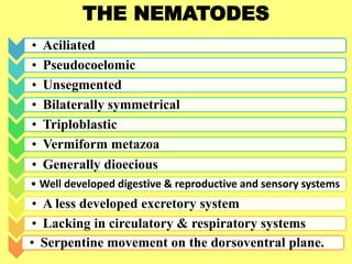
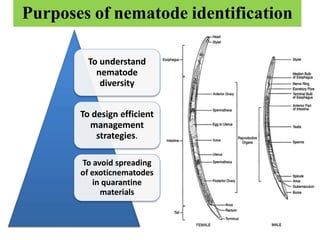
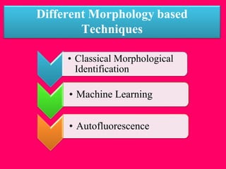
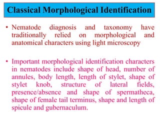
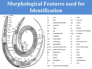
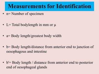
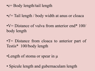
Recent Advancement in Nematode
Identification Techniques
Sabyasachi Ray
MSc. 1st year Second Semester
Dept. of Agricultural entomology
Bidhan Chandra Krishi Viswavidyalaya
Year- 2020
THE NEMATODES
• Aciliated
• Pseudocoelomic
• Unsegmented
• Bilaterally symmetrical
• Triploblastic
• Vermiform metazoa
• Generally dioecious
• Well developed digestive & reproductive and sensory systems
• A less developed excretory system
• Lacking in circulatory & respiratory systems
• Serpentine movement on the dorsoventral plane.
Purposes of nematode identification
To understand
nematode
diversity
To design efficient
management
strategies.
To avoid spreading
of exoticnematodes
in quarantine
materials
Different Morphology based
Techniques
• Classical Morphological
Identification
• Machine Learning
• Autofluorescence
Classical Morphological Identification
• Nematode diagnosis and taxonomy have
traditionally relied on morphological and
anatomical characters using light microscopy
• Important morphological identification characters
in nematodes include shape of head, number of
annules, body length, length of stylet, shape of
stylet knob, structure of lateral fields,
presence/absence and shape of spermatheca,
shape of female tail terminus, shape and length of
spicule and gubernaculum.
Morphological Features used for
Identification
Measurements for Identification
• n= Number of specimen
• L= Total bodylength in mm or µ
• a= Body length/greatest body width
• b= Body length/distance from anterior end to junction of
oesophagous and intestine
• b'= Body length / distance from anterior end to posterior
end of oesophageal glands
•c= Body length/tail length
•c'= Tail length / body width at anus or cloaca
•V= Distance of vulva from anterior end* 100/
body length
•T= Distance from cloaca to anterior part of
Testis* 100/body length
•Length of stoma or spear in µ
• Spicule length and gubernaculam length
The morphology of female perineal patterns has
been a character most frequently used in several
laboratories for the identification of Meloidogyne
species, a character located in the posterior body
region of adult females.
 This area comprises the vulva-anus area
(perineum), tail terminus, phasmids, lateral lines,
and surrounding cuticular striae.
 A more detailed account on root-knot nematode
perineal pattern development was given by Karssen
Comparison of
perineal patterns for 12
major Meloidogyne
spp. A, B: M. arenaria;
C, D: M. hapla; E, F:
M. incognita; G, H: M.
javanica; I: M.
acronea; J: M.
chitwoodi; K, L: M.
enterolobii; M: M.
ethiopica; N, O: M.
exigua; P: M. fallax;
Q, R: M. graminicola;
S, T: M. paranaensis
Advances in machine learning, also referred to as
deep learning or artificial intelligence (AI)
Machine learning for automated detection of
phenotypes takes place in multiple stages.
First, a large number of images (of nematodes, their
eggs, or cysts) is taken and independently annotated
by a group of experts to reduce subjectivity.
Then used to build an algorithm that learns (captures)
the salient features of the objects from the images in a
layer-wise hierarchy while masking (rejecting) the
noise in the background.
Machine learning
Cont.
The pattern of interest in the in-put images is then
reconstructed using a network model with a
supervised learning scheme.
Akintayo et al. designed a novel end-to-end
Convolutional Selective Autoencoder (CSAE) to
identify soybean cyst nematode (SCN) eggs in
dierent backgrounds to cover for variations in
background noise across samples from different
sources.
Another AI technique developed by Hakim et al.
using Caenorhabditis elegans called WorMachine
Qazi et al. demonstrated that eggs of different
helminths revealed characteristic fluorescence when
illuminated. at dierent wavelengths ranging from
white light to infrared
They also showed that differences in florescence
lifetime values (decay in florescence intensity) were
diagnostic of the species considered, Ascaris
lumbricoides and A. suum.
Qazi et al. concluded that spectroscopic features and
lifetime value measurements of autofluorescence in
nematodes are promising tools in the taxonomy of
these organisms.
Autofluorescence
As shown in Fig. a, the egg of Ascaris lumbricoides can
easily be visualized through light microscopy. Moreover,
they are fluorescent in the visible region when illuminated
with 390 nm (Fig. b), and 560 nm laser light (Fig. c).
A) Confocal map of Ascaris suum juvenile, showing red
fluorescence from outer membranes after 532 nm laser
excitation (B) Confocal image of Ascaris
lumbricoides juvenile, showing red fluorescence after
excitation with 561 nm.
LIMITATIONS
• Lack of clear variation among closely related taxa
• Differences of some of these morphological and morphometric
characters are subtle, subjective, and have overlapping characters
• Show intraspecific variation
• Requires well trained and experienced nematode
taxonomists
• Lack of interest of young scientists in classical taxonomy
Isozyme Analysis
 First non-morphology-based method.
 Involves the extraction of soluble proteins from whole nematodes
in buffer solutions, resolving the resulting extracts by starch or
polyacrylamide gel electrophoresis followed by staining for
specific enzymes.
 Multi-locus Enzyme Electrophoresis (MEE), relies on the
migration patterns of isozymes, owing to differences in electrical
charge, molecular weight, and conformation stemming from slight
variations in amino acid compositions.
 Several isozyme systems have been used, nonetheless,
carboxylesterase/esterase EST proved to be the most useful in
discriminating Meloidogyne species.
Two-Dimensional Gel Analyses
 The technique allows resolution of complex protein mixtures by
charge using isoelectric focusing in one-dimension followed by
mass-based resolution in a dimension perpendicular to the first.
 The resolution pattern is then compared among isolates to determine
similarities/dierences,
 which can be scored as presence/absence for phenetic and/or
cladistic analyses of the resulting data matrix.
 Navas et al. used 2-DGE to show proteomic variations among 18
root-knot nematodes representing four species.
 They demonstrated that some of these variations were species-
specific,while other variations revealed evolutionary relationships
among the different species.
Mass Spectral Analysis
 Matrix-assisted laser desorption/ionization (MALDI) is an ionization
technique, which uses laser energy-absorbing matrix to generate
gaseous ions from large molecules in solid state.
 Time of flight mass spectrometer (ToF-MS) measures the time taken
by these ions to reach the detector as determined by the mass/charge
(m/z) values, with smaller and/or more charged ions travelling faster.
 MALDI-ToF-MS is able to detect protein/peptide ions or protein
profiles that are diagnostic to the taxa being considered.
 Perera et al. used intact second stage juveniles (J2s) and/or proteins
extracted from these using various organic solvents and discriminated
between Anguina tritici, A. funesta and M. javanica based on unique
peaks in their spectra and/or the spectral profiles.
Serological Analyses
 The development of the hybridoma technique by Kohler and Milstein
raised the hope of the nematology community to develop mAbs for
diagnostic purposes.
 The technique involved isolating mature B-cells from animals
immunized with nematode antigens, fusing these B-cells with mouse
lymphoid tumor cells to produce hybridomas that can be maintained
indefinitely in vitro for continuous production of the antibodies.
 mAbs provide more specificity depending on the immunogen the
antibodies were raised against.
 mAbs were raised against a variety of agriculturally important
nematodes including H. glycines, M. incognita, G. rostochiensis and
G. pallida using the hybridoma technique.
 Schots et al. reported that some mAbs differentiated between G.
rostochiensis and G. pallida isolates.
DNA Based Analysis
Ribosomal DNA
A vast amount of examples of nematode diagnosis has
mostly been based on amplification of target DNA by
PCR using species-specific primers.
One of the approaches to design DNA-based markers that
can aided diagnosis of nematodes has been based usually
on conserved regions in the ribosomal DNA (rDNA)
cistron, i.e., the external transcribed spacer (ETS),
internal transcribed spacers 1 and 2 (ITS1 and ITS2), and
the intergenic spacer regions 1 and 2 (IGS1 and IGS2)
Cont.
 Sequences that are divergent among nematode
species and conserved within several isolates
of a same species make ideal target for
designing species-specific primers.
Ribosomal DNA regions are multicopy genes
and provide sequences with enough variation
that can be used for diagnosis and
phylogenetic relationships among species.
Mitochondrial and satellite DNA
 Small circular molecules ranging from 12 to 20 kilobases .
 Divergences in mtDNA sequences due to insertions, deletions, and
accelerated ratio of mutations compared with nuclear DNA have
provided target markers suitable for discriminating nematode
species.
 Satellite DNAs (satDNAs) are highly repeated tandem arrays of
short sequences ranging from 70 to 2000 bp.
 It has different signature sequences, copy numbers, length, and
polymorphic regions that can be explored to find species-specific
markers.
 Such PCR-based detection using satDNA markers in nematode
diagnosis represents a target option for designing diagnostic
primers.
RFLP, AFLP, RAPD, SCAR
(RFLP), a method that uses restriction enzymes to digest
whole genomic DNA or an amplified segment of it.
 It generate DNA banding patterns according to divergences
in sequences among isolates.
This technique can also be coupled with DNA hybridization
with radioactive or nonradioactive labeled probes.
 This method is less used nowadays due to technical
complexity and the need for a large amount of target DNA.
Cont.
DNA band obtained from random amplified
polymorphic DNA (RAPD) or amplified fragment
length polymorphism (AFLP) gels, with posterior
cloning and sequencing of bands differentiate across
related species and their conversion into species-
specific sequence characterized amplified region
(SCAR) markers.
 SCAR-based markers and rDNA-based specific
primers have been used to diagnose nematodes with
either conventional or real-time PCR (q-PCR) .
Cont.
Numerous primers and approaches used for diagnosis of
nematodes using conventional and quantitative PCR
were designed based on several target regions in the
nematode genome(e.g., SCAR, rDNA, ITS, D2-D3
segment, IGS, among others).
In particular, successful SCAR markers have been
developed for diagnosing some of the major tropical
Meloidogyne spp. associated with important crops such
as coffee, guava, and grapevine, including M. arenaria ,
M. incognita , M. paranaensis, M. exigua , M.
enterolobii ,M. arabicida, M. izalcoensis , and M.
ethiopica etc.
Nematode species Target region Method
Meloidogyne spp. PCR
M. arabicida and M. izalcoensis SCAR PCR
M. arenaria SCAR PCR
M. chitwoodi IGS, SCAR PCR
M. exigua SCAR PCR
M. enterolobii mtDNA, SCAR PCR
M. ethiopica SCAR PCR
M. fallax IGS, SCAR PCR
M. graminis ITS PCR
M. hapla satDNA, SCAR, IGS PCR
M. incognita SCAR PCR
M. javanica SCAR PCR
M. marylandi 28S D2-D3 PCR
M. naasi ITS, 28S D2-D3 PCR
M. paranaensis SCAR PCR
Other Plant Parasitic Nematodes
Nematode species Target region Method
Bursaphelencus xylophilus satDNA PCR
B. xylophilus satDNA qPCR
B. xylophilus heat shock protein qPCR
Ditylenchus destructor D.
dipsaci
rDNA PCR/qPCR
H. glycines rDNA qPCR
H. schachtii ITS PCR
H. glycines SCAR qPCR
Pratylenchus penetrans rDNA qPCR
SCAR—sequence characterized amplified region; IGS—intergenic
spacer region; ITS—internal transcribed spacer; mtDNA—
mitochondrial DNA; satDNA—satellite DNA; PCR—polymerase
chain reaction; qPCR—quantitative real-time PCR.
qPCR and barcoding
• Quantitative PCR (q-PCR) is a technique that amplifies and quantifies
nucleic acids simultaneously
• Its advantage over conventional PCR is that it is fast, sensitive and does
not need post amplification processing of samples normally seen in
conventional PCR
• Application of q-PCR in nematode diagnosis using rDNA target or other
marker has been showed for major nematode species, including M.
incognita, M. chitwoodi, M. fallax, M. javanica, Bursaphelenchus
xylophilus, Globodera rostochiensis, and G. pallida.
• DNA barcode for nematode taxonomy related to a DNA sequence of a
particular region in the genome as a mean to give unique signature
(barcode) for the identification of nematode species .
• This approach has not been widely accepted since there has not been an
unique DNA locus that can define the limits of species boundary.
Concluding Remarks
 The accurate identification of nematodes to species and
subspecies levels is essential for their control and is a
prerequisite to meaningful research.
 Many nematode species are easily identified based on distinct
morphological characters and restricted host ranges.
 Several species are difficult to identify due to their similarity to
other species or poor taxonomic descriptions.
 The future prospects in nematode taxonomy and diagnostics are
dependent on molecular-based and biochemical based methods
and tools that will discriminate not only at the species level but
also at the level of host races, thereby opening up opportunities
for more focused management strategies.
References
• Carneiro, R. M. D. G., Lima, F. S. O. and Correia, V. R. Methods and
Tools Currently Used for the Identification of Plant Parasitic
Nematodes. Nematology - Concepts, Diagnosis and Control. Chapter 2;
19-35
• DiGennaro, P., Baniya, A. and Bogale, M. 2020. Nematode
Identification Techniques and Recent Advances. Plants 2020, 9, 1260;1-
15.
• Introductory Plant Nematology- P Parvatha Reddy
• Qazi, F., Khalid, A., Poddar, A., Tetienne, J. P., Nadarajah, A., Medina,
A. A., Shahsavari, I., Shukla, R., Prawer, S., Ball, A. S. and Hanic, S.
T.2020. Real-time detection and identification of nematode eggs genus
and species through optical imaging. Scientific Reports | (2020) 10:7219
• Textbook of Introductory Plant Nematology- Raman k. Walia & Harish
K. Bajaj
THANK YOU

Nematode identification techniques.pptx

  • 1.
    Recent Advancement inNematode Identification Techniques Sabyasachi Ray MSc. 1st year Second Semester Dept. of Agricultural entomology Bidhan Chandra Krishi Viswavidyalaya Year- 2020
  • 2.
    THE NEMATODES • Aciliated •Pseudocoelomic • Unsegmented • Bilaterally symmetrical • Triploblastic • Vermiform metazoa • Generally dioecious • Well developed digestive & reproductive and sensory systems • A less developed excretory system • Lacking in circulatory & respiratory systems • Serpentine movement on the dorsoventral plane.
  • 3.
    Purposes of nematodeidentification To understand nematode diversity To design efficient management strategies. To avoid spreading of exoticnematodes in quarantine materials
  • 4.
    Different Morphology based Techniques •Classical Morphological Identification • Machine Learning • Autofluorescence
  • 5.
    Classical Morphological Identification •Nematode diagnosis and taxonomy have traditionally relied on morphological and anatomical characters using light microscopy • Important morphological identification characters in nematodes include shape of head, number of annules, body length, length of stylet, shape of stylet knob, structure of lateral fields, presence/absence and shape of spermatheca, shape of female tail terminus, shape and length of spicule and gubernaculum.
  • 6.
    Morphological Features usedfor Identification
  • 7.
    Measurements for Identification •n= Number of specimen • L= Total bodylength in mm or µ • a= Body length/greatest body width • b= Body length/distance from anterior end to junction of oesophagous and intestine • b'= Body length / distance from anterior end to posterior end of oesophageal glands
  • 8.
    •c= Body length/taillength •c'= Tail length / body width at anus or cloaca •V= Distance of vulva from anterior end* 100/ body length •T= Distance from cloaca to anterior part of Testis* 100/body length •Length of stoma or spear in µ • Spicule length and gubernaculam length
  • 9.
    The morphology offemale perineal patterns has been a character most frequently used in several laboratories for the identification of Meloidogyne species, a character located in the posterior body region of adult females.  This area comprises the vulva-anus area (perineum), tail terminus, phasmids, lateral lines, and surrounding cuticular striae.  A more detailed account on root-knot nematode perineal pattern development was given by Karssen
  • 10.
    Comparison of perineal patternsfor 12 major Meloidogyne spp. A, B: M. arenaria; C, D: M. hapla; E, F: M. incognita; G, H: M. javanica; I: M. acronea; J: M. chitwoodi; K, L: M. enterolobii; M: M. ethiopica; N, O: M. exigua; P: M. fallax; Q, R: M. graminicola; S, T: M. paranaensis
  • 11.
    Advances in machinelearning, also referred to as deep learning or artificial intelligence (AI) Machine learning for automated detection of phenotypes takes place in multiple stages. First, a large number of images (of nematodes, their eggs, or cysts) is taken and independently annotated by a group of experts to reduce subjectivity. Then used to build an algorithm that learns (captures) the salient features of the objects from the images in a layer-wise hierarchy while masking (rejecting) the noise in the background. Machine learning
  • 12.
    Cont. The pattern ofinterest in the in-put images is then reconstructed using a network model with a supervised learning scheme. Akintayo et al. designed a novel end-to-end Convolutional Selective Autoencoder (CSAE) to identify soybean cyst nematode (SCN) eggs in dierent backgrounds to cover for variations in background noise across samples from different sources. Another AI technique developed by Hakim et al. using Caenorhabditis elegans called WorMachine
  • 13.
    Qazi et al.demonstrated that eggs of different helminths revealed characteristic fluorescence when illuminated. at dierent wavelengths ranging from white light to infrared They also showed that differences in florescence lifetime values (decay in florescence intensity) were diagnostic of the species considered, Ascaris lumbricoides and A. suum. Qazi et al. concluded that spectroscopic features and lifetime value measurements of autofluorescence in nematodes are promising tools in the taxonomy of these organisms. Autofluorescence
  • 14.
    As shown inFig. a, the egg of Ascaris lumbricoides can easily be visualized through light microscopy. Moreover, they are fluorescent in the visible region when illuminated with 390 nm (Fig. b), and 560 nm laser light (Fig. c).
  • 15.
    A) Confocal mapof Ascaris suum juvenile, showing red fluorescence from outer membranes after 532 nm laser excitation (B) Confocal image of Ascaris lumbricoides juvenile, showing red fluorescence after excitation with 561 nm.
  • 16.
    LIMITATIONS • Lack ofclear variation among closely related taxa • Differences of some of these morphological and morphometric characters are subtle, subjective, and have overlapping characters • Show intraspecific variation • Requires well trained and experienced nematode taxonomists • Lack of interest of young scientists in classical taxonomy
  • 17.
    Isozyme Analysis  Firstnon-morphology-based method.  Involves the extraction of soluble proteins from whole nematodes in buffer solutions, resolving the resulting extracts by starch or polyacrylamide gel electrophoresis followed by staining for specific enzymes.  Multi-locus Enzyme Electrophoresis (MEE), relies on the migration patterns of isozymes, owing to differences in electrical charge, molecular weight, and conformation stemming from slight variations in amino acid compositions.  Several isozyme systems have been used, nonetheless, carboxylesterase/esterase EST proved to be the most useful in discriminating Meloidogyne species.
  • 18.
    Two-Dimensional Gel Analyses The technique allows resolution of complex protein mixtures by charge using isoelectric focusing in one-dimension followed by mass-based resolution in a dimension perpendicular to the first.  The resolution pattern is then compared among isolates to determine similarities/dierences,  which can be scored as presence/absence for phenetic and/or cladistic analyses of the resulting data matrix.  Navas et al. used 2-DGE to show proteomic variations among 18 root-knot nematodes representing four species.  They demonstrated that some of these variations were species- specific,while other variations revealed evolutionary relationships among the different species.
  • 19.
    Mass Spectral Analysis Matrix-assisted laser desorption/ionization (MALDI) is an ionization technique, which uses laser energy-absorbing matrix to generate gaseous ions from large molecules in solid state.  Time of flight mass spectrometer (ToF-MS) measures the time taken by these ions to reach the detector as determined by the mass/charge (m/z) values, with smaller and/or more charged ions travelling faster.  MALDI-ToF-MS is able to detect protein/peptide ions or protein profiles that are diagnostic to the taxa being considered.  Perera et al. used intact second stage juveniles (J2s) and/or proteins extracted from these using various organic solvents and discriminated between Anguina tritici, A. funesta and M. javanica based on unique peaks in their spectra and/or the spectral profiles.
  • 20.
    Serological Analyses  Thedevelopment of the hybridoma technique by Kohler and Milstein raised the hope of the nematology community to develop mAbs for diagnostic purposes.  The technique involved isolating mature B-cells from animals immunized with nematode antigens, fusing these B-cells with mouse lymphoid tumor cells to produce hybridomas that can be maintained indefinitely in vitro for continuous production of the antibodies.  mAbs provide more specificity depending on the immunogen the antibodies were raised against.  mAbs were raised against a variety of agriculturally important nematodes including H. glycines, M. incognita, G. rostochiensis and G. pallida using the hybridoma technique.  Schots et al. reported that some mAbs differentiated between G. rostochiensis and G. pallida isolates.
  • 21.
    DNA Based Analysis RibosomalDNA A vast amount of examples of nematode diagnosis has mostly been based on amplification of target DNA by PCR using species-specific primers. One of the approaches to design DNA-based markers that can aided diagnosis of nematodes has been based usually on conserved regions in the ribosomal DNA (rDNA) cistron, i.e., the external transcribed spacer (ETS), internal transcribed spacers 1 and 2 (ITS1 and ITS2), and the intergenic spacer regions 1 and 2 (IGS1 and IGS2)
  • 22.
    Cont.  Sequences thatare divergent among nematode species and conserved within several isolates of a same species make ideal target for designing species-specific primers. Ribosomal DNA regions are multicopy genes and provide sequences with enough variation that can be used for diagnosis and phylogenetic relationships among species.
  • 23.
    Mitochondrial and satelliteDNA  Small circular molecules ranging from 12 to 20 kilobases .  Divergences in mtDNA sequences due to insertions, deletions, and accelerated ratio of mutations compared with nuclear DNA have provided target markers suitable for discriminating nematode species.  Satellite DNAs (satDNAs) are highly repeated tandem arrays of short sequences ranging from 70 to 2000 bp.  It has different signature sequences, copy numbers, length, and polymorphic regions that can be explored to find species-specific markers.  Such PCR-based detection using satDNA markers in nematode diagnosis represents a target option for designing diagnostic primers.
  • 24.
    RFLP, AFLP, RAPD,SCAR (RFLP), a method that uses restriction enzymes to digest whole genomic DNA or an amplified segment of it.  It generate DNA banding patterns according to divergences in sequences among isolates. This technique can also be coupled with DNA hybridization with radioactive or nonradioactive labeled probes.  This method is less used nowadays due to technical complexity and the need for a large amount of target DNA.
  • 25.
    Cont. DNA band obtainedfrom random amplified polymorphic DNA (RAPD) or amplified fragment length polymorphism (AFLP) gels, with posterior cloning and sequencing of bands differentiate across related species and their conversion into species- specific sequence characterized amplified region (SCAR) markers.  SCAR-based markers and rDNA-based specific primers have been used to diagnose nematodes with either conventional or real-time PCR (q-PCR) .
  • 26.
    Cont. Numerous primers andapproaches used for diagnosis of nematodes using conventional and quantitative PCR were designed based on several target regions in the nematode genome(e.g., SCAR, rDNA, ITS, D2-D3 segment, IGS, among others). In particular, successful SCAR markers have been developed for diagnosing some of the major tropical Meloidogyne spp. associated with important crops such as coffee, guava, and grapevine, including M. arenaria , M. incognita , M. paranaensis, M. exigua , M. enterolobii ,M. arabicida, M. izalcoensis , and M. ethiopica etc.
  • 27.
    Nematode species Targetregion Method Meloidogyne spp. PCR M. arabicida and M. izalcoensis SCAR PCR M. arenaria SCAR PCR M. chitwoodi IGS, SCAR PCR M. exigua SCAR PCR M. enterolobii mtDNA, SCAR PCR M. ethiopica SCAR PCR M. fallax IGS, SCAR PCR M. graminis ITS PCR M. hapla satDNA, SCAR, IGS PCR M. incognita SCAR PCR M. javanica SCAR PCR M. marylandi 28S D2-D3 PCR M. naasi ITS, 28S D2-D3 PCR M. paranaensis SCAR PCR
  • 28.
    Other Plant ParasiticNematodes Nematode species Target region Method Bursaphelencus xylophilus satDNA PCR B. xylophilus satDNA qPCR B. xylophilus heat shock protein qPCR Ditylenchus destructor D. dipsaci rDNA PCR/qPCR H. glycines rDNA qPCR H. schachtii ITS PCR H. glycines SCAR qPCR Pratylenchus penetrans rDNA qPCR SCAR—sequence characterized amplified region; IGS—intergenic spacer region; ITS—internal transcribed spacer; mtDNA— mitochondrial DNA; satDNA—satellite DNA; PCR—polymerase chain reaction; qPCR—quantitative real-time PCR.
  • 29.
    qPCR and barcoding •Quantitative PCR (q-PCR) is a technique that amplifies and quantifies nucleic acids simultaneously • Its advantage over conventional PCR is that it is fast, sensitive and does not need post amplification processing of samples normally seen in conventional PCR • Application of q-PCR in nematode diagnosis using rDNA target or other marker has been showed for major nematode species, including M. incognita, M. chitwoodi, M. fallax, M. javanica, Bursaphelenchus xylophilus, Globodera rostochiensis, and G. pallida. • DNA barcode for nematode taxonomy related to a DNA sequence of a particular region in the genome as a mean to give unique signature (barcode) for the identification of nematode species . • This approach has not been widely accepted since there has not been an unique DNA locus that can define the limits of species boundary.
  • 30.
    Concluding Remarks  Theaccurate identification of nematodes to species and subspecies levels is essential for their control and is a prerequisite to meaningful research.  Many nematode species are easily identified based on distinct morphological characters and restricted host ranges.  Several species are difficult to identify due to their similarity to other species or poor taxonomic descriptions.  The future prospects in nematode taxonomy and diagnostics are dependent on molecular-based and biochemical based methods and tools that will discriminate not only at the species level but also at the level of host races, thereby opening up opportunities for more focused management strategies.
  • 31.
    References • Carneiro, R.M. D. G., Lima, F. S. O. and Correia, V. R. Methods and Tools Currently Used for the Identification of Plant Parasitic Nematodes. Nematology - Concepts, Diagnosis and Control. Chapter 2; 19-35 • DiGennaro, P., Baniya, A. and Bogale, M. 2020. Nematode Identification Techniques and Recent Advances. Plants 2020, 9, 1260;1- 15. • Introductory Plant Nematology- P Parvatha Reddy • Qazi, F., Khalid, A., Poddar, A., Tetienne, J. P., Nadarajah, A., Medina, A. A., Shahsavari, I., Shukla, R., Prawer, S., Ball, A. S. and Hanic, S. T.2020. Real-time detection and identification of nematode eggs genus and species through optical imaging. Scientific Reports | (2020) 10:7219 • Textbook of Introductory Plant Nematology- Raman k. Walia & Harish K. Bajaj
  • 32.