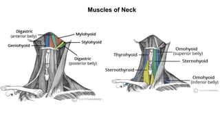
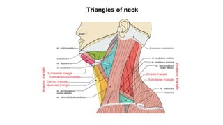
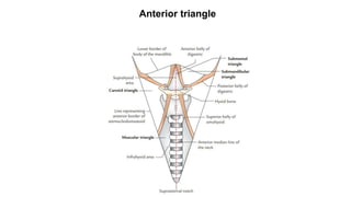
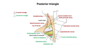
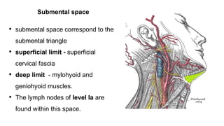
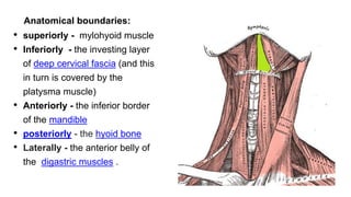
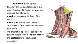
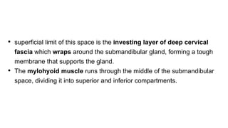
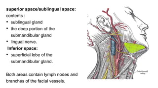
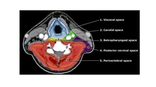
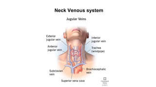
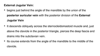
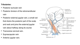
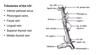
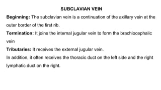
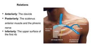
Various Spaces in neck
Neck venous system
Presentor - Dr. M Hema Bindu
Junior resident 2nd yr
Moderator - Dr.Rajesh Kumar
Professor
Muscles of Neck
Triangles of neck
Anterior triangle
Posterior triangle
Various spaces in neck
• Submental space
• Submandibular space
• Peritonsillar space
• Parapharyngeal space
• Retropharyngeal space
• Prevertebral space
• Carotid space
• Parotid space
• Visceral space
• Between the fascial layers of the neck are several anatomical
compartments, containing loose areolar tissue.
• Fascial layers limit the spread of infection, these inter-connected
spaces can act as routes of transmission of infection or neoplasia.
Submental space
• submental space correspond to the
submental triangle
• superficial limit - superficial
cervical fascia
• deep limit - mylohyoid and
geniohyoid muscles.
• The lymph nodes of level Ia are
found within this space.
Anatomical boundaries:
• superiorly - mylohyoid muscle
• Inferiorly - the investing layer
of deep cervical fascia (and this
in turn is covered by the
platysma muscle)
• Anteriorly - the inferior border
of the mandible
• posteriorly - the hyoid bone
• Laterally - the anterior belly of
the digastric muscles .
Submandibular space
• It has key clinical significance as it is a
route of spread of infection between the
neck and floor of mouth.
• superior - mucosa of the floor of the
mouth
• Inferiorly - investing layer of deep
cervical fascia from the mandible and the
hyoid bone.
• The anterior and posterior bellies of the
digastric muscle form the anteroinferior
and posteroinferior boundaries
respectively.
• superficial limit of this space is the investing layer of deep cervical
fascia which wraps around the submandibular gland, forming a tough
membrane that supports the gland.
• The mylohyoid muscle runs through the middle of the submandibular
space, dividing it into superior and inferior compartments.
superior space/sublingual space:
contents :
• sublingual gland
• the deep portion of the
submandibular gland
• lingual nerve.
Inferior space:
• superficial lobe of the
submandibular gland.
Both areas contain lymph nodes and
branches of the facial vessels.
Peritonsillar space
• Space between the palatine tonsil and the
superior constrictor muscle
• Condensation of the pharyngobasillar
which forms the tonsillar capsule may be
breached by bacterial infections of the
tonsil, resulting in abscess formation in the
peritonsillar space, also known as a quinsy.
• Untreated peritonsillar infections may
spread to involve the parapharyngeal
space.
Parapharyngeal space
• Inverted pyramid shaped
• Superiorly: petrous temporal bone
• Inferiorly : level of the hyoid
• Medially : superior constrictor muscle
• Laterally : the pterygoid muscles, the
parotid salivary gland and the
mandible.
• The styloid process divides it into pre-
styloid and post-styloid compartments.
pre-styloid compartment :
• fat
• connective tissue
• branch of maxillary artery
• inferior alveolar nerve
• lingual nerve
• Auriculotemporal nerve.
post-styloid compartment:
• carotid sheath and it contents
• glossopharyngeal
• hypoglossal nerves
• Cervical sympathetic chain
• lymph nodes.
• The determination between pre and post-styloid involvement and
displacement of the parapharyngeal fat pad are key radiological
features which aid in diagnosis of parapharyngeal space neoplasm
• Pre-styloid lesions are most commonly associated with the deep lobe
of the parotid gland and will deflect the carotid sheath and
parapharyngeal fat posteromedially.
• Lesions in the post-styloid compartment are frequently of
neuroendocrine origin, arising from the carotid sheath as carotid
body tumours or vagal schwannomas, or neuromas of the
sympathetic chain.
• Post-styloid lesions displace the parapharyngeal fat pad anteriorly.
Retropharyngeal space
• Between the two parapharyngeal
spaces is the retropharyngeal space
which is continuous with both
• The superior limit is the skull base
and it is continuous inferiorly with the
posterior mediastinum, down to the
level of the carina.
• The anterior boundary in the neck is
the buccopharyngeal fascia which
encases the pharyngeal constrictors,
the posterior limit is the alar fascial
component of the prevertebral fascia.
• The only contents of this space are the
retropharyngeal lymph nodes, which
typically regress by adulthood but can
represent a route of metastatic nodal
spread from midline or posterior
tumours of the pharynx.
• Radiological evidence of
retropharyngeal lymphadenopathy in
adults should therefore raise the
suspicion of head and neck malignancy.
• Infection of the retropharyngeal space may occur indirectly, tracking
from the parapharyngeal space or directly secondary to perforation of
the pharynx or cervical oesophagus.
• Retropharyngeal abscesses are more commonly seen in children than
in adults, secondary to bacterial infection of the upper aerodigestive
tract and may result in airway obstruction due to anterior displacement
of the airway.
Danger space
• The alar space, known as the ‘danger
space’ is a further potential space
located posterior to the retro-
pharyngeal space, between the alar
fascial component of the prevertebral
fascia and the prevertebral fascia itself,
extending to the level of the diaphragm
• This space is only visible radiologically if
distended due to collection within this
area. The name ‘danger space’ refers to
the potential for neck infections to
readily spread into the thorax and
cause mediastinitis.
Prevertebral space
• The prevertebral space is the potential
area posterior to the prevertebral
fascia and anterior to the vertebral
column and para-spinal musculature.
• Spread of infection to this area may
occur as a result of traumatic perforation
of the pharynx or oesophagus, or
because of a breach of prevertebral
fascia from retropharyngeal infection.
Infection in the prevertebral space can
cause spinal osteomyelitis and spinal
cord compression.
Carotid space
• The carotid space is a
potential space within the
carotid sheath, which is itself
formed from a condensation of
all three layers of deep
cervical fascia.
• It contains the common carotid
artery, the IJV and the vagus
nerve.
Parotid space
• The parotid space is formed by
the investing layer of deep
cervical fascia that splits to
encompass the parotid gland and
therefore contains the facial
nerve, the retromandibular vein
and the terminal branches of the
external carotid artery.
Visceral space
• The visceral space is bounded by
the middle layer of deep cervical
fascia that envelops the thyroid
and the trachea anteriorly and
posteriorly by the pretracheal
fascia. It contains the larynx,
hypopharynx, cervical oesophagus,
proximal trachea, thyroid and
parathyroid glands and lymphatics
of level VI.
Neck Venous system
External Jugular Vein:
• begins just behind the angle of the mandible by the union of the
posterior auricular vein with the posterior division of the External
Jugular Vein
• It descends obliquely across the sternocleidomastoid muscle and, just
above the clavicle in the posterior triangle, pierces the deep fascia and
drains into the subclavian vein.
• Its course extends from the angle of the mandible to the middle of the
clavicle.
Tributaries:
• Posterior auricular vein
• Posterior division of the retromandibular
vein
• Posterior external jugular vein, a small vein
that drains the posterior part of the scalp
and neck and joins the external jugular
vein about halfway along its course
• Transverse cervical vein.
• Suprascapular vein
• Anterior jugular vein.
Anterior Jugular Vein
• Begins just below the chin, by the union of several small veins.
• It runs down the neck close to the midline. Just above the suprasternal
notch, the veins of the two sides are united by a transverse trunk called
the jugular arch.
• The vein then turns sharply laterally and passes deep to the
sternocleidomastoid muscle to drain into the external jugular vein
Internal Jugular Vein
• The internal jugular vein is a large vein that receives blood from the
brain, face, and neck.
• Beginning It starts as a continuation of the sigmoid sinus and leaves the
skull through the jugular foramen.
• Course It then descends through the neck in the carotid sheath lateral to
the vagus nerve and the internal and common carotid arteries.
Throughout its course, it is closely related to the deep cervical lymph
nodes
• Termination It ends by joining
the subclavian vein behind
the medial end of the clavicle
to form the brachiocephalic
vein.
• The vein has a dilatation at its
upper end called the superior
bulb and another near its
termination inferior bulb.
• The internal jugular ends by joining the subclavian vein behind the
medial end of the clavicle to form the brachiocephalic vein.
Relations of IJV
• Anterolaterally: The skin, the fascia, the sternocleidomastoid, and the
parotid salivary gland.
• Its lower part is covered by the sternothyroid, sternohyoid, omohyoid and
the sternocleidomastoid muscles . Higher up, it is crossed by the
stylohyoid, the posterior belly of the digastric, and the spinal part of the
accessory nerve.
• The chain of deep cervical lymph nodes runs alongside the vein.
• Posteriorly: The transverse processes of the cervical vertebrae, the
levator scapulae, the scalenus medius, the scalenus anterior, the
cervical plexus, the phrenic nerve.
• The thyrocervical trunk, the vertebral vein, and the first part of the
subclavian artery. On the left side it passes in front of the thoracic duct.
• Medially: Above lie the internal carotid artery and the 9th, 10th, 11th,
and 12th cranialnerves. Below lie the common carotid artery and the
vagus nerve
Tributaries of the IJV:
• Inferior petrosal sinus
• Pharyngeal veins.
• Facial vein
• Lingual vein
• Superior thyroid vein
• Middle thyroid vein
SUBCLAVIAN VEIN
Beginning: The subclavian vein is a continuation of the axillary vein at the
outer border of the first rib.
Termination: It joins the internal jugular vein to form the brachiocephalic
vein
Tributaries: It receives the external jugular vein.
In addition, it often receives the thoracic duct on the left side and the right
lymphatic duct on the right.
Relations
• Anteriorly: The clavicle
• Posteriorly: The scalenus
anterior muscle and the phrenic
nerve
• Inferiorly: The upper surface of
the first rib
THANK YOU

Neck spaces (1).pptx

  • 1.
    Various Spaces inneck Neck venous system Presentor - Dr. M Hema Bindu Junior resident 2nd yr Moderator - Dr.Rajesh Kumar Professor
  • 2.
  • 3.
  • 4.
  • 5.
  • 6.
    Various spaces inneck • Submental space • Submandibular space • Peritonsillar space • Parapharyngeal space • Retropharyngeal space • Prevertebral space • Carotid space • Parotid space • Visceral space
  • 7.
    • Between thefascial layers of the neck are several anatomical compartments, containing loose areolar tissue. • Fascial layers limit the spread of infection, these inter-connected spaces can act as routes of transmission of infection or neoplasia.
  • 8.
    Submental space • submentalspace correspond to the submental triangle • superficial limit - superficial cervical fascia • deep limit - mylohyoid and geniohyoid muscles. • The lymph nodes of level Ia are found within this space.
  • 9.
    Anatomical boundaries: • superiorly- mylohyoid muscle • Inferiorly - the investing layer of deep cervical fascia (and this in turn is covered by the platysma muscle) • Anteriorly - the inferior border of the mandible • posteriorly - the hyoid bone • Laterally - the anterior belly of the digastric muscles .
  • 10.
    Submandibular space • Ithas key clinical significance as it is a route of spread of infection between the neck and floor of mouth. • superior - mucosa of the floor of the mouth • Inferiorly - investing layer of deep cervical fascia from the mandible and the hyoid bone. • The anterior and posterior bellies of the digastric muscle form the anteroinferior and posteroinferior boundaries respectively.
  • 11.
    • superficial limitof this space is the investing layer of deep cervical fascia which wraps around the submandibular gland, forming a tough membrane that supports the gland. • The mylohyoid muscle runs through the middle of the submandibular space, dividing it into superior and inferior compartments.
  • 12.
    superior space/sublingual space: contents: • sublingual gland • the deep portion of the submandibular gland • lingual nerve. Inferior space: • superficial lobe of the submandibular gland. Both areas contain lymph nodes and branches of the facial vessels.
  • 13.
    Peritonsillar space • Spacebetween the palatine tonsil and the superior constrictor muscle • Condensation of the pharyngobasillar which forms the tonsillar capsule may be breached by bacterial infections of the tonsil, resulting in abscess formation in the peritonsillar space, also known as a quinsy. • Untreated peritonsillar infections may spread to involve the parapharyngeal space.
  • 14.
    Parapharyngeal space • Invertedpyramid shaped • Superiorly: petrous temporal bone • Inferiorly : level of the hyoid • Medially : superior constrictor muscle • Laterally : the pterygoid muscles, the parotid salivary gland and the mandible. • The styloid process divides it into pre- styloid and post-styloid compartments.
  • 15.
    pre-styloid compartment : •fat • connective tissue • branch of maxillary artery • inferior alveolar nerve • lingual nerve • Auriculotemporal nerve. post-styloid compartment: • carotid sheath and it contents • glossopharyngeal • hypoglossal nerves • Cervical sympathetic chain • lymph nodes.
  • 16.
    • The determinationbetween pre and post-styloid involvement and displacement of the parapharyngeal fat pad are key radiological features which aid in diagnosis of parapharyngeal space neoplasm • Pre-styloid lesions are most commonly associated with the deep lobe of the parotid gland and will deflect the carotid sheath and parapharyngeal fat posteromedially.
  • 17.
    • Lesions inthe post-styloid compartment are frequently of neuroendocrine origin, arising from the carotid sheath as carotid body tumours or vagal schwannomas, or neuromas of the sympathetic chain. • Post-styloid lesions displace the parapharyngeal fat pad anteriorly.
  • 18.
    Retropharyngeal space • Betweenthe two parapharyngeal spaces is the retropharyngeal space which is continuous with both • The superior limit is the skull base and it is continuous inferiorly with the posterior mediastinum, down to the level of the carina. • The anterior boundary in the neck is the buccopharyngeal fascia which encases the pharyngeal constrictors, the posterior limit is the alar fascial component of the prevertebral fascia.
  • 19.
    • The onlycontents of this space are the retropharyngeal lymph nodes, which typically regress by adulthood but can represent a route of metastatic nodal spread from midline or posterior tumours of the pharynx. • Radiological evidence of retropharyngeal lymphadenopathy in adults should therefore raise the suspicion of head and neck malignancy.
  • 20.
    • Infection ofthe retropharyngeal space may occur indirectly, tracking from the parapharyngeal space or directly secondary to perforation of the pharynx or cervical oesophagus. • Retropharyngeal abscesses are more commonly seen in children than in adults, secondary to bacterial infection of the upper aerodigestive tract and may result in airway obstruction due to anterior displacement of the airway.
  • 21.
    Danger space • Thealar space, known as the ‘danger space’ is a further potential space located posterior to the retro- pharyngeal space, between the alar fascial component of the prevertebral fascia and the prevertebral fascia itself, extending to the level of the diaphragm • This space is only visible radiologically if distended due to collection within this area. The name ‘danger space’ refers to the potential for neck infections to readily spread into the thorax and cause mediastinitis.
  • 22.
    Prevertebral space • Theprevertebral space is the potential area posterior to the prevertebral fascia and anterior to the vertebral column and para-spinal musculature. • Spread of infection to this area may occur as a result of traumatic perforation of the pharynx or oesophagus, or because of a breach of prevertebral fascia from retropharyngeal infection. Infection in the prevertebral space can cause spinal osteomyelitis and spinal cord compression.
  • 23.
    Carotid space • Thecarotid space is a potential space within the carotid sheath, which is itself formed from a condensation of all three layers of deep cervical fascia. • It contains the common carotid artery, the IJV and the vagus nerve.
  • 24.
    Parotid space • Theparotid space is formed by the investing layer of deep cervical fascia that splits to encompass the parotid gland and therefore contains the facial nerve, the retromandibular vein and the terminal branches of the external carotid artery. Visceral space • The visceral space is bounded by the middle layer of deep cervical fascia that envelops the thyroid and the trachea anteriorly and posteriorly by the pretracheal fascia. It contains the larynx, hypopharynx, cervical oesophagus, proximal trachea, thyroid and parathyroid glands and lymphatics of level VI.
  • 26.
  • 27.
    External Jugular Vein: •begins just behind the angle of the mandible by the union of the posterior auricular vein with the posterior division of the External Jugular Vein • It descends obliquely across the sternocleidomastoid muscle and, just above the clavicle in the posterior triangle, pierces the deep fascia and drains into the subclavian vein. • Its course extends from the angle of the mandible to the middle of the clavicle.
  • 28.
    Tributaries: • Posterior auricularvein • Posterior division of the retromandibular vein • Posterior external jugular vein, a small vein that drains the posterior part of the scalp and neck and joins the external jugular vein about halfway along its course • Transverse cervical vein. • Suprascapular vein • Anterior jugular vein.
  • 29.
    Anterior Jugular Vein •Begins just below the chin, by the union of several small veins. • It runs down the neck close to the midline. Just above the suprasternal notch, the veins of the two sides are united by a transverse trunk called the jugular arch. • The vein then turns sharply laterally and passes deep to the sternocleidomastoid muscle to drain into the external jugular vein
  • 31.
    Internal Jugular Vein •The internal jugular vein is a large vein that receives blood from the brain, face, and neck. • Beginning It starts as a continuation of the sigmoid sinus and leaves the skull through the jugular foramen. • Course It then descends through the neck in the carotid sheath lateral to the vagus nerve and the internal and common carotid arteries. Throughout its course, it is closely related to the deep cervical lymph nodes
  • 32.
    • Termination Itends by joining the subclavian vein behind the medial end of the clavicle to form the brachiocephalic vein. • The vein has a dilatation at its upper end called the superior bulb and another near its termination inferior bulb.
  • 33.
    • The internaljugular ends by joining the subclavian vein behind the medial end of the clavicle to form the brachiocephalic vein.
  • 34.
    Relations of IJV •Anterolaterally: The skin, the fascia, the sternocleidomastoid, and the parotid salivary gland. • Its lower part is covered by the sternothyroid, sternohyoid, omohyoid and the sternocleidomastoid muscles . Higher up, it is crossed by the stylohyoid, the posterior belly of the digastric, and the spinal part of the accessory nerve. • The chain of deep cervical lymph nodes runs alongside the vein.
  • 35.
    • Posteriorly: Thetransverse processes of the cervical vertebrae, the levator scapulae, the scalenus medius, the scalenus anterior, the cervical plexus, the phrenic nerve. • The thyrocervical trunk, the vertebral vein, and the first part of the subclavian artery. On the left side it passes in front of the thoracic duct. • Medially: Above lie the internal carotid artery and the 9th, 10th, 11th, and 12th cranialnerves. Below lie the common carotid artery and the vagus nerve
  • 37.
    Tributaries of theIJV: • Inferior petrosal sinus • Pharyngeal veins. • Facial vein • Lingual vein • Superior thyroid vein • Middle thyroid vein
  • 38.
    SUBCLAVIAN VEIN Beginning: Thesubclavian vein is a continuation of the axillary vein at the outer border of the first rib. Termination: It joins the internal jugular vein to form the brachiocephalic vein Tributaries: It receives the external jugular vein. In addition, it often receives the thoracic duct on the left side and the right lymphatic duct on the right.
  • 39.
    Relations • Anteriorly: Theclavicle • Posteriorly: The scalenus anterior muscle and the phrenic nerve • Inferiorly: The upper surface of the first rib
  • 40.