Download to read offline

![International Research Journal of Engineering and Technology (IRJET) e-ISSN: 2395-0056
Volume: 06 Issue: 06 | June 2019 www.irjet.net p-ISSN: 2395-0072
© 2019, IRJET | Impact Factor value: 7.211 | ISO 9001:2008 Certified Journal | Page 2963
place the accurate 3D model of the patient over the target to
use it as a scale.
2.3 Object Tracking
Object tracking is intended topredict the cameraormarker's
spatial position on surgical instruments and is an essential
component of a medical AR scheme. In AR monitoring, the
relative location of an object is usually calculated based on
the camera position. A calibrated camera with recognized
intrinsic parameters can be used to determine the
comparative position as a collection of three or more
combined points between the 3D and the predicted 2D
coordinates [1,2,3-5].
A mixture of these technologies can be used to introduce an
AR scheme that overlays endoscopic or surgical microscope
virtual items.
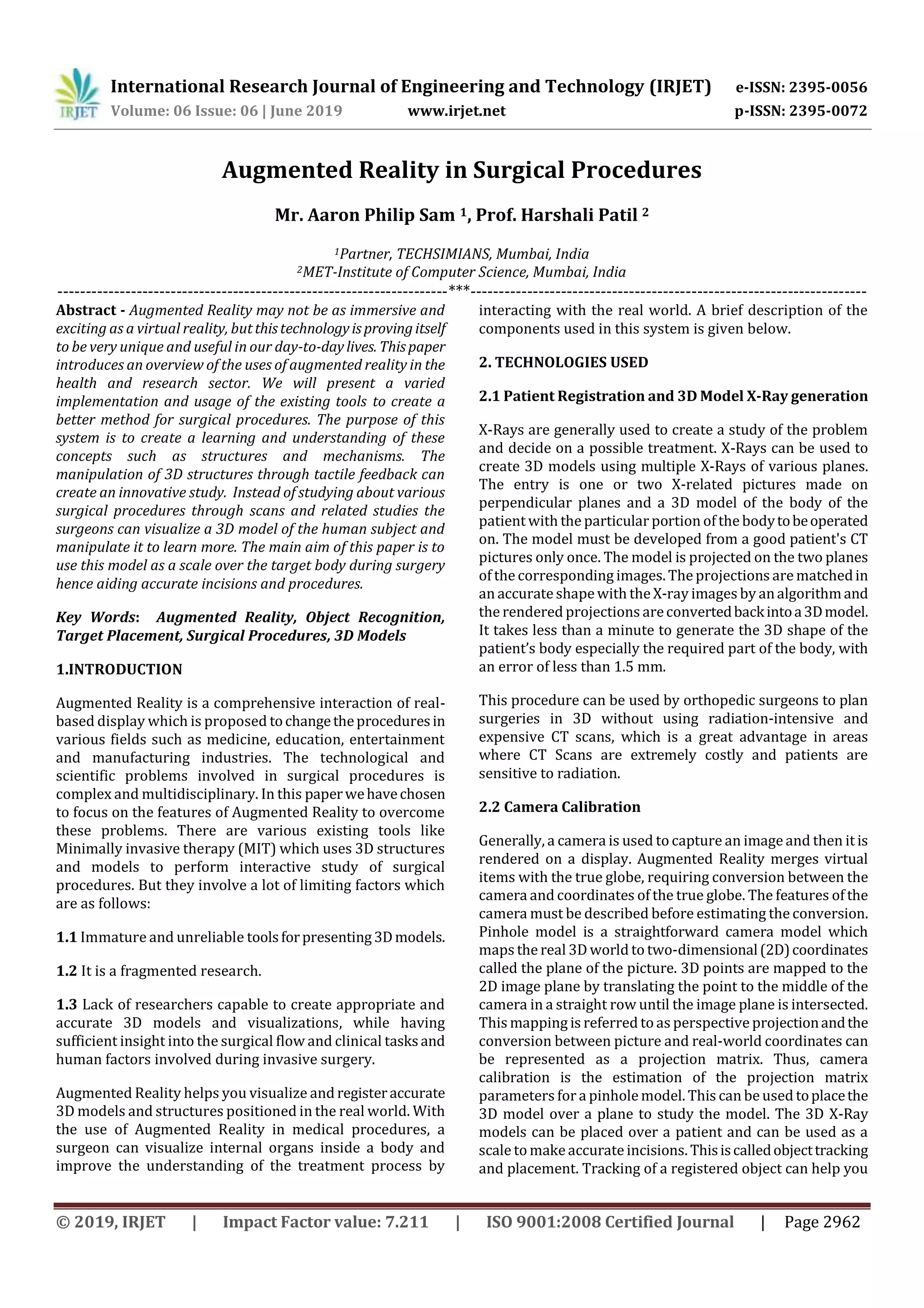
Fig -1: Calculated incisions during surgery
Fig -2: Camera calibration to place model of patient
3. MEDICAL APPLICATIONS
In this section we will work with and optimize the various
surgical procedures in which augmented reality can play a
stellar role. Some of which include spinal surgery, cardiac,
sinus. We briefly explain the attributes of each surgery and
how augmented reality can help in the same.
3.1. Spinal Surgery
For spinal surgery, correctly localizing the surgical
instrument inside the patient’s body is the most important
consideration. Therefore, surgical navigation systemsbased
on Augmented Reality are commonly recognized and have
become a very significant study subject in this sector,
helping the surgeon to acknowledge patient anatomical
structures [6]. Although AR offers a visual representation
that is very intuitive, incorrect perception of depth is a
severe problem. Toenhance depth perception,anintegration
of a VR and AR scheme was suggested to show the distance
between surgical instruments and target organs in a single
window with aligned view axes [5].
3.1.1 Corresponding AR configuration
We can use a combination of VR and AR switchable
navigation procedures which consist of visualization and
position tracking. In position tracking the transformation
coefficient between the camera and the patient is calculated
by an optical tracker and is updated in real time. The
visualization can be any open source visualization library
and graphical processing unit (GPU) to display translucent
objects based on depth peeling technique.
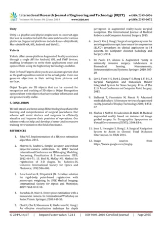
3.1.2 Corresponding Results
By moving the virtual camera around targetobjects,theuser
will move from AR to VR, giving image depth of patient
anatomy. Fig. 3 shows the scheduled surgical navigation
scheme VR and AR switchable. The surgical navigation
system is operated in AR mode once the virtual camera is
placed within the varying camera picture. It is operated in
VR mode otherwise. Additionally, the depth which
suggests the minimum distance between the tipofa medical
instrument and also the nearest purpose of the target is
additionally displayed on the screen.
Fig -3: Proposed AR and VR system for spinal surgery
3.2. Sinus Surgery
Sinus operation is an endoscopic operation. The primary
issue is the difficulty of findinga surgical instrumentthrough
the endoscope to a particular object [7]. Due to the
complexity of the access path to paranasal sinuses,problems
such as blindness and cerebrospinal fluid leakage may
happen owing to orbit and skull base harm.
An embedded system composed of an AR-
based surgical navigation system and endoscope holder was
created to fix these issues [8].
3.2.1 Corresponding AR configuration
The suggested sinus surgery AR navigation system was
comparable to those used in standard surgery. The
suggested scheme consists of three procedures: registration](https://image.slidesharecdn.com/irjet-v6i6602-191216060427/85/IRJET-Augmented-Reality-in-Surgical-Procedures-2-320.jpg)
![International Research Journal of Engineering and Technology (IRJET) e-ISSN: 2395-0056
Volume: 06 Issue: 06 | June 2019 www.irjet.net p-ISSN: 2395-0072
© 2019, IRJET | Impact Factor value: 7.211 | ISO 9001:2008 Certified Journal | Page 2964
of patient images, camera calibration, and camera-based
monitoring supplied by paired point registration, pinhole-
based calibration, and perspective n-points algorithm,
respectively. The endoscope holder scheme comprises of a
stackable parallel mechanism of 3 degrees of liberty (DOF)
and an end-effector of 2-DOF. The parallel stackable 3-DOF
system coupled a five bar with two parallelograms, and the
end-effector 2-DOF governed the position of the endoscope.
A brake was also included in the scheme to keep the
endoscope anywhere the surgeon wanted [8].
3.2.2 Corresponding Results
Fig. 4, the suggested AR navigation system, displays 2D
multi-planar reconstruction (MPR) pictures (axial, coronal
and sagittal planes) along with views of the AR and VR.
Functions for adjusting warningandautomatic transparency
were also introduced. If a goal is too near to the tip of the
surgical instrument, an alert noise is produced.
Fig -4: AR based model can help detect accuracy and
precision of the instrument used
3.3. Cardiac Surgery
To guide chronic complete occlusion intervention,a surgical
navigationscheme wassuggested.Conventional intervention
for the coronary artery's permanent complete occlusion is
extremely dependent on 2D X-rayimagesandtheexperience
of the surgeon. Large displacements between the hand-eye
coordination of the surgeon can therefore be caused by
discrepancies in the patient's position or orientationandthe
acquired images [9]. This may result in the coronary artery
being misidentified or the stenosis being incorrectly
positioned on the coronary artery [10]. The scheme
suggested combined the 3D CT angiography model with X-
ray images to provide the surgeon with 3D anatomical data
[11].
3.3.1 Corresponding AR configuration
The position of markers connected to the patient and C-arm
device was tracked using a commercial optical monitoring
scheme. Using a transformation matrix, the various system
coordinates were unified.
3.3.2 Corresponding Results
The angiograph of the CT is superimposed on the X-ray
picture and the pictures of the VR are placed next to it. We
expect surgeons to be able to readily comprehend
anatomical data contained in the initial X-raypicture,aswell
as the vascular anatomy and relative place of the tool using
the suggested prototype scheme. As fluoroscopy is less
needed than standard surgery,theschemecanalsominimize
X-ray exposure and injection of the contrast medium.
4. PROPOSED SYSTEM
The scheme proposed is intended to create high-quality
manuals for 3D video using AR. A camera is used to track
fiducial objects (Image Targets) and the 3D models are
mapped to the target. These 3D models represent a product
or idea that can be seen on your screen by the user. The user
can communicate with these 3D models and watch the
animations to see the processes.
4.1 Smartphone / Holo Lens / Computer
4.2 Vuforia
4.3 Unity
4.4 Storage and processing server
Fig -5: System Architecture](https://image.slidesharecdn.com/irjet-v6i6602-191216060427/85/IRJET-Augmented-Reality-in-Surgical-Procedures-3-320.jpg)

This document discusses the use of augmented reality in surgical procedures. It begins by introducing augmented reality and how it can help visualize 3D models and structures overlaid on a patient's body to aid surgeons. Examples of its applications discussed include using it to help with spinal, sinus and cardiac surgeries by visualizing internal organs and tracking surgical instruments for improved accuracy and reduced risks. A proposed system is described that would use technologies like Vuforia and Unity to create 3D interactive manuals overlaid during operations through devices like smartphones or HoloLens to enhance surgical training and comprehension.


![Welcome to the New-Era in Automation]](https://cdn.slidesharecdn.com/ss_thumbnails/a73cde01-449f-4ab4-b012-7731e75ab889-160705040211-thumbnail.jpg?width=640&height=640&fit=bounds)


