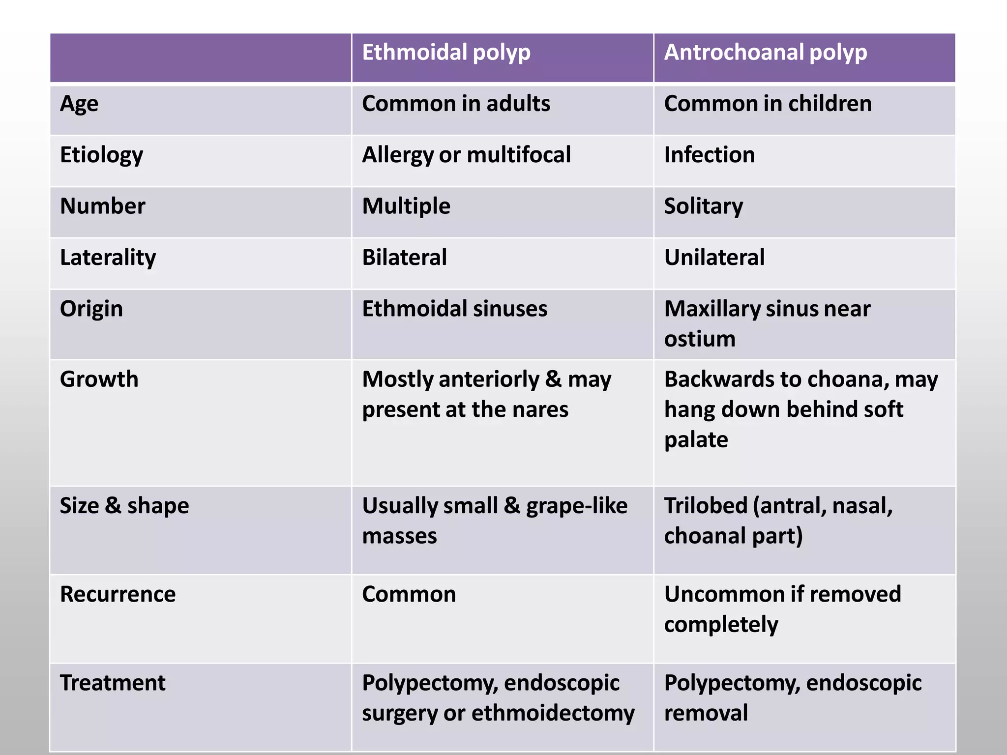
NASAL POLYPS
EMAN ZAYED
SUPERVISED BY
PROF DR AHMED ALI
NON-NEOPLASTIC
MASSES OF
OEDEMATOUS
NASAL OR SINUS
TYPES
NASAL POLYPS
BILATERAL
ETHMOIDAL
POLYP
ANTROCHOANAL
POLYP
BILATERAL ETHMOIDAL POLYP
AETIOLOGY
• Inflammatory conditions of nasal mucosa
– Rhinosinusitis
• Disorders of ciliary motility
– Kartagener’s syndrome
• Abnormal composition of nasal mucus
– Cystic fibrosis
AETIOLOGY
• Various disease associated with the formation of nasal
polyps are:
– Chronic rhinosinusitis
– Kartagener syndrome
– Cystic fibrosis
– Asthma
– Aspirin tolerance
– Allergic fungal sinusitis
– Young syndrome
– Churg-Strauss syndrome
– Nasal mastocytosis
PATHOGENESIS
Nasal mucosa  becomes edematous due to
collection of ECF
polypoidal change
Sessile  pedunculated
(due to gravity and excessive sneezing)
PATHOLOGY
Early stage  Nasal polyp (surface covered by
ciliated columnar epithelium)
Transitional & squamous epithelium
Submucosa  large ICS filled with serous fluid
+ infiltration with eosinophils and
round cells
Metaplastic change
in exposure to
atmospheric irritation
SITE OF ORIGIN
• Multiple nasal polyps always arise from the
lateral wall of nose, usually from the middle
meatus
• Common sites:
– Uncinate process
– Bulla ethmoidalis
– Ostia of sinuses
– Medial surface & edge of middle turbinate
SYMPTOMS
• Mostly seen in adults
• Nasal stuffiness  leading to
total nasal obstruction
• Partial/total loss of smell
• Headache (associated sinusitis)
• Sneezing and watery nasal
discharge (associated allergy)
• Protruding mass
SIGNS
• On anterior rhinoscopy, polyps appear as
– Smooth, glistening
– Grape-like masses
– Often pale in color
– May be sessile or pedunculated
– Insensitive to probing
– Do not bleed on touch
– Often multiple and bilateral
• Broadening of nose
• Increase intercanthal distance
• May protrude from the nostril and appear pink and vascular, simulating
neoplasm
• Purulent discharge (associated sinusitis)
long standing case
DIAGNOSIS
• Clinical examination
• CT scan of paranasal sinuses
– exclude neoplasia
– plan surgery
• Histological examination
– especially in people >40 years
TREATMENT
CONSERVATIVE
• Antihistaminics & control of allergy
– may revert early polypoidal changes with
oedematous mucosa to normal
• Short course steroids
– in people who cannot tolerate antihistaminics or
with asthma
TREATMENT
SURGICAL
• Polypectomy
• Intranasal ethmoidectomy
• Extranasal ethmoidectomy
• Transantral ethmoidectomy
• Endoscopic sinus surgery
ANTROCHOANAL POLYP
SITE OF ORIGIN
• Arise from the mucosa of
maxillary antrum near its
accessory ostium, comes out of
it, and grows in the choana
and nasal cavity
• Thus, it has 3 parts:
1. Antral: thin stalk
2. Choanal: round and globular
3. Nasal: flat from side to side
AETIOLOGY
• Exact cause is unknown
• Nasal allergy coupled with sinus infection is
incriminated
• Seen in children and young adults
• Usually single and unilateral
SYMPTOMS
• Unilateral nasal obstruction
• Bilateral nasal obstruction
– when polyp grows into the nasopharynx
– starts obstructing the opposite choana
• Thick and dull voice – hyponasality
• Nasal discharge – mostly mucoid
SIGNS
• Anterior rhinoscopy - may be
missed as it grows posteriorly
• Large, smooth, greyish mass
covered with nasal discharge
• Soft, can be moved up and
down with the probe
• May protrude from nostril
– shows pink, congested loop
SIGNS
• POSTERIOR RHINOSCOPY
– GLOBULAR MASS FILLING THE CHOANA OR THE NA
– A LARGE POLYP MAY HANG DOWN BEHINDSOFT P
• AND PRESENT IN OROPHARYNX
INVESTIGATIONS
• Nasal endoscopy
• May reveal choanal or antrochoanal polyp
hidden posteriorly in the nasal cavity
• Xrays of paranasal sinuses
• May show opacity of the involved antrum
• Lateral view xray:
• globular swelling in postnasal space
• column of air behind the polyp
DIFFERENTIAL DIAGNOSIS
–
1. A blob of mucus
disappear on blowing nose
–
–
2. Hypertrophied middle turbinate
pink appearance
hard feel of bone on probe testing
–
–
–
3. Angiofibroma
history of profuse recurrent epistaxis
firm in consistency
easily bleed on touch
–
–
–
4. Neoplasms
fleshy pink appearance
friable nature
tendency to bleed)
TREATMENT
• Avulsion (nasal/oral route)
• Recurrence is uncommon after complete removal
• In case of reccurence, Caldwell-Luc operation
– Complete removal of polyp from site of origin
• Endoscopic sinus surgery is now preferred
Ethmoidal polyp Antrochoanal polyp
Age Common in adults Common in children
Etiology Allergy or multifocal Infection
Number Multiple Solitary
Laterality Bilateral Unilateral
Origin Ethmoidal sinuses Maxillary sinus near
ostium
Growth Mostly anteriorly & may
present at the nares
Backwards to choana, may
hang down behind soft
palate
Size & shape Usually small & grape-like
masses
Trilobed (antral, nasal,
choanal part)
Recurrence Common Uncommon if removed
completely
Treatment Polypectomy, endoscopic
surgery or ethmoidectomy
Polypectomy, endoscopic
removal
REFERENCE
• Diseases of Ear, Nose and Throat & Head and
Neck Surgery, 6th Edition, PL Dhingra, Elsevier
THANK YOU

nasalpolyps-151224095805.pptx

  • 1.
  • 2.
  • 3.
  • 4.
  • 5.
    AETIOLOGY • Inflammatory conditionsof nasal mucosa – Rhinosinusitis • Disorders of ciliary motility – Kartagener’s syndrome • Abnormal composition of nasal mucus – Cystic fibrosis
  • 6.
    AETIOLOGY • Various diseaseassociated with the formation of nasal polyps are: – Chronic rhinosinusitis – Kartagener syndrome – Cystic fibrosis – Asthma – Aspirin tolerance – Allergic fungal sinusitis – Young syndrome – Churg-Strauss syndrome – Nasal mastocytosis
  • 7.
    PATHOGENESIS Nasal mucosa becomes edematous due to collection of ECF polypoidal change Sessile  pedunculated (due to gravity and excessive sneezing)
  • 8.
    PATHOLOGY Early stage Nasal polyp (surface covered by ciliated columnar epithelium) Transitional & squamous epithelium Submucosa  large ICS filled with serous fluid + infiltration with eosinophils and round cells Metaplastic change in exposure to atmospheric irritation
  • 9.
    SITE OF ORIGIN •Multiple nasal polyps always arise from the lateral wall of nose, usually from the middle meatus • Common sites: – Uncinate process – Bulla ethmoidalis – Ostia of sinuses – Medial surface & edge of middle turbinate
  • 10.
    SYMPTOMS • Mostly seenin adults • Nasal stuffiness  leading to total nasal obstruction • Partial/total loss of smell • Headache (associated sinusitis) • Sneezing and watery nasal discharge (associated allergy) • Protruding mass
  • 11.
    SIGNS • On anteriorrhinoscopy, polyps appear as – Smooth, glistening – Grape-like masses – Often pale in color – May be sessile or pedunculated – Insensitive to probing – Do not bleed on touch – Often multiple and bilateral • Broadening of nose • Increase intercanthal distance • May protrude from the nostril and appear pink and vascular, simulating neoplasm • Purulent discharge (associated sinusitis) long standing case
  • 12.
    DIAGNOSIS • Clinical examination •CT scan of paranasal sinuses – exclude neoplasia – plan surgery • Histological examination – especially in people >40 years
  • 13.
    TREATMENT CONSERVATIVE • Antihistaminics &control of allergy – may revert early polypoidal changes with oedematous mucosa to normal • Short course steroids – in people who cannot tolerate antihistaminics or with asthma
  • 14.
    TREATMENT SURGICAL • Polypectomy • Intranasalethmoidectomy • Extranasal ethmoidectomy • Transantral ethmoidectomy • Endoscopic sinus surgery
  • 15.
  • 16.
    SITE OF ORIGIN •Arise from the mucosa of maxillary antrum near its accessory ostium, comes out of it, and grows in the choana and nasal cavity • Thus, it has 3 parts: 1. Antral: thin stalk 2. Choanal: round and globular 3. Nasal: flat from side to side
  • 17.
    AETIOLOGY • Exact causeis unknown • Nasal allergy coupled with sinus infection is incriminated • Seen in children and young adults • Usually single and unilateral
  • 18.
    SYMPTOMS • Unilateral nasalobstruction • Bilateral nasal obstruction – when polyp grows into the nasopharynx – starts obstructing the opposite choana • Thick and dull voice – hyponasality • Nasal discharge – mostly mucoid
  • 19.
    SIGNS • Anterior rhinoscopy- may be missed as it grows posteriorly • Large, smooth, greyish mass covered with nasal discharge • Soft, can be moved up and down with the probe • May protrude from nostril – shows pink, congested loop
  • 20.
    SIGNS • POSTERIOR RHINOSCOPY –GLOBULAR MASS FILLING THE CHOANA OR THE NA – A LARGE POLYP MAY HANG DOWN BEHINDSOFT P • AND PRESENT IN OROPHARYNX
  • 21.
    INVESTIGATIONS • Nasal endoscopy •May reveal choanal or antrochoanal polyp hidden posteriorly in the nasal cavity • Xrays of paranasal sinuses • May show opacity of the involved antrum • Lateral view xray: • globular swelling in postnasal space • column of air behind the polyp
  • 22.
    DIFFERENTIAL DIAGNOSIS – 1. Ablob of mucus disappear on blowing nose – – 2. Hypertrophied middle turbinate pink appearance hard feel of bone on probe testing – – – 3. Angiofibroma history of profuse recurrent epistaxis firm in consistency easily bleed on touch – – – 4. Neoplasms fleshy pink appearance friable nature tendency to bleed)
  • 23.
    TREATMENT • Avulsion (nasal/oralroute) • Recurrence is uncommon after complete removal • In case of reccurence, Caldwell-Luc operation – Complete removal of polyp from site of origin • Endoscopic sinus surgery is now preferred
  • 25.
    Ethmoidal polyp Antrochoanalpolyp Age Common in adults Common in children Etiology Allergy or multifocal Infection Number Multiple Solitary Laterality Bilateral Unilateral Origin Ethmoidal sinuses Maxillary sinus near ostium Growth Mostly anteriorly & may present at the nares Backwards to choana, may hang down behind soft palate Size & shape Usually small & grape-like masses Trilobed (antral, nasal, choanal part) Recurrence Common Uncommon if removed completely Treatment Polypectomy, endoscopic surgery or ethmoidectomy Polypectomy, endoscopic removal
  • 26.
    REFERENCE • Diseases ofEar, Nose and Throat & Head and Neck Surgery, 6th Edition, PL Dhingra, Elsevier
  • 27.