Nasal septal perforations
•Download as DOC, PDF•
16 likes•2,173 views

Nasal septal perforations are defects through the nasal septum, most commonly involving the anterior quadrilateral cartilage. The majority of cases in the UK are due to trauma, though recreational drug use can also be a cause. Symptoms include crusting, epistaxis, and whistling noises. Management options include prevention techniques during surgery, nonsurgical approaches like saline irrigation to reduce drying, and surgical repair using grafts or flaps to close the perforation.

More Related Content
What's hot
What's hot (20)
Similar to Nasal septal perforations
Similar to Nasal septal perforations (20)
More from Shekhar Krishna Debnath
More from Shekhar Krishna Debnath (20)
Nasal septal perforations
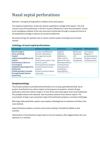
- 1. Nasal septal perforations Definition : through & through defect or defects of the nasal septum. The majority of perforations involve the anterior quadrilateral cartilage of the septum. The most common cause of the perforation in the UK is trauma( selfinduced or nasal instrumentation). Injuries to the cartilaginous skeleton of the nose may result in perforation through a compound fracture of the quadrilateral cartilage in absence of any external deformity. Recreational drugs, for example crack or cocaine snorted nasally increasingly cause of septal ncerosis. Aetiology of nasal septal perforations: Traumatic Surface irritants Infections Neoplastic Inflammatory Nasal surgery Cocaine insufflations, Syphilis Melanoma, Nose pricking Heroine inhalation, Typhoid Squamous Septal Decongestant nasal Diphtheria cell cauterization(bilateral) sprays, Tuberculosis carcinoma, Septal Lime,cement, glass, salt, Rhinoscleroma, Lymphoma haematoma/abscess dust,tar, paste, Rhinosporidiosis Crysurgery Fumes(chromic/sulfuric Leishmaniasis Intubation(nasogastric) Arsenicals,mercurials, Actinomycosis Radiation calcium Candidiasis Stab& gunshot wounds nitrate,cyanide,sodium Aspergillosis. Foreign body( button carbonate. batteries) Sarcoidosis Wegener’s granulomatosis, Systemic lupus erythematosus Symptomatology The principle symptoms associated with perforations are crusting ,epistaxis& whistling. Size & position of perforation has a direct relation on the presence of symptoms. Anterior & large perforations where the anterior margin is in front of the nasal valve appear to be most troublesome. This probably relates to the relatively slow mucociliary clearance from anterior septum. The accumulation of large crusts around the margin of the perforation produces a sensation of blockage. With large stable perforation, patient may complain of blockage due to turbulence of airflow in the nasal vestibule. Huge perforations produce a common nasal cavity resulting in rhinolalia & inability to clear secretion. Inflammation in the perforation margin leads to recurrent epistaxis, often triggered when the crust separates or is removed.
- 2. Clinical assessment An overall assessment of the external & internal nose skeleton should be made together with nasal endoscopy. Endoscopy is the best method to assess the margin & state of residual septum. Special investigations Full blood count & ESR Urea & electrolytes Urine analysis C-ANCA Treponemal antibodies ACE titre X-Ray chest A nasal swab Routine biopsy of septal perforation to cxclude vasculitis & malignancy(irregular margin). Management The majority of septal perforation are asymptomatic & require no specific treatment. The management can be divided into three groups. 1)Prevention Meticulous attention to technique is required in elevation of mucoperichondrial flap in correct plain, particularly avoiding overlapping bilateral mucosal tears. Starting the dissection on the easier (usually the concave) side to raise one intact flap first &using an autograft of cartilage or ethmoid plate to support any tears, is good practice. Septal ulcer or inflammation should be treated by withdrawing the source of the irritant, eradication of pathogens, avoidance of aggressive cleaning & use of mucosal protectants(petroleum jelly for 6 to 8 weeks) 2)Nonsurgical This , essentially is aimed at reducing the drying effect in the nasal mucosa to alleviate crusts & epistaxis. Alkaline nasal douches(twice daily), normal saline sprays & petroleum-based ointments are commonly used for 6 to 8 weeks will be effective in maturing the margins of a perforation. Obturation: Inert sheet(usually silastic sheet) is placed to prevent drying & encourage epitheliazation over bone or cartilage septum to create a mature mucosal edge. The main benefit of
- 3. obturator appears to be the control of whistling &epistaxis. The main disadvantage are patient interference & movement of the mobile membranous septum against the edge of the obturator can lead to granuloma formation. 3)Surgical repair of septal perforation The varity of repair may be classified as: a)Free grafts: simple or composite autografts. b)Pedicle flaps: local nasal mucosal, buccal mucosal, composite septal cartilage& mucosa, c)Rotation /advancement of mucoperichondrial or mucoperiosteal flaps: When there is sufficient residual mucosa in the nasal fossa which can be mobilized & transported allowing direct suturing of the mucosal defect. The second is the routine use of a connective tissue interposition graft to support the repair & epitheliazation. Bilateral mucosal flaps with the main blood supply derived from the sphenopalatine vessels form the basis of most techniques. Interposition grafts using temporal fascia, mastoid periosteum, cartilage(septal, auricular, rib cartilage) bone(either locally or from rib or iliac crest). Small defects can be closed with bipedical flaps, by making relieving incisions either superiorly or inferiorly. Large defects upto 2 cm require larger flaps which are pedicled only posteriorly based on sphenopalatine vessels & are effectively transposition/rotation flaps.