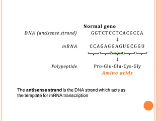
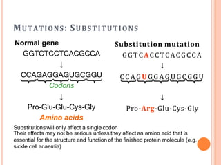
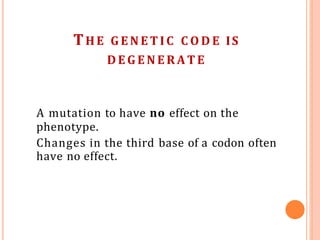
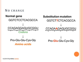
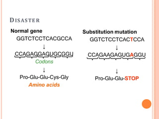
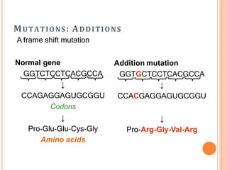
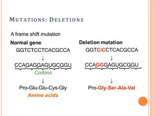
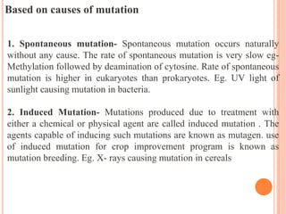
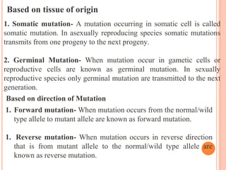
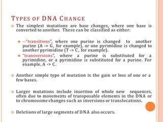
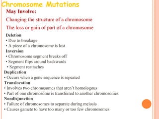
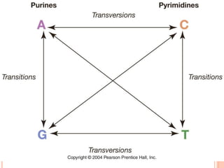
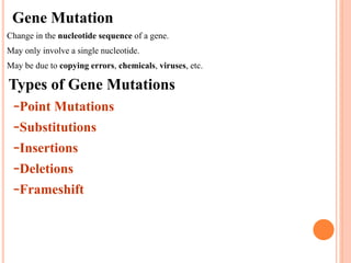
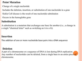
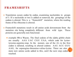
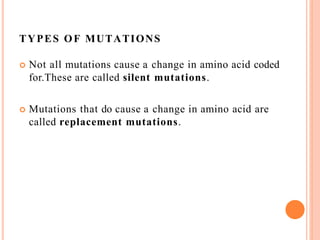
G E N E MUTATIONS
MUTATIONS
Any change in the DNA sequence of an organism is a
mutation.
Mutations are the source of the altered versions of genes that
provide the raw material for evolution.
Most mutations have no effect on the organism, especially
among the eukaryotes, because a large portion of the DNA is
not in genes and thus does not affect the organism’s phenotype.
Of the mutations that do affect the phenotype, the most
common effect of mutations is lethality, because most genes are
necessary for life.
Only a small percentage of mutations causes a visible but non-
lethal change in the phenotype.
Mutation which causes changes in base sequence of a gene are known
as gene mutation.
Changes in the nucleotide sequence of DNA
May occur in somatic cells (aren’t passed to offspring).
May occur in gametes (eggs & sperm) and be passed to offspring.
Factor or agents causing mutation are known as mutagens.
HISTORY
• English farmer Seth Wright recorded case of mutation first time in 1791 in male
lamb with unusual short legs.
• The term mutation is coined by Hugo de Vries in 1900 by his observation in
Oenothera
• Systematic study of mutation was started in 1910 when Morgan genetically
analyzed white eye mutant of Drosophila
• H. J. Muller induced mutation in Drosophila by using X- rays in 1927 ; he was
awarded with Nobel prize in 1946
CHARACTERISTICS OF MUTATION
• Generally mutant alleles are recessive to their wild type or normal alleles.
• Most mutations have harmful effect, but some mutations are beneficial
• Spontaneous mutations occurs at very low rate.
• Some genes shows high rate of mutation such genes are called as mutable
gene.
• Highly mutable sites within a gene are known as hot spots.
• Mutation can occur in any tissue/cell (somatic or germinal) of an organism.
Classification of Mutation
Based on the survival of an individual
1. Lethal mutation – when mutation causes death of all individuals
undergoing mutation are known as lethal.
2. Sub lethal mutation - causes death of 90% individuals.
3. Sub vital mutation– such mutation kills less than 90% individuals.
4. Vital mutation -when mutation don’t affect the survival of an
individual are known as vital.
5. Supervital mutation – This kind of mutation enhances the survival
of individual.
Are Mutations Helpful or Harmful?
Mutations happen regularly
Almost all mutations are neutral
Chemicals & UV radiation cause mutations
Many mutations are repaired by enzymes Some type of skin cancers and
leukemia result from somatic mutations
Some mutations may improve an organism’s survival (beneficial)
GE N E MUTATIONS WHICH AFFECT O NLY
O N E G E N E
DNA sequence
↓
mRNA sequence
↓
Polypeptide
Transcription
Translation
Quick Review: What is a chromosome?
A chromosome is a DNA molecule that is tightly coiled around
proteins called histones, which support its structure, to form a
thread-like structures.
DNA (antisense strand)
mR NA
Polypeptide
Normal gene
GGTCTCCTCACGCCA
↓
C C A G A G G A G U G C G G U
Codons
↓
Pro-Glu-Glu-Cys-Gly
Amino acids
The antisense strand is the DNA strand which acts as
the template for mRNA transcription
MUTATIONS: SUBSTITUTIONS
Substitution mutation
GGTCACCTCACGCCA
↓
C C A G U G G A G U G C G G U
↓
Pro-Arg-Glu-Cys-Gly
Their effects may not be serious unless they affect an amino acid that is
essential for the structure and function of the finished protein molecule (e.g.
sickle cell anaemia)
Normal gene
GGTCTCCTCACGCCA
↓
CCAGAGGAGUGCGGU
Codons
↓
Pro-Glu-Glu-Cys-Gly
Amino acids
Substitutions will only affect a single codon
THE G E N E T I C C O D E IS
D E G E N E R A T E
A mutation to have no effect on the
phenotype.
Changes in the third base of a codon often
have no effect.
NO C H A N G E
Normal gene
GGTCTCCTCACGCCA
↓
CCAGAGGAGUGCGGU
Codons
↓
Pro-Glu-Glu-Cys-Gly
Amino acids
Substitution mutation
GGTCTTCTCACGCCA
↓
CCAGAAGAGUGCGGU
↓
Pro-Glu-Glu-Cys-Gly
© 2010 Paul Billiet ODWS
DISASTE R
Normal gene
GGTCTCCTCACGCCA
↓
CCAGAGGAGUGCGGU
Codons
↓
Pro-Glu-Glu-Cys-Gly
Amino acids
Substitution mutation
GGTCTCCTCACTCCA
↓
CCAGAAGAGUGAGGU
↓
Pro-Glu-Glu-STOP
MUTATIONS: INVERSION
Normal gene
GGTCTCCTCACGCCA
↓
CCAGAGGAGUGCGGU
Codons
↓
Pro-Glu-Glu-Cys-Gly
Amino acids
Inversion mutation
GGTCCTCTCACGCCA
↓
CCAGGAGAGUGCGGU
↓
Pro-Gly-Glu-Cys-Gly
Inversion mutations, also, only affect a small part of the
gene
MUTATIONS: ADDITIONS
A frame shift mutation
Normal gene
GGTCTCCTCACGCCA
↓
CCAGAGGAGUGCGGU
Codons
↓
Pro-Glu-Glu-Cys-Gly
Amino acids
Addition mutation
GGTGCTCCTCACGCCA
↓
CCACGAGGAGUGCGGU
↓
Pro-Arg-Gly-Val-Arg
MUTATIONS: DELETIONS
A frame shift mutation
Normal gene
GGTCTCCTCACGCCA
↓
CCAGAGGAGUGCGGU
Codons
↓
Pro-Glu-Glu-Cys-Gly
Amino acids
Deletion mutation
GGTC/CCTCACGCCA
↓
CCAGGGAGUGCGGU
↓
Pro-Gly-Ser-Ala-Val
MUTATIONS O F HAEMOGLOBIN
 Haemoglobin is a tetramer = 2  and 2 -
chains
 The genes for these polypeptides are found on
different chromosomes
 The -chain gene is found on chromosome 11
 The -chain gene is found on chromosome 16
 The nucleotide sequences have been worked
out
 Several inherited diseases occur on the -
chain, which contains 146 amino acids.
 H A EMOG L OBIN SE N SE STRAN D
C DNA S E Q U E N C E
 cDNA (complementary DNA) is obtained by
back-transcribing the mRNA used to translate
the polypeptide
 So cDNA has no introns
 This is done using reverse transcriptase
enzyme.
Mutation Codon Change to DNA
sense strand
Change in
Amino Acid
S (sickle cell
anaemia)
6 GAG to GTG Glu to Val
C (cooley’s
syndrome)
6 GAG to AAG Glu to Lys
Sickle Cell Anaemia
Blood smear (normal)
Image Credit: http://lifesci.rutgers.edu/~babiarz/
Sickle cell anemia
Image Credit: http://explore.ecb.org/
Based on causes of mutation
1. Spontaneous mutation- Spontaneous mutation occurs naturally
without any cause. The rate of spontaneous mutation is very slow eg-
Methylation followed by deamination of cytosine. Rate of spontaneous
mutation is higher in eukaryotes than prokaryotes. Eg. UV light of
sunlight causing mutation in bacteria.
2. Induced Mutation- Mutations produced due to treatment with
either a chemical or physical agent are called induced mutation . The
agents capable of inducing such mutations are known as mutagen. use
of induced mutation for crop improvement program is known as
mutation breeding. Eg. X- rays causing mutation in cereals
Based on tissue of origin
1. Somatic mutation- A mutation occurring in somatic cell is called
somatic mutation. In asexually reproducing species somatic mutations
transmits from one progeny to the next progeny.
2. Germinal Mutation- When mutation occur in gametic cells or
reproductive cells are known as germinal mutation. In sexually
reproductive species only germinal mutation are transmitted to the next
generation.
Based on direction of Mutation
1. Forward mutation- When mutation occurs from the normal/wild
type allele to mutant allele are known as forward mutation.
1. Reverse mutation- When mutation occurs in reverse direction
that is from mutant allele to the normal/wild type allele are
known as reverse mutation.
Type of trait affected
1. Visible mutation- Those mutation which affects on phenotypic character and can
be detected by normal observation are known as visible mutation.
2. Biochemical mutation- Mutation which affect the production of biochemicals
and which does not show any phenotypic character are known as biochemical
mutation.
TYPES OF DNA CHA NGE
 The simplest mutations are base changes, where one base is
converted to another. These can be classified as either:
⚫ --“transitions”, where one purine is changed to another
purine (A -> G, for example), or one pyrimidine is changed to
another pyrimidine (T -> C, for example).
⚫ “transversions”, where a purine is substituted for a
pyrimidine, or a pyrimidine is substituted for a purine. For
example, A -> C.
 Another simple type of mutation is the gain or loss of one or a
few bases.
 Larger mutations include insertion of whole new sequences,
often due to movements of transposable elements in the DNA or
to chromosome changes such as inversions or translocations.
 Deletions of large segments of DNA also occurs.
Chromosome Mutations
•May Involve:
•Changing the structure of a chromosome
•The loss or gain of part of a chromosome
•Deletion
•• Due to breakage
•• A piece of a chromosome is lost
•Inversion
•• Chromosome segment breaks off
•• Segment flips around backwards
• • Segment reattaches
Duplication
• Occurs when a gene sequence is repeated
Translocation
• Involves two chromosomes that aren’t homologous
• Part of one chromosome is transferred to another chromosomes
Nondisjunction
• Failure of chromosomes to separate during meiosis
• Causes gamete to have too many or too few chromosomes
Fig 4.4
Gene Mutation
•Change in the nucleotide sequence of a gene.
•May only involve a single nucleotide.
•May be due to copying errors, chemicals, viruses, etc.
Types of Gene Mutations
–Point Mutations
–Substitutions
–Insertions
–Deletions
–Frameshift
•Point Mutation
•Change of a single nucleotide
•Includes the deletion, insertion, or substitution of one nucleotide in a gene
•Sickle Cell disease is the result of one nucleotide substitution
•Occurs in the hemoglobin gene
Substitution
A substitution is a mutation that exchanges one base for another (i.e., a change in
a single "chemical letter" such as switching an A to a G).
Insertion
The addition of one or more nucleotide base pairs into a DNA sequence
•Deletion
A part of a chromosome or a sequence of DNA is lost during DNA replication.
Any number of nucleotides can be deleted, from a single base to an entire piece of
chromosome.
FRAMESHIFTS
•Inserting or deleting one or more nucleotides
•Changes the “reading frame” like changing a sentence
•Proteins built incorrectly
•Original:
–The fat cat ate the wee rat.
•Frame Shift (“a” added):
– The fat caa tet hew eer at.
FRAMESHIFTS
 Translation occurs codon by codon, examining nucleotides in groups
of 3. If a nucleotide or two is added or removed, the groupings of the
codons is altered. This is a “frameshift” mutation, where the reading
frame of the ribosome is altered.
 Frameshift mutations result in all amino acids downstream from the
mutation site being completely different from wild type. These
proteins are generally non-functional.
⚫ example Hb-α Wayne. The final codons of the alpha globin chain
are usually AAA UAC CGU UAA, which code for lysine-
tyrosine-arginine-stop. In the mutant, one of the A’s in the first
codon is deleted, resulting in altered codons: AAU ACC GUU
AAG, for asparagine-threonine-valine-lysine. There are also 5
more new amino acids added to this, until the next stop codon is
reached.
TYPES OF MUTATIONS
 Not all mutations cause a change in amino acid coded
for.These are called silent mutations.
 Mutations that do cause a change in amino acid are
called replacement mutations.
TYPES OF MUTATION
 Mutations can be classified according to their effects on the protein
or mRNA) produced by the gene that is mutated.
1. Silent mutations (synonymous mutations). Since the genetic code is
degenerate, several codons produce the same amino acid. Especially,
third base changes often have no effect on the amino acid sequence of
the protein. These mutations affect the DNA but not the protein.
Therefore, they have no effect on the organism’s phenotype.
2. Missense mutations. Missense mutations substitute one amino acid for
another. Some missense mutations have very large effects, while others
have minimal or no effect. It depends on where the mutation occurs in
the protein’s structure, and how big a change in the type of amino acid
it is.
⚫ Example: HbS, sickle cell hemoglobin, is a change in the beta-
globin gene, where a GAG codon is converted to GUG. GAG
codes for glutamic acid, which is a hydrophilic amino acid that
carries a -1 charge, and GUG codes for valine, a hydrophobic
amino acid. This amino acid is on the surface of the globin
molecule, exposed to water. Under low oxygen conditions, valine’s
affinity for hydrophobic environments causes the hemoglobin to
crystallize out of solution.
TYPES OF MUTATION
3. Nonsense mutations convert an amino acid into a stop codon. The effect is
to shorten the resulting protein. Sometimes this has only a little effect, as
the ends of proteins are often relatively unimportant to function. However,
often nonsense mutations result in completely non-functional proteins.
⚫ an example: Hb-β McKees Rock. Normal beta-globin is 146 amino
acids long. In this mutation, codon 145 UAU (codes for tyrosine) is
mutated to UAA (stop). The final protein is thus 143 amino acids
long. The clinical effect is to cause overproduction of red blood cells,
resulting in thick blood subject to abnormal clotting and bleeding.
4. Sense mutations are the opposite of nonsense mutations. Here, a stop codon
is converted into an amino acid codon. Since DNA outside of protein-coding
regions contains an average of 3 stop codons per 64, the translation process
usually stops after producing a slightly longer protein.
⚫ Example: Hb-α Constant Spring. alpha-globin is normally 141 amino
acids long. In this mutation, the stop codon UAA is converted to
CAA (glutamine). The resulting protein gains 31 additional amino
acids before it reaches the next stop codon. This results in
thalassemia, a severe form of anemia.
A “reversion” is a second mutation that reverse the effects of an
initial mutation, bringing the phenotype back to wild type (or
almost).
Frameshift mutations sometimes have “second site reversions”,
where a second frameshift downstream from the first frameshift
reverses the effect.
Example: consider Hb Wayne above. If another mutation
occurred that added a G between the 2 C’s in the second
codon, the resulting codons would be: AAU ACG CGU
UAA, or asparagine-threonine-arginine-stop. Note that the
last 2 codons are back to the original. Two amino acids are
still altered, but the main mutational effect has been reverted to
wild type.
Reversions
mRNA PROBLEMS
 Although many mutations affect the protein sequence directly, it is
possible to affect the protein without altering the codons.
 Splicing mutations. Intron removal requires several specific
sequences. Most importantly, introns are expected to start with GT
and end in AG. Several beta globin mutations alter one of these
bases. The result is that one of the 2 introns is not spliced out of the
mRNA. The polypeptide translated from these mRNAs is very
different from normal globin, resulting in severe anemia.
 Polyadenylation site mutations. The primary RNA transcript of a
gene is cleaved at the poly-A addition site, and 100-200 A’s are added
to the 3’ end of the RNA. If this site is altered, an abnormally long
and unstable mRNA results. Several beta globin mutations alter this
site: one example is AATAAA -> AACAAA. Moderate anemia was
the result.
TRINUCLEOTIDE REPEATS
 A fairly new type of mutation has been described, in which a
particular codon is repeated.
 During replication, DNA polymerase can “stutter” when it replicates
several tandem copies of a short sequence. For example,
CAGCAGCAGCAG, 4 copies of CAG, will occasionally be converted to
3 copies or 5 copies by DNA polymerase stuttering.
 Outside of genes, this effect produces useful genetic markers called
SSR (simple sequence repeats).
 Within a gene, this effect can cause certain amino acids to be
repeated many times within the protein. In some cases this causes
disease.
 The Huntington’s disease gene normally has between 11 and 33
copies of CAG (codon for glutamine) in a row. The number
occasionally changes. People with HD have 37 or more copies, up to
200.
 Interestingly, the age of onset of the disease is related to the number
of CAG repeats present: the more repeats, the earlier the onset.
G ERMINALVS. S OMATIC M UTATIONS
 Mutations can occur in any cell. They only affect future generations if
they occur in the cells that produce the gametes: these are “germinal” or
“germ line” mutations.
 Mutations in other cells are rarely noticed, except in the case of cancer,
where the mutated cell proliferates uncontrollably. Mutations in cells
other than germ line cells are “somatic” mutations.
 A human body contains 1013 - 1014 cells approximately. The average
mutation rate for any given nucleotide is about 1 in 109. That is, on the
average 1 cell in 109 has that particular nucleotide altered. This means
that virtually every possible base change mutation occurs repeatedly in
our body cells.
MUTATION RATES
 Most data on mutations comes from analysis of loss-
of-function mutations.
 Loss-of-function mutations cause gene to produce
a non-working protein.
 Examples of loss-of-function mutations include:
insertions and deletions, mutation to a stop codon
and insertion of jumping genes.
MUTATION RATES
 Some mutations
phenotypic changes.
cause readily identified
 E.g. Achrondoplastic dwarfism is a dominant
disorder. An Achrondoplastic individual’s
condition must be the result of a mutation, if his
parents do not have the condition.
MUTATION RATES
 Human estimate
mutations/genome/generation.
is 1.6
 In Drosophila rate is only 0.14 m/g/g, but
when corrected for number of cell
divisions needed to produce sperm (400 in
humans 25 in Drosophila) mutation rates
per cell division are very similar.

Mutation.pptx

  • 1.
    G E NE MUTATIONS
  • 2.
    MUTATIONS Any change inthe DNA sequence of an organism is a mutation. Mutations are the source of the altered versions of genes that provide the raw material for evolution. Most mutations have no effect on the organism, especially among the eukaryotes, because a large portion of the DNA is not in genes and thus does not affect the organism’s phenotype. Of the mutations that do affect the phenotype, the most common effect of mutations is lethality, because most genes are necessary for life. Only a small percentage of mutations causes a visible but non- lethal change in the phenotype. Mutation which causes changes in base sequence of a gene are known as gene mutation. Changes in the nucleotide sequence of DNA May occur in somatic cells (aren’t passed to offspring). May occur in gametes (eggs & sperm) and be passed to offspring. Factor or agents causing mutation are known as mutagens.
  • 3.
    HISTORY • English farmerSeth Wright recorded case of mutation first time in 1791 in male lamb with unusual short legs. • The term mutation is coined by Hugo de Vries in 1900 by his observation in Oenothera • Systematic study of mutation was started in 1910 when Morgan genetically analyzed white eye mutant of Drosophila • H. J. Muller induced mutation in Drosophila by using X- rays in 1927 ; he was awarded with Nobel prize in 1946 CHARACTERISTICS OF MUTATION • Generally mutant alleles are recessive to their wild type or normal alleles. • Most mutations have harmful effect, but some mutations are beneficial • Spontaneous mutations occurs at very low rate. • Some genes shows high rate of mutation such genes are called as mutable gene. • Highly mutable sites within a gene are known as hot spots. • Mutation can occur in any tissue/cell (somatic or germinal) of an organism.
  • 4.
    Classification of Mutation Basedon the survival of an individual 1. Lethal mutation – when mutation causes death of all individuals undergoing mutation are known as lethal. 2. Sub lethal mutation - causes death of 90% individuals. 3. Sub vital mutation– such mutation kills less than 90% individuals. 4. Vital mutation -when mutation don’t affect the survival of an individual are known as vital. 5. Supervital mutation – This kind of mutation enhances the survival of individual.
  • 5.
    Are Mutations Helpfulor Harmful? Are Mutations Helpful or Harmful? Mutations happen regularly Almost all mutations are neutral Chemicals & UV radiation cause mutations Many mutations are repaired by enzymes Some type of skin cancers and leukemia result from somatic mutations Some mutations may improve an organism’s survival (beneficial)
  • 6.
    GE N EMUTATIONS WHICH AFFECT O NLY O N E G E N E DNA sequence ↓ mRNA sequence ↓ Polypeptide Transcription Translation
  • 7.
    Quick Review: Whatis a chromosome? A chromosome is a DNA molecule that is tightly coiled around proteins called histones, which support its structure, to form a thread-like structures.
  • 8.
    DNA (antisense strand) mRNA Polypeptide Normal gene GGTCTCCTCACGCCA ↓ C C A G A G G A G U G C G G U Codons ↓ Pro-Glu-Glu-Cys-Gly Amino acids The antisense strand is the DNA strand which acts as the template for mRNA transcription
  • 9.
    MUTATIONS: SUBSTITUTIONS Substitution mutation GGTCACCTCACGCCA ↓ CC A G U G G A G U G C G G U ↓ Pro-Arg-Glu-Cys-Gly Their effects may not be serious unless they affect an amino acid that is essential for the structure and function of the finished protein molecule (e.g. sickle cell anaemia) Normal gene GGTCTCCTCACGCCA ↓ CCAGAGGAGUGCGGU Codons ↓ Pro-Glu-Glu-Cys-Gly Amino acids Substitutions will only affect a single codon
  • 10.
    THE G EN E T I C C O D E IS D E G E N E R A T E A mutation to have no effect on the phenotype. Changes in the third base of a codon often have no effect.
  • 11.
    NO C HA N G E Normal gene GGTCTCCTCACGCCA ↓ CCAGAGGAGUGCGGU Codons ↓ Pro-Glu-Glu-Cys-Gly Amino acids Substitution mutation GGTCTTCTCACGCCA ↓ CCAGAAGAGUGCGGU ↓ Pro-Glu-Glu-Cys-Gly © 2010 Paul Billiet ODWS
  • 12.
    DISASTE R Normal gene GGTCTCCTCACGCCA ↓ CCAGAGGAGUGCGGU Codons ↓ Pro-Glu-Glu-Cys-Gly Aminoacids Substitution mutation GGTCTCCTCACTCCA ↓ CCAGAAGAGUGAGGU ↓ Pro-Glu-Glu-STOP
  • 13.
    MUTATIONS: INVERSION Normal gene GGTCTCCTCACGCCA ↓ CCAGAGGAGUGCGGU Codons ↓ Pro-Glu-Glu-Cys-Gly Aminoacids Inversion mutation GGTCCTCTCACGCCA ↓ CCAGGAGAGUGCGGU ↓ Pro-Gly-Glu-Cys-Gly Inversion mutations, also, only affect a small part of the gene
  • 14.
    MUTATIONS: ADDITIONS A frameshift mutation Normal gene GGTCTCCTCACGCCA ↓ CCAGAGGAGUGCGGU Codons ↓ Pro-Glu-Glu-Cys-Gly Amino acids Addition mutation GGTGCTCCTCACGCCA ↓ CCACGAGGAGUGCGGU ↓ Pro-Arg-Gly-Val-Arg
  • 15.
    MUTATIONS: DELETIONS A frameshift mutation Normal gene GGTCTCCTCACGCCA ↓ CCAGAGGAGUGCGGU Codons ↓ Pro-Glu-Glu-Cys-Gly Amino acids Deletion mutation GGTC/CCTCACGCCA ↓ CCAGGGAGUGCGGU ↓ Pro-Gly-Ser-Ala-Val
  • 16.
    MUTATIONS O FHAEMOGLOBIN  Haemoglobin is a tetramer = 2  and 2 - chains  The genes for these polypeptides are found on different chromosomes  The -chain gene is found on chromosome 11  The -chain gene is found on chromosome 16  The nucleotide sequences have been worked out  Several inherited diseases occur on the - chain, which contains 146 amino acids.
  • 17.
     H AEMOG L OBIN SE N SE STRAN D C DNA S E Q U E N C E  cDNA (complementary DNA) is obtained by back-transcribing the mRNA used to translate the polypeptide  So cDNA has no introns  This is done using reverse transcriptase enzyme.
  • 18.
    Mutation Codon Changeto DNA sense strand Change in Amino Acid S (sickle cell anaemia) 6 GAG to GTG Glu to Val C (cooley’s syndrome) 6 GAG to AAG Glu to Lys
  • 19.
    Sickle Cell Anaemia Bloodsmear (normal) Image Credit: http://lifesci.rutgers.edu/~babiarz/ Sickle cell anemia Image Credit: http://explore.ecb.org/
  • 20.
    Based on causesof mutation 1. Spontaneous mutation- Spontaneous mutation occurs naturally without any cause. The rate of spontaneous mutation is very slow eg- Methylation followed by deamination of cytosine. Rate of spontaneous mutation is higher in eukaryotes than prokaryotes. Eg. UV light of sunlight causing mutation in bacteria. 2. Induced Mutation- Mutations produced due to treatment with either a chemical or physical agent are called induced mutation . The agents capable of inducing such mutations are known as mutagen. use of induced mutation for crop improvement program is known as mutation breeding. Eg. X- rays causing mutation in cereals
  • 21.
    Based on tissueof origin 1. Somatic mutation- A mutation occurring in somatic cell is called somatic mutation. In asexually reproducing species somatic mutations transmits from one progeny to the next progeny. 2. Germinal Mutation- When mutation occur in gametic cells or reproductive cells are known as germinal mutation. In sexually reproductive species only germinal mutation are transmitted to the next generation. Based on direction of Mutation 1. Forward mutation- When mutation occurs from the normal/wild type allele to mutant allele are known as forward mutation. 1. Reverse mutation- When mutation occurs in reverse direction that is from mutant allele to the normal/wild type allele are known as reverse mutation.
  • 22.
    Type of traitaffected 1. Visible mutation- Those mutation which affects on phenotypic character and can be detected by normal observation are known as visible mutation. 2. Biochemical mutation- Mutation which affect the production of biochemicals and which does not show any phenotypic character are known as biochemical mutation.
  • 23.
    TYPES OF DNACHA NGE  The simplest mutations are base changes, where one base is converted to another. These can be classified as either: ⚫ --“transitions”, where one purine is changed to another purine (A -> G, for example), or one pyrimidine is changed to another pyrimidine (T -> C, for example). ⚫ “transversions”, where a purine is substituted for a pyrimidine, or a pyrimidine is substituted for a purine. For example, A -> C.  Another simple type of mutation is the gain or loss of one or a few bases.  Larger mutations include insertion of whole new sequences, often due to movements of transposable elements in the DNA or to chromosome changes such as inversions or translocations.  Deletions of large segments of DNA also occurs.
  • 24.
    Chromosome Mutations •May Involve: •Changingthe structure of a chromosome •The loss or gain of part of a chromosome •Deletion •• Due to breakage •• A piece of a chromosome is lost •Inversion •• Chromosome segment breaks off •• Segment flips around backwards • • Segment reattaches Duplication • Occurs when a gene sequence is repeated Translocation • Involves two chromosomes that aren’t homologous • Part of one chromosome is transferred to another chromosomes Nondisjunction • Failure of chromosomes to separate during meiosis • Causes gamete to have too many or too few chromosomes
  • 25.
  • 26.
    Gene Mutation •Change inthe nucleotide sequence of a gene. •May only involve a single nucleotide. •May be due to copying errors, chemicals, viruses, etc. Types of Gene Mutations –Point Mutations –Substitutions –Insertions –Deletions –Frameshift
  • 27.
    •Point Mutation •Change ofa single nucleotide •Includes the deletion, insertion, or substitution of one nucleotide in a gene •Sickle Cell disease is the result of one nucleotide substitution •Occurs in the hemoglobin gene Substitution A substitution is a mutation that exchanges one base for another (i.e., a change in a single "chemical letter" such as switching an A to a G). Insertion The addition of one or more nucleotide base pairs into a DNA sequence •Deletion A part of a chromosome or a sequence of DNA is lost during DNA replication. Any number of nucleotides can be deleted, from a single base to an entire piece of chromosome.
  • 28.
    FRAMESHIFTS •Inserting or deletingone or more nucleotides •Changes the “reading frame” like changing a sentence •Proteins built incorrectly •Original: –The fat cat ate the wee rat. •Frame Shift (“a” added): – The fat caa tet hew eer at.
  • 29.
    FRAMESHIFTS  Translation occurscodon by codon, examining nucleotides in groups of 3. If a nucleotide or two is added or removed, the groupings of the codons is altered. This is a “frameshift” mutation, where the reading frame of the ribosome is altered.  Frameshift mutations result in all amino acids downstream from the mutation site being completely different from wild type. These proteins are generally non-functional. ⚫ example Hb-α Wayne. The final codons of the alpha globin chain are usually AAA UAC CGU UAA, which code for lysine- tyrosine-arginine-stop. In the mutant, one of the A’s in the first codon is deleted, resulting in altered codons: AAU ACC GUU AAG, for asparagine-threonine-valine-lysine. There are also 5 more new amino acids added to this, until the next stop codon is reached.
  • 30.
    TYPES OF MUTATIONS Not all mutations cause a change in amino acid coded for.These are called silent mutations.  Mutations that do cause a change in amino acid are called replacement mutations.
  • 31.
    TYPES OF MUTATION Mutations can be classified according to their effects on the protein or mRNA) produced by the gene that is mutated. 1. Silent mutations (synonymous mutations). Since the genetic code is degenerate, several codons produce the same amino acid. Especially, third base changes often have no effect on the amino acid sequence of the protein. These mutations affect the DNA but not the protein. Therefore, they have no effect on the organism’s phenotype. 2. Missense mutations. Missense mutations substitute one amino acid for another. Some missense mutations have very large effects, while others have minimal or no effect. It depends on where the mutation occurs in the protein’s structure, and how big a change in the type of amino acid it is. ⚫ Example: HbS, sickle cell hemoglobin, is a change in the beta- globin gene, where a GAG codon is converted to GUG. GAG codes for glutamic acid, which is a hydrophilic amino acid that carries a -1 charge, and GUG codes for valine, a hydrophobic amino acid. This amino acid is on the surface of the globin molecule, exposed to water. Under low oxygen conditions, valine’s affinity for hydrophobic environments causes the hemoglobin to crystallize out of solution.
  • 32.
    TYPES OF MUTATION 3.Nonsense mutations convert an amino acid into a stop codon. The effect is to shorten the resulting protein. Sometimes this has only a little effect, as the ends of proteins are often relatively unimportant to function. However, often nonsense mutations result in completely non-functional proteins. ⚫ an example: Hb-β McKees Rock. Normal beta-globin is 146 amino acids long. In this mutation, codon 145 UAU (codes for tyrosine) is mutated to UAA (stop). The final protein is thus 143 amino acids long. The clinical effect is to cause overproduction of red blood cells, resulting in thick blood subject to abnormal clotting and bleeding. 4. Sense mutations are the opposite of nonsense mutations. Here, a stop codon is converted into an amino acid codon. Since DNA outside of protein-coding regions contains an average of 3 stop codons per 64, the translation process usually stops after producing a slightly longer protein. ⚫ Example: Hb-α Constant Spring. alpha-globin is normally 141 amino acids long. In this mutation, the stop codon UAA is converted to CAA (glutamine). The resulting protein gains 31 additional amino acids before it reaches the next stop codon. This results in thalassemia, a severe form of anemia.
  • 33.
    A “reversion” isa second mutation that reverse the effects of an initial mutation, bringing the phenotype back to wild type (or almost). Frameshift mutations sometimes have “second site reversions”, where a second frameshift downstream from the first frameshift reverses the effect. Example: consider Hb Wayne above. If another mutation occurred that added a G between the 2 C’s in the second codon, the resulting codons would be: AAU ACG CGU UAA, or asparagine-threonine-arginine-stop. Note that the last 2 codons are back to the original. Two amino acids are still altered, but the main mutational effect has been reverted to wild type. Reversions
  • 34.
    mRNA PROBLEMS  Althoughmany mutations affect the protein sequence directly, it is possible to affect the protein without altering the codons.  Splicing mutations. Intron removal requires several specific sequences. Most importantly, introns are expected to start with GT and end in AG. Several beta globin mutations alter one of these bases. The result is that one of the 2 introns is not spliced out of the mRNA. The polypeptide translated from these mRNAs is very different from normal globin, resulting in severe anemia.  Polyadenylation site mutations. The primary RNA transcript of a gene is cleaved at the poly-A addition site, and 100-200 A’s are added to the 3’ end of the RNA. If this site is altered, an abnormally long and unstable mRNA results. Several beta globin mutations alter this site: one example is AATAAA -> AACAAA. Moderate anemia was the result.
  • 35.
    TRINUCLEOTIDE REPEATS  Afairly new type of mutation has been described, in which a particular codon is repeated.  During replication, DNA polymerase can “stutter” when it replicates several tandem copies of a short sequence. For example, CAGCAGCAGCAG, 4 copies of CAG, will occasionally be converted to 3 copies or 5 copies by DNA polymerase stuttering.  Outside of genes, this effect produces useful genetic markers called SSR (simple sequence repeats).  Within a gene, this effect can cause certain amino acids to be repeated many times within the protein. In some cases this causes disease.  The Huntington’s disease gene normally has between 11 and 33 copies of CAG (codon for glutamine) in a row. The number occasionally changes. People with HD have 37 or more copies, up to 200.  Interestingly, the age of onset of the disease is related to the number of CAG repeats present: the more repeats, the earlier the onset.
  • 36.
    G ERMINALVS. SOMATIC M UTATIONS  Mutations can occur in any cell. They only affect future generations if they occur in the cells that produce the gametes: these are “germinal” or “germ line” mutations.  Mutations in other cells are rarely noticed, except in the case of cancer, where the mutated cell proliferates uncontrollably. Mutations in cells other than germ line cells are “somatic” mutations.  A human body contains 1013 - 1014 cells approximately. The average mutation rate for any given nucleotide is about 1 in 109. That is, on the average 1 cell in 109 has that particular nucleotide altered. This means that virtually every possible base change mutation occurs repeatedly in our body cells.
  • 37.
    MUTATION RATES  Mostdata on mutations comes from analysis of loss- of-function mutations.  Loss-of-function mutations cause gene to produce a non-working protein.  Examples of loss-of-function mutations include: insertions and deletions, mutation to a stop codon and insertion of jumping genes.
  • 38.
    MUTATION RATES  Somemutations phenotypic changes. cause readily identified  E.g. Achrondoplastic dwarfism is a dominant disorder. An Achrondoplastic individual’s condition must be the result of a mutation, if his parents do not have the condition.
  • 39.
    MUTATION RATES  Humanestimate mutations/genome/generation. is 1.6  In Drosophila rate is only 0.14 m/g/g, but when corrected for number of cell divisions needed to produce sperm (400 in humans 25 in Drosophila) mutation rates per cell division are very similar.