This case report describes an extremely rare set of vascular anomalies found during a routine cadaver dissection. Specifically:
1) Instead of the celiac trunk, superior mesenteric artery, and inferior mesenteric artery branching from the abdominal aorta as normal, a single arterial trunk arose and supplied the digestive tract.
2) The inferior phrenic and ovarian arteries were absent bilaterally.
3) The portal vein was absent (congenital absence of portal vein), and the digestive tract drained via a single vein into the left renal vein.
4) The right adrenal gland was also absent.
5) A persistent ductus arteriosus was found connecting the pulmonary
![Case Report
Corresponding author:
Shahriar Ahmadpour
Department of Anatomy and Pathobiology, School of Medicine, North
Khorasan University of Medical Sciences,Bojnurd,Iran
Tel: +98-584-2220133, Fax: +98-584-2247124, E-mail: Shahahmadpour@
gmail.com
This is an Open Access article distributed under the terms of the Creative Commons Attribution Non-Commercial License (http://creativecommons.org/licenses/by-nc/3.0/)
which permits unrestricted non-commercial use, distribution, and reproduction in any medium, provided the original work is properly cited.
Copyright © 2014. Anatomy & Cell Biology
http://dx.doi.org/10.5115/acb.2014.47.4.274
pISSN 2093-3665 eISSN 2093-3673
branches of main arteries accompanied with other develop
mental abnormalities is an extremely rare variation. The
Abdominal aorta (AA) is the largest and main artery in
abdominal cavity. Classically its pattern of branching has been
described as paired and single branches. Celiac trunk (CT),
superior mesenteric artery (SMA), and inferior mesenteric
artery (IMA) provide oxygenated blood to embryonic gut and
its derivatives, while the paired visceral branches of AA supply
diaphragm and retroperitoneal glands including kidney and
ovary [2]. The pattern of branching of AA exhibits diverse
variations both in level and types of branching. Various
publications have reported diverse and multiple variations
of the pattern of branching of AA including different
arising levels of arterial branches, separated branches of CT,
combined or fused arterial trunks, absence of one branch and
arterial duplication [3]. In spite of a vast number of reported
cases on variations of the branches of AA, absence of its
multiple branches accompanied with other vascular, heart and
Introduction
Knowledge of vascular variations is of clinical and sur
gical importance. Indeed knowledge about the vascular
variations as an essential pre-requisite in relevant invasive
interventional diagnostic procedure could reduce possible
morbidity and mortality [1]. Most of the vascular variations
are asymptomatic and usually detected during the surgical
procedures such as angiography or morbid anatomy studies.
Among the vascular variations, arterial ones have been
reported in various forms, but multiple absences of the
Multiple absences of the branches of abdominal
aorta with congenital absence of the portal
vein,unilateral adrenal agenesis and persistent
ductus arteriosus in a female cadaver
Shahriar Ahmadpour, Khadijeh Foghi
Department of Anatomy and Pathobiology, School of Medicine, North Khorasan University of Medical Sciences, Bojnurd, Iran
Abstract: We report on an extremely rare case of multiple absences of the branches of abdominal aorta with congenital absence
of the portal vein, unilateral adrenal agenesis and persistent ductus arteriosus in an adult female cadaver. Specifically, instead
of celiac trunk, superior and inferior mesenteric arteries, solely a single arterial trunk aroused from the anterior aspect of
abdominal aorta, inferior phrenic and ovarian arteries were absent in both sides. Left kidneys drained by two veins. There were
not superior, splenic and mesenteric veins, while left renal vein received an additional vein, which run downward and drained
primarily all parts of digestive tract and its associated glands (portal vein did not exist). Right adrenal gland was absent. To the
best of our knowledge, it is the only reported case with such widespread anomalies. We think the importance of this case is
beyond the surgical consideration and needs more profound developmental studies.
Key words: Abdominal aorta, Portal vein, Adrenal, Congenital, Agenesis, Persistent ductus arteriosus
Received October 10, 2014; Revised October 13, 2014; Accepted November 25, 2014](https://image.slidesharecdn.com/f0850889-6d62-4811-b2b6-136204d93d67-170201102512/85/multiple-absence-1-320.jpg)
![Anat Cell Biol 2014;47:274-278 Shahriar Ahmadpour and Khadijeh Foghi276
www.acbjournal.orghttp://dx.doi.org/10.5115/acb.2014.47.4.274
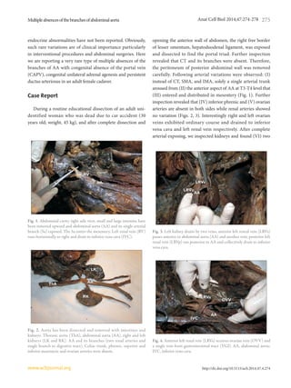
veins drained left kidney as follow; anterior renal vein run
horizontally to right side in front of aorta and other behind
AA and drained to inferior vena cava (Fig. 4). (VII) There
were not superior, splenic and mesenteric veins, while left
renal vein received an additional vein, which run downward
and drained primarily all parts of digestive tract and its
associated gland (portal vein did not exist) (Fig. 5). After
meticulous inspection of renal vessels, we examined both
kidneys and noticed that (VIII) right adrenal gland was absent
(unilateral agenesis). Other findings included two pair’s small
lumbar arteries. Due to the extensive arterial variations of the
branches of AA, we suspected to possible heart anomalies,
so after opening the chest wall we removed and dissected the
heart of cadaver. An interesting finding was persistent ductus
arteriosus (PDA) (Fig. 6).
Discussion
We presented here the most uncommon multiple ar
terial variations accompanied with CAPV, unilateral right
congenital adrenal gland agenesis (UCAA) and persistent
ductus arteriosus which have not been reported before. The
stomach, liver, spleen, pancreas, small and large intestines
were supplied by a single artery which stemmed from the
anterior aspect of AA at level of T3-T4. Additionally the
right and left ovarian arteries were absent, while their venous
pattern showed normal course. Interestingly the portal
vein was absent and venous return of digestive tract were
drained by a single vein which itself ended in the left renal
vein (CAPV). Actually, these extensive vascular variations
(multiple asence of the arterial branches of AA and CAPV)
with heart anomaly and UCAA suggest a cardiovascular/
endocrine syndrome which has not been reported. The
embryological basis for such multiple variations can be
explained as follow. Each primitive dorsal aorta gives off
ventral splanchnic arteries (to the embryonic guts), lateral
splanchnic (to the mesonephric ridge) and somatic arteries
(supply the body wall). CT, SMA, and IMA are derived
from embryonic ventral splanchnic arteries and provide
blood supply to three primitive embryonic guts. The ventral
splanchnic branches undergo a series of developmental
changes including migration (descending), fusion and
regression. In case of CT, in addition to descending from
cervical region to subdiapharagmatic position, three separate
branches (left gastric, splenic and common hepatic) are
united by a series of anastomoses and finally CT is formed.
The lateral splanchnic arteries including suprarenal,
testicular and ovarian arteries persist on each side and retain
their position [4]. In our presented case, the predominant
embryonic scenario has probably been “regression.” In case of
absence ovarian arteries, the only reasonable explanation may
be that after ovaries descending to pelvic cavity, their arteries
(the lateral splanchnic arteries) regressed. In tandem with
arterial scenario, venous system develops during embryonic
period. Portal vein is formed by vetilline venous loop around
the duodenum of the digestive tract, and then enters the
septum transversum during 5-10 embryonic weeks [5].
With respect to the embryological description, it seems the
CAPV in our presented case was secondary to the arterial
variations and the portal vein was replaced with a vein which
was drained to left renal vein. CAPV is an extremely rare
variation (abnormality) and there are few reports on such
abnormalities. Venkat-Raman et al. [6] reported CAPV in
Fig. 5. Left adrenal (LA) gland and left kidney (LK). Adrenal gland
was absent in right side.
LALA
LKLK
Fig. 6. Persistent ductus arteriosus. Pulmonary trunk (PT) connects
to aorta by persistent ductus arteriosus (arrow). BC, brachiocephalic
trunk; LCc, left common carotid; LSc, left subsabclavian.
LScLSc
LCcLCc
AortaAorta
BCBC
PTPT](https://image.slidesharecdn.com/f0850889-6d62-4811-b2b6-136204d93d67-170201102512/85/multiple-absence-3-320.jpg)
![Multipleabsencesofthebranchesofabdominalaorta
http://dx.doi.org/10.5115/acb.2014.47.4.274
Anat Cell Biol 2014;47:274-278 277
www.acbjournal.org
a fetus. Another case of CAPV was reported by Northrup
et al. [7]. Based on previous studies (totally reported 18
cases) there are 2 subtypes of CAPV. In type Ia, the superior
mesenteric vein (SMV) and the splenic vein (SV) do not
join; therefore, no portal vein is formed anatomically. In type
Ib, the SMV and SV do join in a normal manner, but this
confluence drains to the IVC, not the liver. Type Ia CAPV is
associated with atrial septal defect, ventricular sepal defect,
and PDA [7]. Given the findings in our study, we propose a
third subtype a single vein unit left renal vein with PDA and
UCAA. Another set of our findings were multiple arterial
variations. Based on the examination of abdominal organs
(liver, spleen, small intestine and kidney; unpublished data)
in one hand and multiple absence of the branches of AA in
the other hand, we prefer to call these arterial variation as
arterial anomalies. One of these anomalies was the absence
of ovarian arteries. Although there are many reports on the
variations of ovarian arteries [8], bilateral absent of ovarian
arteries is a rare variation and only in one case bilateral absent
of ovarian arteries has been reported [9]. With respect to the
extensive absence of the arterial branches of AA, our reported
case could be considered as one of the most uncommon
arterial variation has been reported so far, because most of the
previous literatures have reported cases with variations of the
pattern of branching of AA including variations in origin of
branches, level of branching, unusual course, common origin
of arteries and single absent of one of the visceral branches
of AA [3]. Among the reported variations, CT variations
has been reported and studied extensively. According to
previous reports CT shows a wide range of variations which
have been analyzed and classified in detail based on the
patterns of branching. For instance, Lipshutz [10] described
different types of CT variations including hepatosplenic,
gastrosplenic and celiacomesenteric trunk, but absent CT was
not described. Complete absent of CT is a rare variation and
its prevalence has been estimated between 0 to maximum 2%
[11]. Morita [12] put forwarded a classification method of CT
variation. According to Morita [12] four types of CT has been
describes as follow as: 1) CT (textbook type), 2) hepatosplenic
trunk, 3) gastrosplenic trunk, 4) hepatogastric trunk, and 5)
absent CT [12]. Yi et al. [13] reported a rare variation of the
absent of the CT in a Japanese cadaver, with the left gastric,
splenic, common hepatic, and superior mesenteric arteries
branching independently from the AA. Matusz et al. [14]
described a case with absent of the CT, while the left gastric
artery, common hepatic artery, and splenic artery originated
directly and independently from the AA. Wu et al. [15]
reported a 69-year-old female complete absence of SMA and
compensatory dilation of the IMA. Accordingly he proposed
a new classification method of superior-inferior mesenteric
arterial variation (SIMAV). In this method SIMAV has been
divided into four types as follow: type I, the normal type;
type II, absent SMA; type III, absent IMA; and type IV, there
is an aberrant middle mesenteric artery [15]. As we have
seen in the present case, many visceral branches of AA were
absent and additionally absence of portal system with other
anomalies recommend a new revision in classification of such
variations. The combination of multiple variation of arterial
branches of AA with CAPV, PDA, and UCAA presented in
our study suggest a new syndrome in living bodies. To the
best of our knowledge it is the only reported case with such
widespread anomalies. We think the importance of such
case is beyond the surgical consideration and needs more
profound developmental studies.
References
1. Dogan NU, Fazliogullari Z, Yilmaz MT, Uysal II, Cicekcibasi
AE, Ulusoy M, Gunaslan P. A complex variation of the parietal
and visceral branches of the abdominal aorta. Int J Morphol
2011;29:90-3.
2. Songür A, Toktaş M, Alkoç O, Acar T, Uzun I, Baş O, Özen OA.
Abdominal aorta and its branches: morphometry. Variations in
autopsy cases. Eur J Gen Med 2010;7:321-5.
3. Shivarama CH, Bhat S, Shetty RK, Avadhani R. Multiple vari
ations of branches of abdominal aorta: a case study. Nitte Univ J
Health Sci 2012;2:48-50.
4. Kalthur SG, Sarda R, Bankar M. Multiple vascular variations of
abdominal vessels in a male cadaver: embryological perspective
and clinical importance. J Morphol Sci 2011;28:152-6.
5. Morse SS, Taylor KJ, Strauss EB, Ramirez E, Seashore JH. Con
genital absence of the portal vein in oculoauriculovertebral
dysplasia (Goldenhar syndrome). Pediatr Radiol 1986;16:437-9.
6. Venkat-Raman N, Murphy KW, Ghaus K, Teoh TG, Higham
JM, Carvalho JS. Congenital absence of portal vein in the fetus: a
case report. Ultrasound Obstet Gynecol 2001;17:71-5.
7. Northrup M, Mendez-Castillo A, Sethi Y, Churchill R. Conge
nital absence of the portal vein with an intrahepatic inferior
vena cava branch showing hepatopetal flow. J Ultrasound Med
2002;21:569-72.
8. Gupta A, Singal R, Singh D. Variations of gonadal artery: em
bryological basis and clinical significance. Int J Biol Med Res
2011;2:1006-10.
9. Kasindye GU, Mwasunga AS, Fabian FM. Bilateral absence of
ovarian artery in a Tanzanian female cadaver: a rare variation.
Int J Anat Var 2012;5:73-5.](https://image.slidesharecdn.com/f0850889-6d62-4811-b2b6-136204d93d67-170201102512/85/multiple-absence-4-320.jpg)