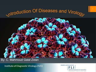
Introduction of Diseases and Virology
•Download as PPTX, PDF•
1 like•726 views

Diseases , symptoms / Virus: Origins, size , shape , properties and classification , Viral cycle , capsid , viral envelope

Recommended
More Related Content
What's hot
What's hot (16)
Similar to Introduction of Diseases and Virology
Similar to Introduction of Diseases and Virology (20)
More from FLI
More from FLI (20)
Recently uploaded
Recently uploaded (20)
Introduction of Diseases and Virology
- 1. By: C. Mahmoud Galal Zidan 1
- 2. Content Diseases and Symptoms Symptoms Presentation Medical Symptoms Types of Diseases and Causes Introduction of Pathogens Introduction of Virology Viruses Origins structure Size Genes Viral life cycle Capsid Viral Envelope Virus classification 2
- 3. What’s Disease ? And How may the disease be caused ? A disease is a particular abnormal condition that negatively affect the structure or function of all or part of an organism, and that is not due to any immediate external injury. Diseases are often known to be medical conditions that are associated with specific symptoms and signs. A disease may be caused by external factors such as pathogens or by internal dysfunctions. 3
- 4. What’s Symptoms ? And What’s the types of symptoms A Symptom is a departure from normal function or feeling which is apparent to a patient, reflecting the presence of an unusual state, or of a disease. A symptom can be subjective or objective. Symptoms may be briefly acute or a more prolonged but acute or chronic, relapsing or remitting . Asymptomatic conditions also exist (e.g. subclinical infections and silent diseases like sometimes, high blood pressure). Constitutional or general symptoms are those related to the systemic effects of a disease (e.g., fever, malaise, anorexia, and weight loss). They affect the entire body rather than a specific organ or location. The symptom that ultimately leads to a diagnosis is called a "cardinal symptom". Non- Specific Positive Negative Positive and Negative 4
- 5. Non- Specific Symptoms Non-specific symptoms are self-reported symptoms that do not indicate a specific disease process or involve an isolated body system. For example, fatigue is a feature of many acute and chronic medical conditions, which may or may not be mental, and may be either a primary or secondary symptom. Fatigue is also a normal, healthy condition when experienced after exertion or at the end of a day. 5
- 6. Positive and Negative Symptoms Positive symptoms are symptoms that are present in the disorder and are not normally experienced by most individuals. It reflects an excess or distortion of normal functions . Examples are hallucinations, delusions, and bizarre behavior. For somatic sensory symptoms , the positive ones are tingling, itching and various sensations described as pricking, bandlike, lightning-like shooting feelings (lancinations), aching, knifelike, twisting, drawing, pulling, tightening, burning, searing, electrical, or raw feelings. The terms used to describe positive sensory symptoms are paresthesia and dysesthesia. Negative symptoms are functions that are normally found in healthy persons, but that are diminished or not present in affected persons. Thus, it is something that has disappeared from a person's normal way of functioning. Examples are social withdrawal, apathy, inability to experience pleasure and defects in attention control. The negative sensory symptoms are diminished or missing sensations. The most common one is numbness. The following terms are used for negative symptoms: Hypoesthesia (hypesthesia) is a partial loss of sensitivity to moderate stimuli, such as pressure, touch, warmness, coldness, etc. Anesthesia is the complete loss of sensitivity to stronger stimuli, such as pinprick. Hypalgesia (analgesia) is loss of sensation to painful stimuli. 6
- 7. Symptoms Presentation ( Can’t and Feeling) Breathe normally Hear normally: losing hearing , sounds are too loud , ringing or hissing in my ears Move one side – arm and/or leg Pass a bowel action normally Pass urine normally Remember normally See properly: Blindness , blurred vision, double vision Sleep normally Smell things normally Speak normally Stop passing watery bowel actions Stop scratching Stop sweating Swallow normally Taste properly Walk normally Write normally Chills Fever Paresthesia (numbness, tingling, electric tweaks) Light-headed Dizzy Dizzy – about to black out Dizzy – with the room spinning around me My mouth is dry Nauseated Sick like I have the flu like I have to vomit Short of breath Sleepy Sweaty Thirsty Tired Weak 7
- 8. Symptoms Presentations (Common Pain Expressions ) abdomen back chest ear head pelvis tooth rectum skin Extremities Chronic pain 8
- 9. Types of Medical Symptoms General Cardiovascular Ear, Nose and Throat Gastrointestinal Integumentary Neurological Obstetric / Gyneacological Ocular Psychiatric Pulmonary Rheumatologic Urologic 9
- 10. General Anorexia (R63.0) Fatigue (R53) Cachexia (R64) Hypothermia (T68) Chills and shivering Jaundice (P58, P59, R17) Convulsions (R56) Muscle weakness (M62.8) Deformity Pyrexia (R50) Discharge Sweats Dizziness / Vertigo (R42) swelling swollen or painful lymph node(s) (I88, L04, R59.1) weight gain (R63.5) 10
- 11. Medical Symptoms Neurological Abnormal posturing Acalculia Insomnia (F51.0, G47.0) Agnosia Anosognosia Lhermitte's sign Alexia Confusion loss of consciousness Amnesia Dysarthria Neck stiffness Anomia Dysdiadochokinesia Paralysis and paresis Aphasia and Apraxia Dysgraphia Paresthesia (R20.2) Ataxia Hallucination Prosopagnosia Cataplexy (G47.4 ) Headache Somnolence (R40.0) Hypokinetic movement disorder Hyperkinetic movement disorder Opisthotonus 11
- 12. Medical Symptoms Obstetric / Gyneacological Ocular Psychiatric Abnormal vaginal bleeding Amaurosis fugax (G45.3) and Amaurosis Amusia Apathy Amenorrhea Blurred vision Anhedonia Confabulation Infertility Dalrymple's sign Anxiety Depression Painful intercourse (N94.1) Double vision (H53.2) Euphoria Delusion Pelvic pain Exophthalmos (H05.2) Irritability Homicidal ideation Vaginal discharge Mydriasis /miosis (H570) Mania (F30) Paranoid ideation Nystagmus Phobia Suicidal ideation 12
- 13. Cardiovascular Ear , Nose and Throat Gastrointestinal Arrhythmia Dry mouth (R68.2) Abdominal pain Nausea Bradycardia (R00.1) Epistaxis (R04.0) Bloating Odynophagia Claudication Halitosis Belching proctalgia fugax Chest pain (R07) Hearing loss Bleeding Pyrosis Palpitations (R00.2) Nasal discharge Constipation Rectal tenesmus Tachycardia (R00.0) Otalgia (H92.0) Diarrhea Steatorrhea Otorrhea (H92.1) Dysphagia Vomiting Sore throat Dyspepsia Flatulence Toothache Fecal incontinence Heartburn Tinnitus Trismus 13
- 14. Medical Symptoms Urologic Pulmonary Integumentary Dysuria Apnea and Hypopnea Alopecia Rash Hematospermia Cough Hirsutism Urticaria Hematuria Dyspnea Hypertrichosis Itching Retrograde ejaculation Hemoptysis Abrasion Nail disease Impotence Pleuritic chest pain Anasarca Laceration Polyuria Sputum Blister Edema Retrograde ejaculation Rheumatologic Bleeding into the skin : Petechia , Purpura , ecchymosis Janeway lesions and Osler’s nodeStrangury Arthralgia Urethral discharge Back pain Urinary frequency Sciatica Urinary incontinence Urinary retention 14
- 15. Main Types and Classification of Diseases There are four main types of disease: Infectious diseases Deficiency diseases Hereditary diseases (including both genetic diseases and non-genetic hereditary diseases), Physiological diseases. Diseases can also be classified in other ways, such as : Communicable diseases . Non-communicable diseases. Diseases may be classified by : cause, pathogenesis (mechanism by which the disease is caused), symptom(s). Alternatively, diseases may be classified according to the organ system involved. A chief difficulty in nosology is that diseases often cannot be defined and classified clearly, especially when cause or pathogenesis are unknown. Thus diagnostic terms often only reflect a symptom or set of symptoms (syndrome). Classical classification of human disease derives from observational correlation between pathological analysis and clinical syndromes. Today it is preferred to classify them by their cause if it is known. The most known and used classification of diseases is the World Health Organization's ICD. This is periodically updated. Currently the last publication is the ICD-10 . 15
- 16. Types of Causes Airborne An airborne disease is any disease that is caused by pathogens and transmitted through the air. Foodborne Foodborne illness or food poisoning is any illness resulting from the consumption of food contaminated with pathogenic bacteria, toxins, viruses, prions or parasites. Infectious Infectious diseases, also known as transmissible diseases or communicable diseases, comprise clinically evident illness resulting from the infection, presence and growth of pathogenic biological agents in an individual host organism. Lifestyle A lifestyle disease is any disease that appears to increase in frequency as countries become more industrialized and people live longer, especially if the risk factors include behavioral choices like a sedentary lifestyle or a diet high in unhealthful foods Non-communicable disease is a medical condition or disease that is non-transmissible. Non-communicable diseases cannot be spread directly from one person to another. 16
- 17. Pathogens Pathogens is used to describe an infectious microorganism or agent, such as Virus Bacterium Protozoan Prion Fungus Small animals, such as certain kinds of worms and insect larvae, can also produce disease. However, these animals are usually, in common parlance, referred to as parasites rather than pathogens. The scientific study of microscopic organisms, including microscopic pathogenic organisms, is called microbiology, while the study of disease that may include these pathogens is called pathology. There are several pathways through which pathogens can invade a host. The principal pathways have different episodic time frames, but soil has the longest or most persistent potential for harboring a pathogen. Diseases in humans that are caused by infectious agents are known as pathogenic diseases, though not all diseases are caused by pathogens. Some diseases, such as Huntington's disease, are caused by inheritance of abnormal genes. 17
- 18. Introduction of Virology Virology is the study of viral submicroscopic, parasitic particles of genetic material contained in a protein coat and virus-like agents. It focuses on the following aspects of viruses: their structure, classification and evolution, their ways to infect and exploit host cells for reproduction, their interaction with host organism physiology and immunity, the diseases they cause, the techniques to isolate and culture them, and their use in research and therapy. Virology is considered to be a subfield of microbiology or of medicine. Viruses are small particles, typically between 20 and 300 nanometers in length, containing RNA or DNA. Viruses require a host cell to replicate. Some of the diseases that are caused by viral pathogens include smallpox , influenza , mumps , measles , chickenpox , ebola , HIV , and rubella . Pathogenic viruses are mainly from the families: Adenoviridae Picornaviridae Herpesviridae Hepadnaviridae Flaviviridae Retroviridae Orthomyxoviridae Paramyxoviridae Papovaviridae Polyomavirus Rhabdoviridae Togaviridae . 18
- 19. Origins Viruses co-exist with life wherever it occurs. They have probably existed since living cells first evolved. The origin of viruses remains unclear because they do not form fossils, so molecular techniques have been the most useful means of hypothesizing how they arose. These techniques rely on the availability of ancient viral DNA or RNA but most of the viruses that have been preserved and stored in laboratories are less than 90 years old. Molecular methods have only been successful in tracing the ancestry of viruses that evolved in the 20th century. New groups of viruses might have repeatedly emerged at all stages of the evolution of life. Three main theories speculate on the origins of viruses: 1. Regressive theory 2. Cellular origin theory 3. Coevolution theory There are problems with all of these hypotheses: the regressive hypothesis does not explain why even the smallest of cellular parasites do not resemble viruses in any way. The escape hypothesis does not explain the structures of virus particles. The coevolution, or virus-first hypothesis, contravenes the definition of viruses, in that they are dependent on host cells. But viruses are recognized as ancient and to have origins that pre-date the divergence of life into the three domains . This discovery has led modern virologists to reconsider and re-evaluate these three classical hypotheses. 19
- 20. Regressive theory Viruses may have once been small cells that parasitised larger cells. Over time, genes not required by their parasitism were lost. The bacteria rickettsia and chlamydia are living cells that, like viruses, can reproduce only inside host cells. They lend credence to this theory, as their dependence on parasitism is likely to have caused the loss of genes that enabled them to survive outside a cell. Cellular origin theory Some viruses may have evolved from bits of DNA or RNA that "escaped" from the genes of a larger organism. The escaped DNA could have come from plasmids pieces of DNA that can move between cells while others may have evolved from bacteria. Coevolution theory Viruses may have evolved from complex molecules of protein and DNA at the same time as cells first appeared on earth and would have depended on cellular life for many millions of years. 20
- 21. Structure A virus particle, also known as a virion, consists of genes made from DNA or RNA which are surrounded by a protective coat of protein called a capsid. The capsid is made of many smaller, identical protein molecules which are called capsomers. The arrangement of the capsomers can either be icosahedral (20-sided), helical or more complex . There is an inner shell around the DNA or RNA called the nucleocapsid, which is formed by proteins. Some viruses are surrounded by a bubble of lipid (fat) called an envelope, which makes them vulnerable to soap and alcohol. 21
- 22. Size Viruses are among the smallest infectious agents and are too small to be seen by light microscopy (in other words, they are sub- microscopic). Most of them can only be seen by electron microscopy. Their sizes range from 20 to 300 nanometers; it would take 30,000 to 750,000 of them, side by side, to stretch to one centimeter (0.39 in). Bacteria are typically around 1 micrometer (1000 nm) in diameter, and the cells of higher organisms a few tens of micrometers. Some viruses such as megaviruses and pandoraviruses are relatively large. At around 1 micrometer, these viruses, which infect amoebae, were discovered in 2003 and 2013. They are around a thousand times larger than influenza viruses and the discovery of these "giant" viruses astonished scientists. 22
- 23. Genes The genes of viruses are made from DNA (deoxyribonucleic acid) and, in many viruses, RNA (ribonucleic acid). The biological information contained in an organism is encoded in its DNA or RNA. Most organisms use DNA, but many viruses have RNA as their genetic material. The DNA or RNA of viruses consists of either a single strand or a double helix. Viruses reproduce rapidly because they have only a few genes compared to humans who have 20,000–25,000. For example, influenza virus has only eight genes and rotavirus has eleven. These genes encode structural proteins that form the virus particle, or non-structural proteins, that are only found in cells infected by the virus. All cells, and many viruses, produce proteins that are enzymes called DNA polymerase and RNA polymerase which make new copies of DNA and RNA. A virus's polymerase enzymes are often much more efficient at making DNA and RNA than the equivalent enzymes of the host cells, but viral RNA polymerase enzymes are error-prone, and this is one of the ways why RNA viruses can mutate to form new strains. In some species of RNA virus, the genes are not on a continuous molecule of RNA, but are separated. The influenza virus, for example, has eight separate genes made of RNA. When two different strains of influenza virus infect the same cell, these genes can mix and produce new strains of the virus in a process called reassortment. 23
- 24. Protein synthesis Proteins are essential to life. Cells produce new protein molecules from amino acid building blocks based on information coded in DNA. Each type of protein is a specialist that usually only performs one function, so if a cell needs to do something new, it must make a new protein. Viruses force the cell to make new proteins that the cell does not need, but are needed for the virus to reproduce. Protein synthesis consists of two major steps: transcription and translation. Transcription is the process where information in DNA, called the genetic code, is used to produce RNA copies called messenger RNA (mRNA). These migrate through the cell and carry the code to ribosomes where it is used to make proteins. This is called translation because the protein's amino acid structure is determined by the mRNA's code. Information is hence translated from the language of nucleic acids to the language of amino acids. 24
- 25. Transcription is the process where information in DNA, called the genetic code, is used to produce RNA copies called messenger RNA (mRNA). These migrate through the cell and carry the code to ribosomes where it is used to make proteins. This is called translation because the protein's amino acid structure is determined by the mRNA's code. Information is hence translated from the language of nucleic acids to the language of amino acids. Some nucleic acids of RNA viruses function directly as mRNA without further modification. For this reason, these viruses are called positive- sense RNA viruses. In other RNA viruses, the RNA is a complementary copy of mRNA and these viruses rely on the cell's or their own enzyme to make mRNA. These are called negative-sense RNA viruses. In viruses made from DNA, the method of mRNA production is similar to that of the cell. The species of viruses called retroviruses behave completely differently: they have RNA, but inside the host cell a DNA copy of their RNA is made with the help of the enzyme reverse transcriptase. This DNA is then incorporated into the host's own DNA, and copied into mRNA by the cell's normal pathways. 25
- 26. Diagram of a typical Eukaryotic cell showing subcellular components. Organelles: 1. Nucleolus 2. Nucleus 3. Ribosome 4. Vesicle 5. Rough endoplasmic reticulum (ER) 6. Golgi apparatus 7. Cytoskeleton 8. smooth ER 9. Mitochondria 10.Vacuole 11.Cytoplasm 12.Lysosome 13.Centrioles within Centrosome 14.Virus particle shown to approximate scale 26
- 27. Viral Life-cycle When a virus infects a cell, the virus forces it to make thousands more viruses. It does this by making the cell copy the virus's DNA or RNA, making viral proteins, which all assemble to form new virus particles. There are six basic, overlapping stages in the life cycle of viruses in living cells: 1. Attachment 2. Penetration 3. Uncoating 4. Replication 5. Assembly 6. Release 27
- 28. Stages in the life cycle of viruses in living cell 1. Attachment is the binding of the virus to specific molecules on the surface of the cell. This specificity restricts the virus to a very limited type of cell. For example, the human immunodeficiency virus (HIV) infects only human T cells, because its surface protein, gp120, can only react with CD4 and other molecules on the T cell's surface. Plant viruses can only attach to plant cells and cannot infect animals. This mechanism has evolved to favour those viruses that only infect cells in which they are capable of reproducing. 2. Penetration follows attachment; viruses penetrate the host cell by endocytosis or by fusion with the cell. 3. Uncoating happens inside the cell when the viral capsid is removed and destroyed by viral enzymes or host enzymes, thereby exposing the viral nucleic acid. 4. Replication of virus particles is the stage where a cell uses viral messenger RNA in its protein synthesis systems to produce viral proteins. The RNA or DNA synthesis abilities of the cell produce the virus's DNA or RNA. 5. Assembly takes place in the cell when the newly created viral proteins and nucleic acid combine to form hundreds of new virus particles. 6. Release occurs when the new viruses escape or are released from the cell. Most viruses achieve this by making the cells burst, a process called lysis. Other viruses such as HIV are released more gently by a process called budding. 28
- 29. Effects on the host cell Viruses have an extensive range of structural and biochemical effects on the host cell. These are called cytopathic effects. Most virus infections eventually result in the death of the host cell. The causes of death include cell lysis (bursting), alterations to the cell's surface membrane and apoptosis (cell "suicide"). Often cell death is caused by cessation of its normal activity due to proteins produced by the virus, not all of which are components of the virus particle. Some viruses cause no apparent changes to the infected cell. Cells in which the virus is latent (inactive) show few signs of infection and often function normally. This causes persistent infections and the virus is often dormant for many months or years. This is often the case with herpes viruses. Some viruses, such as Epstein-Barr virus, often cause cells to proliferate without causing malignancy; but some other viruses, such as papillomavirus, are an established cause of cancer. When a cell's DNA is damaged by a virus such that the cell cannot repair itself, this often triggers apoptosis. One of the results of apoptosis is destruction of the damaged DNA by the cell itself. Some viruses have mechanisms to limit apoptosis so that the host cell does not die before progeny viruses have been produced; HIV, for example, does this. 29
- 30. Viruses and diseases There are many ways in which viruses spread from host to host but each species of virus uses only one or two. Many viruses that infect plants are carried by organisms; such organisms are called vectors. Some viruses that infect animals, including humans, are also spread by vectors, usually blood-sucking insects. However, direct transmission is more common. Some virus infections, such as norovirus and rotavirus , are spread by contaminated food and water, hands and communal objects and by intimate contact with another infected person, while others are airborne (influenza virus). Viruses such as HIV, hepatitis B and hepatitis C are often transmitted by unprotected sex or contaminated hypodermic needles . In humans Common human diseases caused by viruses include the common cold, the flu, chickenpox and cold sores. Serious diseases such as Ebola and AIDS are also caused by viruses. Many viruses cause little or no disease and are said to be "benign". The more harmful viruses are described as virulent. Viruses cause different diseases depending on the types of cell that they infect. Some viruses can cause lifelong or chronic infections where the viruses continue to reproduce in the body despite the host's defense mechanisms. This is common in hepatitis B virus and hepatitis C virus infections. People chronically infected with a virus are known as carriers. They serve as important reservoirs of the virus. Endemic If there is a high proportion of carriers in a given population, a disease is said to be endemic. Before the advent of vaccination, infections with viruses were common and outbreaks occurred a regularly. In countries with a temperate climate viral diseases are usually seasonal. Poliomyelitis, cause by poliovirus often occurred in the summer months. By contrast colds, influenza and rotavirus infections are usually a problem during the winter months. Other viruses, such as measles virus caused outbreaks regularly every third year. In developing countries, viruses that cause respiratory and enteric infections are common throughout the year. Viruses carried by insects are a common cause of diseases in these settings. Zika and dengue viruses for example are transmitted by the female Aedes mosquitoes, which bite humans particularly during their breeding season. 30
- 31. Pandemic and emergent Although viral pandemics are rare events, HIV—which evolved from viruses found in monkeys and chimpanzees—has been pandemic since at least the 1980s. During the 20th century there were four pandemics caused by influenza virus and those that occurred in 1918, 1957 and 1968 were severe. Before its eradication, smallpox was a cause of pandemics for more than 3,000 years. Throughout history, human migration has aided the spread of pandemic infections; initially by ships and in modern times by air travel. With the exception of smallpox, most pandemics are caused by newly-evolved viruses. These "emergent“ viruses are usually mutants of less harmful viruses that have circulated previously either in humans or other animals. Severe acute respiratory syndrome (SARS) and Middle East respiratory syndrome (MERS) are caused by new types of coronaviruses. Other coronaviruses are known to cause mild infections in humans, so the virulence and rapid spread of SARS infections—that by July 2003 had caused around 8,000 cases and 800 deaths was unexpected and most countries were not prepared. A related coronavirus emerged in Wuhan , China in November 2019 and spread rapidly around the world. Thought to have originated in bats and subsequently named severe acute respiratory syndrome coronavirus 2 , infections with the virus caused a pandemic in 2020. Measures to curtail the impact of the pandemic have been hampered by fear and prejudice and the stigmatization of infected people. Unprecedented restrictions in peacetime have been placed on international travel, and curfews imposed in several major cities worldwide. 31
- 32. Origin and Evolution All the viruses came from bats as coronavirus-related viruses before mutating and adapting to intermediate hosts and the to humans and causing the diseases SARS, MERS and COVID-19. 32
- 33. Host Resistance Innate immunity of animals Animals, including humans, have many natural defences against viruses. Some are non-specific and protect against many viruses regardless of the type. This innate immunity is not improved by repeated exposure to viruses and does not retain a "memory" of the infection. The skin of animals, particularly its surface, which is made from dead cells, prevents many types of viruses from infecting the host. The acidity of the contents of the stomach destroys many viruses that have been swallowed. When a virus overcomes these barriers and enters the host, other innate defences prevent the spread of infection in the body. A special hormone called interferon is produced by the body when viruses are present, and this stops the viruses from reproducing by killing the infected cells and their close neighbours. Inside cells, there are enzymes that destroy the RNA of viruses. This is called RNA interference. Some blood cells engulf and destroy other virus-infected cells. Adaptive immunity of animals Two rotavirus particles: the one on the right is coated with antibodies which stop its attaching to cells and infecting them Specific immunity to viruses develops over time and white blood cells called lymphocytes play a central role. Lymphocytes retain a "memory" of virus infections and produce many special molecules called antibodies. These antibodies attach to viruses and stop the virus from infecting cells. Antibodies are highly selective and attack only one type of virus. The body makes many different antibodies, especially during the initial infection. After the infection subsides, some antibodies remain and continue to be produced, usually giving the host lifelong immunity to the virus. 33
- 34. Two rotavirus particles: the one on the right is coated with antibodies which stop its attaching to cells and infecting them 34
- 35. Prevention and treatment of viral disease Vaccination is a way of preventing diseases caused by viruses. Vaccines simulate a natural infection and its associated immune response, but do not cause the disease. Their use has resulted in the eradication of smallpox and a dramatic decline in illness and death caused by infections such as polio, measles, mumps and rubella. Vaccines are available to prevent over fourteen viral infections of humans and more are used to prevent viral infections of animals. Vaccines may consist of either live or killed viruses. Live vaccines contain weakened forms of the virus, but these vaccines can be dangerous when given to people with weak immunity. In these people, the weakened virus can cause the original disease.[Biotechnology and genetic engineering techniques are used to produce "designer" vaccines that only have the capsid proteins of the virus. Hepatitis B vaccine is an example of this type of vaccine . These vaccines are safer because they can never cause the disease. Antiviral drugs Since the mid 1980s, the development of antiviral drugs has increased rapidly, mainly driven by the AIDS pandemic. Antiviral drugs are often nucleoside analogues, which are molecules very similar, but not identical to DNA building blocks. When the replication of virus DNA begins, some of these fake building blocks are incorporated. As soon as that happens, replication stops prematurely the fake building blocks lack the essential features that allow the addition of further building blocks. Thus, DNA production is halted, and the virus can no longer reproduce. Examples of nucleoside analogues are acyclovir for herpes virus infections and lamivudine for HIV and hepatitis B virus infections. Aciclovir is one of the oldest and most frequently prescribed antiviral drugs. Other antiviral drugs target different stages of the viral life cycle. HIV is dependent on an enzyme called the HIV-1 protease for the virus to become infectious. There is a class of drugs called protease inhibitors, which bind to this enzyme and stop it from functioning. Hepatitis C is caused by an RNA virus. In 80% of people infected, the disease becomes chronic, and they remain infectious for the rest of their lives unless they are treated. There is an effective treatment that uses the nucleoside analogue drug ribavirin. Treatments for chronic carriers of the hepatitis B virus have been developed by a similar strategy, using lamivudine and other anti-viral drugs. In both diseases, the drugs stop the virus from reproducing and the interferon kills any remaining infected cells. HIV infections are usually treated with a combination of antiviral drugs, each targeting a different stage in the virus's life-cycle. There are drugs that prevent the virus from attaching to cells, others that are nucleoside analogues and some poison the virus's enzymes that it needs to reproduce. The success of these drugs is proof of the importance of knowing how viruses reproduce. 35 35
- 36. The structure of the DNA base guanosine and the antiviral drug Aciclovir 36
- 37. Role in Ecology Viruses are the most abundant biological entity in aquatic environments there are about ten million of them in a teaspoon of seawater and they are essential to the regulation of saltwater and freshwater ecosystems. Most of these viruses are bacteriophages, which are harmless to plants and animals. They infect and destroy the bacteria in aquatic microbial communities and this is the most important mechanism of recycling carbon in the marine environment. The organic molecules released from the bacterial cells by the viruses stimulate fresh bacterial and algal growth. Microorganisms constitute more than 90% of the biomass in the sea. It is estimated that viruses kill approximately 20% of this biomass each day and that there are fifteen times as many viruses in the oceans as there are bacteria and archaea. Viruses are mainly responsible for the rapid destruction of harmful algal blooms, which often kill other marine life. The number of viruses in the oceans decreases further offshore and deeper into the water, where there are fewer host organisms. Their effects are far-reaching; by increasing the amount of respiration in the oceans, viruses are indirectly responsible for reducing the amount of carbon dioxide in the atmosphere by approximately 3 gigatonnes of carbon per year. Marine mammals are also susceptible to viral infections. In 1988 and 2002, thousands of harbour seals were killed in Europe by phocine distemper virus. Many other viruses, including caliciviruses, herpesviruses, adenoviruses and parvoviruses, circulate in marine mammal populations. 37
- 38. Capsid A capsid is the protein shell of a virus. It consists of several oligomeric structural subunits made of protein called protomers. The observable 3-dimensional morphological subunits, which may or may not correspond to individual proteins, are called capsomeres. The capsid encloses the genetic material of the virus. Capsids are broadly classified according to their structure. The majority of viruses have capsids with either helical or icosahedral structure. Some viruses are enveloped, meaning that the capsid is coated with a lipid membrane known as the viral envelope. The envelope is acquired by the capsid from an intracellular membrane in the virus' host; examples include the inner nuclear membrane, the Golgi membrane, and the cell's outer membrane. 38 38
- 39. Illustration of geometric model changing between two possible capsids. A similar change of size has been observed as the result of a single amino-acid mutation 39 39
- 40. Specific shapes Icosahedral Prolate Helical 40
- 41. Functions and Origin and evolution The functions of the capsid are to: protect the genome, deliver the genome, and interact with the host. The virus must assemble a stable, protective protein shell to protect the genome from lethal chemical and physical agents. These include forms of natural radiation, extremes of pH or temperature and proteolytic and nucleolytic enzymes. For non-enveloped viruses, the capsid itself may be involved in interaction with receptors on the host cell, leading to penetration of the host cell membrane and internalization of the capsid. Delivery of the genome occurs by subsequent uncoating or disassembly of the capsid and release of the genome into the cytoplasm, or by ejection of the genome through a specialized portal structure directly into the host cell nucleus. It has been suggested that many viral capsid proteins have evolved on multiple occasions from functionally diverse cellular proteins. The recruitment of cellular proteins appears to have occurred at different stages of evolution, so that some cellular proteins were captured and refunctionalized prior to the divergence of cellular organisms into the three contemporary domains of life, whereas others were hijacked relatively recently. As a result, some capsid proteins are widespread in viruses infecting distantly related organisms (e.g., capsid proteins with the jelly-roll fold), whereas others are restricted to a particular group of viruses (e.g., capsid proteins of alphaviruses). 41
- 42. Viral envelope Some viruses (e.g. HIV and many animal viruses) have viral envelopes as their outer layer at the stage of their life-cycle when they are between host cells. Some enveloped viruses also have a protein layer called a capsid between the envelope and their genome . The envelopes are typically derived from portions of the host cell membranes (phospholipids and proteins), but include some viral glycoproteins. They may help viruses avoid the host immune system. Glycoproteins on the surface of the envelope serve to identify and bind to receptor sites on the host's membrane. The viral envelope then fuses with the host's membrane, allowing the capsid and viral genome to enter and infect the host. The cell from which the virus itself buds will often die or be weakened and shed more viral particles for an extended period. The lipid bilayer envelope of these viruses is relatively sensitive to desiccation, heat, and detergents, therefore these viruses are easier to sterilize than non-enveloped viruses, have limited survival outside host environments, and typically must transfer directly from host to host. Enveloped viruses possess great adaptability and can change in a short time in order to evade the immune system. Enveloped viruses can cause persistent infections. 42
- 43. Classes of Enveloped and Nonenveloped Viruses 1. Enveloped Viruses i. DNA viruses ii. RNA viruses iii.Retroviruses 2. Nonenveloped Viruses i. DNA viruses ii. RNA viruses 43
- 44. Enveloped Viruses Examples Herpesviruses Poxviruses Hepadnaviruses Asfarviridae DNA viruses Flavivirus Alphavirus Togavirus Coronavirus Hepatitis D Orthomyxovirus Paramyxovirus Rhabdovirus Bunyavirus Filovirus RNA viruses Retroviruses Retroviruses Adenoviridae Papillomaviridae DNA viruses Picornaviridae Caliciviridae RNA viruses Nonenveloped Viruses Examples 44
- 45. Virus classification 1. ICTV classification 2. Structure-based virus classification 3. Baltimore classification I. DNA viruses : ( dsDNA , ssDNA ) II. RNA viruses : ( dsRNA , +ssRNA , -SSRNA ) III. Reverse transcribing viruses ( ssRNA-RT , dsDNA-RT ) 4. Holmes classification 5. LHT System of Virus Classification 6. Subviral agents I. Viroids II. Satellites III. Prions IV. Defective interfering particles 45
- 46. Realm (-viria) Subrealm (-vira) Kingdom (-viriae) Subkingdom (-virites) Phylum (-viricota) Subphylum -viricotina Class-viricetes Subclass (-viricetidae) Order (-virales) Suborder (-virineae) Family (-viridae) Subfamily (-virinae) Genus (-virus) Species International Committee on Taxonomy of Viruses Classification (ICTV) Viral classification starts at the level of realm and continues as follows, with the taxon suffixes given in italics 46
- 47. 47
- 49. DNA Viruses Virus family Examples (common names) Virion naked/enveloped Capsid symmetry Nucleic acid type Group 1. Adenoviridae Adenovirus, infectious canine hepatitis virus Naked Icosahedral ds I 2. Papovaviridae Papillomavirus, polyomaviridae, simian vacuolating virus Naked Icosahedral ds circular I 3. Parvoviridae Parvovirus B19, canine parvovirus Naked Icosahedral ss II 4. Herpesviridae Herpes simplex virus, varicella- zoster virus, cytomegalovirus, Epstein–Barr virus Enveloped Icosahedral ds I 5. Poxviridae Smallpox virus, cow pox virus, sheep pox virus, orf virus, monkey pox virus, vaccinia virus Complex coats Complex ds I 6. Anelloviridae Torque teno virus Naked Icosahedral ss circular II 7. Pleolipoviridae HHPV1, HRPV1, HGPV1, His2V Enveloped ss/ds linear/circula r I/II 49 49
- 50. RNA Viruses Virus Family Examples (common names) Capsid naked/enveloped Capsid Symmetry NA type Group Reoviridae Reovirus, rotavirus Naked Icosahedral ds III Picornaviridae Enterovirus, rhinovirus, hepatovirus, cardiovirus, aphthovirus, poliovirus, parechovirus, erbovirus kobuvirus, teschovirus, coxsackie Naked Icosahedral ss IV Caliciviridae Norwalk virus Naked Icosahedral ss IV Togaviridae Rubella virus, Eastern equine encephalitis Enveloped Icosahedral ss IV Arenaviridae Lymphocytic choriomeningitis virus, Lassa fever Enveloped Complex ss(-) V Flaviviridae Dengue virus, hepatitis C virus, yellow fever virus, Zika virus Enveloped Icosahedral ss IV Orthomyxoviridae Influenzavirus A, influenzavirus B, influenzavirus C, isavirus, thogotovirus Enveloped Helical ss(-) V Paramyxoviridae Measles virus, mumps virus, respiratory syncytial virus, Rinderpest virus, canine distemper virus Enveloped Helical ss(-) V Bunyaviridae California encephalitis virus, Sin nombre virus Enveloped Helical ss(-) V Rhabdoviridae Rabies virus, Vesicular stomatitis Enveloped Helical ss(-) V Filoviridae Ebola virus, Marburg virus Enveloped Helical ss(-) V Coronaviridae SARS-CoV-2 Enveloped Helical ss IV Astroviridae Astrovirus Naked Icosahedral ss IV Bornaviridae Borna disease virus Enveloped Helical ss(-) V Arteriviridae Arterivirus, equine arteritis virus Enveloped Icosahedral ss IV Hepeviridae Hepatitis E virus Naked Icosahedral ss IV 50
- 51. Reverse Transcribing Viruses Virus Family Examples (common names) Capsid naked/enveloped Capsid Symmetry Nucleic acid type Group Retroviridae HIV Enveloped dimer RNA VI Caulimoviridae Caulimovirus, Cacao swollen- shoot virus (CSSV) Naked VII Hepadnaviridae Hepatitis B virus Enveloped Icosahedral circular, Partially ds VII 51 51
- 52. Holmes classification Group I: Phaginae (attacks bacteria) Group II: Phytophaginae (attacks plants) Group III: Zoophaginae (attacks animals) 52
- 53. LHT System of Virus Classification Phylum Vira (divided into 2 subphyla) Subphylum Deoxyvira (DNA viruses) Class Deoxybinala (dual symmetry) Order Urovirales Family Phagoviridae Class Deoxyhelica (helical symmetry) Order Chitovirales Family Poxviridae Class Deoxycubica (cubical symmetry) Order Peplovirales Family Herpesviridae (162 capsomeres) Order Haplovirales (no envelope) Family Iridoviridae (812 capsomeres) Family Adenoviridae (252 capsomeres) Family Papiloviridae (72 capsomeres) Family Paroviridae (32 capsomeres) Family Microviridae (12 capsomeres) Subphylum Ribovira (RNA viruses) Class Ribocubica Order Togovirales Family Arboviridae Order Tymovirales Family Napoviridae Family Reoviridae Class Ribohelica Order Sagovirales Family Stomataviridae Family Paramyxoviridae Family Myxoviridae Order Rhabdovirales Suborder Flexiviridales Family Mesoviridae Family Peptoviridae Suborder Rigidovirales Family Pachyviridae Family Protoviridae Family Polichoviridae 53
- 54. Subviral agents Viroids Family Avsunviroidae Genus Avsunviroid; type species: Avocado sunblotch viroid Genus Pelamoviroid; type species: Peach latent mosaic viroid Genus Elaviroid; type species: Eggplant latent viroid Family Pospiviroidae Genus Pospiviroid; type species: Potato spindle tuber viroid Genus Hostuviroid; type species: Hop stunt viroid Genus Cocadviroid; type species: Coconut cadang-cadang viroid Genus Apscaviroid; type species: Apple scar skin viroid Genus Coleviroid; type species: Coleus blumei viroid 1 Satellite viruses Single-stranded RNA satellite viruses Subgroup 1: Chronic bee-paralysis satellite virus Subgroup 2: Tobacco necrosis satellite virus Double-stranded DNA satellite viruses (virophages) Satellite nucleic acids Single-stranded satellite DNAs Double-stranded satellite RNAs Single-stranded satellite RNAs Subgroup 1: Large satellite RNAs Subgroup 2: Small linear satellite RNAs Subgroup 3: Circular satellite RNAs (virusoids) 54
- 55. Subviral agents Prions Mammalian prions: Agents of spongiform encephalopathies Fungal prions: PSI+ prion of Saccharomyces cerevisiae URE3 prion of Saccharomyces cerevisiae RNQ/PIN+ prion of Saccharomyces cerevisiae Het-s prion of Podospora anserina Defective interfering particles Defective interfering RNA Defective interfering DNA 55
- 56. I am an academic who believes the role of a researcher is not only to better understand the world but also to improve it and I devote my time equally to both goals. I work as a medical representative then a research chemist . My primary position is at FLI as Researcher Chemist My studies about the role of biochemistry and biotechnology in Virology Science . Current projects studding the mechanism of action of COVID-19 in Human and capsid structure and the prevention of diseases by providing the background for modern control strategies for animal diseases and zoonoses . Mobile : +201272122218 E-Mail : m7moud.zidan@fli.de 56