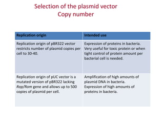
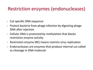
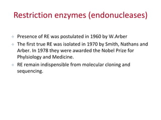
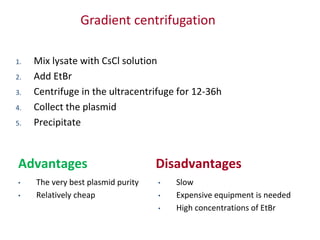
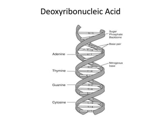
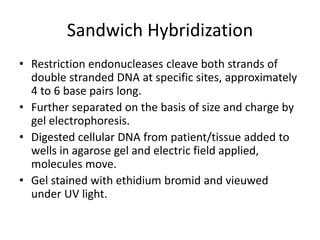
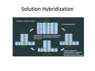
Plasmids, restriction enzymes,
analytics
Plasmid is an extra-chromosomal DNA molecule separate from
the chromosomal DNA which is capable of replicating
independently of the chromosomal DNA.
Vector – a carrier (plasmid or other type) used for bringing target
DNA fragment into a host cell.
Vector types
Vector Target fragment length
Plasmid 0-10 kb (total size up to 15 kb)
Cosmid 10-40 kb
P1 artificial chromosome (PAC) 130-150 kb
Bacterial artificial chromosome (BAC) About 300 kb
Yeast artificial chromosome (YAC) 200 kb to 2 Mb
Plasmids are essential instruments of molecular
biology
 Cloning and sequencing of DNA and cDNA fragments
 Generation of genomic and cDNA libraries
 Expression of recombinant proteins
 Generation of mutant proteins
 Analysis of regulatory sequences
 Gene targeting
Essential vector elements
 Origin of replication
 Antibiotic resistance gene (Amp, Kan, Tet, Chl)
 (Multiple cloning site)
Map of pOTB7 vector
showing Chloramphenicol
resistance gene (CMR),
replication origin (ORI) and
multiple cloning site (MCS)
Optional plasmids elements
 Multiple cloning site
 Promoter for cloned sequence
 Reporter gene
 Tag
 Regulatory sequences
pcDNA3.1(+) EGFP
6131bp
Amp r
Neo
BHG polyA
SV40 poly A
P CMV
SV40 prom
T7
Apa I (1705)
Bam HI (930)
Bgl II (13)
Bst XI (1678)
Eco RI (1656)
Eco RV(1668)
Ehe I (2969)
HindIII (912)
KpnI (922)
Mlu I (229)
Nde I (485)
Not I (1683)
Sca I (5689)
Sma I (2781)
Spe I (250)
Xba I (1695)
Xho I (1689)
Important plasmid information
 Replication origin defines the host bacteria: ColE1
replication origin is required for E.coli
 Replication origin may define the number of plasmid
copies per bacterial cell
 Bacteria may lose recombinant plasmid during cultivation
due to the absence of partitioning system (par). Naturally
occurring plasmids contain par that ensures that every
bacterial cell contains the plasmid.
Selection of the plasmid vector
Copy number
Replication origin Intended use
Replication origin of pBR322 vector
restricts number of plasmid copies per
cell to 30-40.
Expression of proteins in bacteria.
Very useful for toxic protein or when
tight control of protein amount per
bacterial cell is needed.
Replication origin of pUC vector is a
mutated version of pBR322 lacking
Rop/Rom gene and allows up to 500
copies of plasmid per cell.
Amplification of high amounts of
plasmid DNA in bacteria.
Expression of high amounts of
proteins in bacteria.
Selection of the plasmid vector
Purpose of use
Purpose Special vector feature(s) Example
Recombinant protein expression
in bacteria
Regulated bacterial promoter
Tag for protein purification
pGEX4T
Recombinant protein expression
in eukaryotic cells
Eukaryotic promoter
Tag for protein purification or
detection
Eukaryotic selection marker
pcDNA3.1
Analysis of eukaroytic promoter Reporter gene pGL3basic
General cloning - pBluescript KS
Restriction enzymes (endonucleases)
 Cut specific DNA sequence
 Protect bacteria from phage infection by digesting phage
DNA after injection
 Cellular DNA is protected by methylation that blocks
restriction enzyme activity
 Restriction enzyme (RE) means restricts virus replication
 Endonucleases are enzymes that produce internal cut called
as cleavage in DNA molecule
Restriction enzymes (endonucleases)
 Presence of RE was postulated in 1960 by W.Arber
 The first true RE was isolated in 1970 by Smith, Nathans and
Arber. In 1978 they were awarded the Nobel Prize for
Phylsiology and Medicine.
 RE remain indispensible from molecular cloning and
sequencing.
Type I enzymes cut at a site that differs, and is located at least at at least
1000 bp away, from their recognition site.
Type II enzymes recognize sites of 4-8 nucleotides and cleave DNA at the
same site
Type III enzymes recognize two separate non-palindromic sequences that
are inversely oriented. They cut DNA about 20-30 base pairs after the
recognition site.
Restriction enzymes
(endonucleases)
Restriction enzymes (endonucleases)
 Creating genomic and cDNA libraries
 Cloning DNA molecules
 Studying nucleotide sequence
 Generating mutated proteins
Plasmids, restriction enzymes,
analytics
Gel electrophoresis is a technique used for the separation of
deoxyribonucleic acid (DNA), ribonucleic acid (RNA), or
protein molecules using an electric current applied to a gel
matrix.
Ethidium bromide stained agarose gel of
total RNA (1-3) and DNA ladder (M)
Plasmid preparation stage 1
1. Plasmid-containing bacteria are cultivated in liquid media,
supplemented with the antibiotics for 18 h at 37°C with
intensive shaking
2. Cells are harvested by centrifugation
Preparation of the lysate
3 solutions strategy
1. Resuspend in hypotonic buffer with RNase (buffer P1)
2. Lyse bacteria using NaOH/SDS solution (buffer P2)
3. Neutralize NaOH and precipitate proteins using NaAc buffer
(buffer P3)
Plasmid can be isolated from obtained lysate using
various strategies.
Possible methods for isolation
1. Ethanol or Isopropanol precipitation
2. Silica matrix bind-wash-elute procedure
3. Density gradient centrifugation
Precipitation “quick and dirty”
1. Ethanol is added to the lysate
2. Obtained sample incubated for 30 min
3. DNA is collected by centrifugation
• Cheap
• Fast
Advantages Disadvantages
• Small amounts of DNA
• Poor purity, not sufficient for
applications like transfection
and in vitro translation
• Concentration of the plasmid
can not be determined
photometrically
Also known as mini prep
Silica matrix columns
1. Apply lysate on the column
2. Wash the column
3. Elute the plasmid
4. Precipitate
• High purity of the plasmid
• Fast
Advantages Disadvantages
• Expensive
Gradient centrifugation
1. Mix lysate with CsCl solution
2. Add EtBr
3. Centrifuge in the ultracentrifuge for 12-36h
4. Collect the plasmid
5. Precipitate
• The very best plasmid purity
• Relatively cheap
Advantages Disadvantages
• Slow
• Expensive equipment is needed
• High concentrations of EtBr
Concentration measurement
Photometric measurement of DNA concentration
UV 260 nm
Conc=50xOD260
Important! Photometric measurement of DNA concentration can not be
applied for “quick and dirty” plasmids, because of the presence of RNA
rests.
Gel electrophoresis of plasmid DNA
Selection of agarose concentration
Plasmid on an agarose gel
Characteristics of Nucleic Acids
• Two types of nucleic acids: RNA & DNA
• DNA is encoded with four interchangeable
"building blocks", called "bases", Adenine,
Thymine, Cytosine, and Guanine, with Uracil
rarely replacing Thymine
• RNA has five different bases: adenine,
guanine, cytosine, uracil, and more rarely
thymine.
Deoxyribonucleic Acid
DNA Replication
• Replication is performed by splitting (unzipping) the double
strand down the middle via relatively trivial chemical
reactions, and recreating the "other half" of each new single
strand by drowning each half in a "soup" made of the four
bases.
• Each of the "bases" can only combine with one other base,
the base on the old strand dictates which base will be on the
new strand.
• This way, each split half of the strand plus the bases it collects
from the soup will ideally end up as a complete replica of the
original, unless a mutation occurs.
DNA Replication
Nucleic Acid Probes
• Spontaneous pairing of complementary DNA strands forms basis for
techniques used to detect and characterize genes.
• Probe technology used to identify individual genes or DNA sequences.
• Nucleic acid probe short strand of DNA or RNA of known sequence used to
identify presence of complementary single strand of DNA in patient
sample.
• Binding of 2 strands (probe and patient) known as hybridization.
• Two DNA strands must share at least 16 to 20 consecutive bases of perfect
complementarity to form stable hybrid.
• Match occurring as a result of chance less than 1 in a billion.
• Probes labeled with marker: radioisotope, fluorochrome, enzyme or
chemiluminescent substrate.
• Hybridization can take place in solid support medium or liquid.
Dot-Blot
• Dot-blot clinical sample applied to membrane,
heated to denature DNA.
• Labeled probes added,
• Wash to remove unhybridized probe and
measure reactants.
• Qualitative test only.
• May be difficult to interpret.
Dot-Blot Hybridization
• Figure 1 DNA–DNA dot-blot hybridization between maize genomic DNA and a
CaMV p-35S probe. Sample numbers coincide with those in ref. 1. Top row: 1,
100% transgenic; 2, 10% transgenic; 3, 5% transgenic; 4, 1% transgenic, 5, 0.5%
transgenic; 6, historical maize negative control; 7, water negative control; 8,
Diconsa sample K1. Bottom row: 1, criollo sample B1; 2, criollo sample B2; 3,
criollo sample B3; 4, criollo sample A1; 5, criollo sample A2; 6, criollo sample A3; 7,
Peru maize negative control P1; 8, water negative control.
Sandwich Hybridization
• Uses two probes, one bound to membrane and
serves as capture target for sample DNA.
• Second probe anneals to different site on target DNA
and has label for detection.
• Sample nucleic acid sandwiched between the two.
• Two hybridization events occur, increases specificity.
• Can be adapted to microtiter plates.
Sandwich Hybridization
• Restriction endonucleases cleave both strands of
double stranded DNA at specific sites, approximately
4 to 6 base pairs long.
• Further separated on the basis of size and charge by
gel electrophoresis.
• Digested cellular DNA from patient/tissue added to
wells in agarose gel and electric field applied,
molecules move.
• Gel stained with ethidium bromid and vieuwed
under UV light.
Sandwich Hybridization
• Differences in restriction patterns referred to as restriction
fragment length polymorphisms (RFLPs)
• Caused by variations in nucleotides within genes that change
where the restriction enzymes cleave the DNA.
• When such a mutation occurs different size pieces of DNA are
obtained.
• Caused by variations in nucleotides within genes that change
where the restriction enzymes cleave the DNA.
• When such a mutation occurs different size pieces of DNA are
obtained.
Southern Blot
• DNA fragments separated by electrophoresis.
• Pieces denatured and transferred to membrane for
hybridization reaction.
• Place membrane on top of gel and allow buffer plus DNA to
wick up into it.
• Once DNA is on membrane heat or use UV ligth to crosslink
strands onto membrane to immobilize.
• Add labeled probes for hybridization to take place.
• Probes added in excess so target molecules reanneal and
more likely to attach to probe.
Southern Blot
• The Southern Blot takes advantage of the fact that DNA fragments will stick to a
nylon or nitrocellulose membrane. The membrane is laid on top of the agarose gel
and absorbent material (e.g. paper towels or a sponge) is placed on top. With
time, the DNA fragments will travel from the gel to the membrane by capillary
action as surrounding liquid is drawn up to the absorbent material on top. After
the transfer of DNA fragments has occurred, the membrane is washed, then the
DNA fragments are permanently fixed to the membrane by heating or exposing it
to UV light. The membrane is now a mirror image of the agarose gel.
Southern Blot
Southern Blot
• MOM [blue], DAD [yellow], and their four children: D1 (the biological
daughter), D2 (step-daughter, child of Mom and her former husband
[red]), S1 (biological son), and S2 (adopted son,not biologically related [his
parents are light and dark green]).
Northern Blot
• Northern blots allow investigators to determine the molecular weight of an mRNA and to
measure relative amounts of the mRNA present in different samples.
• RNA (either total RNA or just mRNA) is separated by gel electrophoresis, usually an agarose
gel. Because there are so many different RNA molecules on the gel, it usually appears as a
smear rather than discrete bands.
• The RNA is transferred to a sheet of special blotting paper called nitrocellulose, though other
types of paper, or membranes, can be used. The RNA molecules retain the same pattern of
separation they had on the gel.
• The blot is incubated with a probe which is single-stranded DNA. This probe will form base
pairs with its complementary RNA sequence and bind to form a double-stranded RNA-DNA
molecule. The probe cannot be seen but it is either radioactive or has an enzyme bound to it
(e.g. alkaline phosphatase or horseradish peroxidase).
• The location of the probe is revealed by incubating it with a colorless substrate that the
attached enzyme converts to a colored product that can be seen or gives off light which will
expose X-ray film. If the probe was labeled with radioactivity, it can expose X-ray film directly.
Northern Blot
Solution Hybridization
• Both target nucleic acid and probe free to interact in
solution.
• Hybridization of probe to target in solution is more
sensitive than hybridization on solid support
• Requires less sample and is more sensitive.
• Probe must be single-stranded and incapable of self-
annealing.
• Fairly adaptable to automation, especially tose using
chemiluminescent labels.
• Assays performed in a few hours.
Solution Hybridization
In-Situ Hybridization
• Target nucleic acid found in intact cells.
• Provides information about presence of
specific DNA targets and distribution in
tissues.
• Probes must be small enough to reach nucleic
acid.
• Radioactive or fluorescent tags used.
• Requires experience.
Fluorescent In-Situ Hibridization FISH
DNA Chip aka Microarrays
• A DNA chip (DNA microarray) is a biosensor which analyzes gene
information from humans and bacteria.
• This utilizes the complementation of the four bases labeled A (adenine), T
(thymine), G (guanine) and C (cytosine) in which A pairs with T and G pairs
with C through hydrogen bonding.
• A solution of DNA sequences containing known genes called a DNA probe
is placed on glass plates in microspots several microm in diameter
arranged in multiple rows.
• Genes are extracted from samples such as blood, amplified and then
reflected in the DNA chip, enabling characteristics such as the presence
and mutation of genes in the test subject to be determined.
• As gene analysis advances, the field is gaining attention particularly in the
clinical diagnosis of infectious disease, cancer and other maladies.
How DNA Chips Are Made
• Used to examine DNA, RNA and other substances
• Allow thousands of biological reactions to be
performed at once.
Step 1: Make gene probes.
• Using conventional techniques such as polymerase chain
reaction and biochemical synthesis, strands of identified DNA
are made and purified. A variety of probes are available from
commercial sources, many of which also offer custom
production services.
Step 2: Manufacture substrate wafer.
• Companies use photolithography and other
nanomanufacturing techniques to turn glass and
plastic wafers into receptacles for the DNA probes.
Step 3: Deposit genetic sequences.
• Manufacturers use a variety of processes ranging from
electrophoretic bonding to robotic deposition to adhere
genetic material to the substrate. Cleanroom conditions and
standards must be observed to attain the degree of
contamination control needed during the deposition process.
DNA Chip
Drawbacks
• Stringency, or correct pairing, is affected by:
– salt concentration
– Temperature
– concentration of destabilizing agent such as formamide or
urea.
• If conditions not carefully controlled mismatches can
occur.
• Patient nucleic acid may be present in small
amounts, below threshold for probe detection.
• Sensitivity can be increased by amplification: target,
probe and signal
Target Amplification
• In-vitro systems for enzymatic replication of
target molecule to detectable levels.
• Allows target to be identified and further
characterized.
• Examples: Polymerase chain reaction,
transcription mediated amplification,, strand
displacement amplification and nucleic acid
sequence-based amplification.
Polymerase Chain Reaction
• Capable of amplifying tiny quantities of nucleic acid.
• Cells separated and lysed.
• Double stranded DNA separated into single strands.
• Primers, small segments of DNA no more than 20-30 nucleotides long added.
• Primers are complementary to segments of opposite strands of that flank the
target sequence.
• Only the segments of target DNA between the primers will be replicated.
• Each cycle of PCR consists of three cycles:
– denaturation of target DNA to separate 2 strands.
– annealing step in which the reaction mix is cooled to allow primers to anneal to target
sequence
– Extension reaction in which primers initiate DNA synthesis using a DNA polymerase.
– These three steps constitute a thermal cycle
• Each PCR cycle results in a doubling of target sequences and typically allowed to
run through 30 cycles, one cylce takes approximately 60-90 seconds.
Taq
• Taq polymerase ("Taq pol") is a thermostable
polymerase isolated from thermus aquaticus,
a bacterium that lives in hot springs and
hydrothermal vents.
• "Taq polymerase" is an abbreviation of
Thermus Aquaticus Polymerase.
• It is often used in polymerase chain reaction,
since it is reasonably cheap and it can survive
PCR conditions.
PCR
PCR
Transcription Mediated Amplification
• TMA is the next generation of nucleic acid amplification technology.
• TMA is an RNA transcription amplification system using two enzymes to
drive the reaction: RNA polymerase and reverse transcriptase.
• TMA is isothermal; the entire reaction is performed at the same
temperature in a water bath or heat block. This is in contrast to other
amplification reactions such as PCR or LCR that require a thermal cycler
instrument to rapidly change the temperature to drive the reaction.
• TMA can amplify either DNA or RNA, and produces RNA amplicon, in
contrast to most other nucleic acid amplification methods that only
produce DNA.
• TMA has very rapid kinetics resulting in a billion fold amplification within
15-30 minutes.
TMA
QB Replicase
• Uses an RNA directed RNA polymerase that replicates the
genomic RNA of a bacteriophage named QB.
• The RNA genome of QB is essentially the only substrate
recognized by the polymerase.
• Because a short probe can be inserted into the QB RNA this
becomes the system for amplification.
• After the probe has annealed to the target, unbound probe is
treated with RNase and washed away.
• The hybridized probe is RNase resistant.
• When QB replicase is added the probe is enzymatically
replicated to detectable levels.
Ligase Chain Reaction
• The LCR test employs four synthetic oligonucleotide probes to anneal at specific
target sites on the cryptic plasmid.
• Each pair of probes hybridize close together on the target DNA template.
• Once the probes are annealed, the gap is filled by DNA polymerase and close by
the ligase enzyme.
• This two-step process of closing the gap between annealed probes makes the LCR,
in theory, more specific than PCR technology.
• The ligated probe pairs anneal to each other and, upon denaturation, form the
template for successive reaction cycles, thus producing a logarithmic amplification
of the target sequence.
• Like PCR, LCR is made in a thermocycler.
• The LCR product is detected in an automated instrument that uses an
immunocolorimetric bead capture system.
• At the end of the LCR assay, amplified products are inactivated by the automatic
addition of a chelated metal complex and a oxidizing agent.
Drawbacks of Amplification Systems
• Potential for false-positive results due to contaminating
nucleic acids.
• PCR and LCR, DNA products main source of contamination.
• QB replicase and TMA, RNA products are possible
contaminants.
• Must have product inactivation as part of QC program.
• Separate preparation areas from amplification areas and use
of inactivation systems such as UV light help alleviate
contamination.
• Very expensive.
• Closed system, automation will also decrease number of
problems.
Future of Molecular Diagnostic Techniques
• Despite expense may be times that rapid diagnosis will result in decreased
cost.
• Example: Mycobacteria - quick diagnosis no need for expensive respiratory
isolation.
• Detection of multi-drug resistant M. Tuberculosis will lead to more timely
public health measures.
• Incredibly useful in serology and microbiology.
• Increased specificity and sensitivity of molecular testing will become the
standard of practice in immunology and microbiology.
• Testing will continue to become more rapid as assays are automated
which will also bring down the costs.
• Author states will not replace culture for routine organisms, but it already
is, and as DNA chip technology improves, the ability to test for multiple
organisms will become easier
Signal Amplification
• Replicates signal rather than either the target or the probe.
• Based on the reporter group (the labeled tag) being attached in greater numbers to the
probe molecule or increasing the intensity generated by each labeled tag.
• Patient nucleic acid not replicated or amplified technique is less prone to contamination.
• Sensitivity is lower.
• Branched chain signal amplification employs several simultaneous hybridization steps.
• Author states similar to decorating a Christmas tree and involves several sandwich
hybridizations.
– first, target specific oligonucleotide probes captures target sequence to solid support.
– Second set of target specific probes called extenders hybridize to adjoining sequences and act as
binding site for large piece called branched amplification multimer.
– Each branch has multiple side branches capable of binding numerous oligonucleotides.
• Branched chains are well suited to detection of nucleic acid targets with sequence
heterogeneity such as hepatitis C and HIV.
Molecular techniques

Molecular techniques

  • 2.
    Plasmids, restriction enzymes, analytics Plasmidis an extra-chromosomal DNA molecule separate from the chromosomal DNA which is capable of replicating independently of the chromosomal DNA. Vector – a carrier (plasmid or other type) used for bringing target DNA fragment into a host cell.
  • 3.
    Vector types Vector Targetfragment length Plasmid 0-10 kb (total size up to 15 kb) Cosmid 10-40 kb P1 artificial chromosome (PAC) 130-150 kb Bacterial artificial chromosome (BAC) About 300 kb Yeast artificial chromosome (YAC) 200 kb to 2 Mb
  • 4.
    Plasmids are essentialinstruments of molecular biology  Cloning and sequencing of DNA and cDNA fragments  Generation of genomic and cDNA libraries  Expression of recombinant proteins  Generation of mutant proteins  Analysis of regulatory sequences  Gene targeting
  • 5.
    Essential vector elements Origin of replication  Antibiotic resistance gene (Amp, Kan, Tet, Chl)  (Multiple cloning site) Map of pOTB7 vector showing Chloramphenicol resistance gene (CMR), replication origin (ORI) and multiple cloning site (MCS)
  • 6.
    Optional plasmids elements Multiple cloning site  Promoter for cloned sequence  Reporter gene  Tag  Regulatory sequences pcDNA3.1(+) EGFP 6131bp Amp r Neo BHG polyA SV40 poly A P CMV SV40 prom T7 Apa I (1705) Bam HI (930) Bgl II (13) Bst XI (1678) Eco RI (1656) Eco RV(1668) Ehe I (2969) HindIII (912) KpnI (922) Mlu I (229) Nde I (485) Not I (1683) Sca I (5689) Sma I (2781) Spe I (250) Xba I (1695) Xho I (1689)
  • 7.
    Important plasmid information Replication origin defines the host bacteria: ColE1 replication origin is required for E.coli  Replication origin may define the number of plasmid copies per bacterial cell  Bacteria may lose recombinant plasmid during cultivation due to the absence of partitioning system (par). Naturally occurring plasmids contain par that ensures that every bacterial cell contains the plasmid.
  • 8.
    Selection of theplasmid vector Copy number Replication origin Intended use Replication origin of pBR322 vector restricts number of plasmid copies per cell to 30-40. Expression of proteins in bacteria. Very useful for toxic protein or when tight control of protein amount per bacterial cell is needed. Replication origin of pUC vector is a mutated version of pBR322 lacking Rop/Rom gene and allows up to 500 copies of plasmid per cell. Amplification of high amounts of plasmid DNA in bacteria. Expression of high amounts of proteins in bacteria.
  • 9.
    Selection of theplasmid vector Purpose of use Purpose Special vector feature(s) Example Recombinant protein expression in bacteria Regulated bacterial promoter Tag for protein purification pGEX4T Recombinant protein expression in eukaryotic cells Eukaryotic promoter Tag for protein purification or detection Eukaryotic selection marker pcDNA3.1 Analysis of eukaroytic promoter Reporter gene pGL3basic General cloning - pBluescript KS
  • 10.
    Restriction enzymes (endonucleases) Cut specific DNA sequence  Protect bacteria from phage infection by digesting phage DNA after injection  Cellular DNA is protected by methylation that blocks restriction enzyme activity  Restriction enzyme (RE) means restricts virus replication  Endonucleases are enzymes that produce internal cut called as cleavage in DNA molecule
  • 11.
    Restriction enzymes (endonucleases) Presence of RE was postulated in 1960 by W.Arber  The first true RE was isolated in 1970 by Smith, Nathans and Arber. In 1978 they were awarded the Nobel Prize for Phylsiology and Medicine.  RE remain indispensible from molecular cloning and sequencing.
  • 12.
    Type I enzymescut at a site that differs, and is located at least at at least 1000 bp away, from their recognition site. Type II enzymes recognize sites of 4-8 nucleotides and cleave DNA at the same site Type III enzymes recognize two separate non-palindromic sequences that are inversely oriented. They cut DNA about 20-30 base pairs after the recognition site. Restriction enzymes (endonucleases)
  • 13.
    Type I enzymescut at a site that differs, and is located at least at at least 1000 bp away, from their recognition site. Type II enzymes recognize sites of 4-8 nucleotides and cleave DNA at the same site Type III enzymes recognize two separate non-palindromic sequences that are inversely oriented. They cut DNA about 20-30 base pairs after the recognition site. Restriction enzymes (endonucleases)
  • 14.
    Restriction enzymes (endonucleases) Creating genomic and cDNA libraries  Cloning DNA molecules  Studying nucleotide sequence  Generating mutated proteins
  • 15.
    Plasmids, restriction enzymes, analytics Gelelectrophoresis is a technique used for the separation of deoxyribonucleic acid (DNA), ribonucleic acid (RNA), or protein molecules using an electric current applied to a gel matrix. Ethidium bromide stained agarose gel of total RNA (1-3) and DNA ladder (M)
  • 16.
    Plasmid preparation stage1 1. Plasmid-containing bacteria are cultivated in liquid media, supplemented with the antibiotics for 18 h at 37°C with intensive shaking 2. Cells are harvested by centrifugation
  • 17.
    Preparation of thelysate 3 solutions strategy 1. Resuspend in hypotonic buffer with RNase (buffer P1) 2. Lyse bacteria using NaOH/SDS solution (buffer P2) 3. Neutralize NaOH and precipitate proteins using NaAc buffer (buffer P3) Plasmid can be isolated from obtained lysate using various strategies.
  • 18.
    Possible methods forisolation 1. Ethanol or Isopropanol precipitation 2. Silica matrix bind-wash-elute procedure 3. Density gradient centrifugation
  • 19.
    Precipitation “quick anddirty” 1. Ethanol is added to the lysate 2. Obtained sample incubated for 30 min 3. DNA is collected by centrifugation • Cheap • Fast Advantages Disadvantages • Small amounts of DNA • Poor purity, not sufficient for applications like transfection and in vitro translation • Concentration of the plasmid can not be determined photometrically Also known as mini prep
  • 20.
    Silica matrix columns 1.Apply lysate on the column 2. Wash the column 3. Elute the plasmid 4. Precipitate • High purity of the plasmid • Fast Advantages Disadvantages • Expensive
  • 21.
    Gradient centrifugation 1. Mixlysate with CsCl solution 2. Add EtBr 3. Centrifuge in the ultracentrifuge for 12-36h 4. Collect the plasmid 5. Precipitate • The very best plasmid purity • Relatively cheap Advantages Disadvantages • Slow • Expensive equipment is needed • High concentrations of EtBr
  • 22.
    Concentration measurement Photometric measurementof DNA concentration UV 260 nm Conc=50xOD260 Important! Photometric measurement of DNA concentration can not be applied for “quick and dirty” plasmids, because of the presence of RNA rests.
  • 23.
    Gel electrophoresis ofplasmid DNA Selection of agarose concentration Plasmid on an agarose gel
  • 24.
    Characteristics of NucleicAcids • Two types of nucleic acids: RNA & DNA • DNA is encoded with four interchangeable "building blocks", called "bases", Adenine, Thymine, Cytosine, and Guanine, with Uracil rarely replacing Thymine • RNA has five different bases: adenine, guanine, cytosine, uracil, and more rarely thymine.
  • 25.
  • 26.
  • 27.
    DNA Replication • Replicationis performed by splitting (unzipping) the double strand down the middle via relatively trivial chemical reactions, and recreating the "other half" of each new single strand by drowning each half in a "soup" made of the four bases. • Each of the "bases" can only combine with one other base, the base on the old strand dictates which base will be on the new strand. • This way, each split half of the strand plus the bases it collects from the soup will ideally end up as a complete replica of the original, unless a mutation occurs.
  • 28.
  • 29.
    Nucleic Acid Probes •Spontaneous pairing of complementary DNA strands forms basis for techniques used to detect and characterize genes. • Probe technology used to identify individual genes or DNA sequences. • Nucleic acid probe short strand of DNA or RNA of known sequence used to identify presence of complementary single strand of DNA in patient sample. • Binding of 2 strands (probe and patient) known as hybridization. • Two DNA strands must share at least 16 to 20 consecutive bases of perfect complementarity to form stable hybrid. • Match occurring as a result of chance less than 1 in a billion. • Probes labeled with marker: radioisotope, fluorochrome, enzyme or chemiluminescent substrate. • Hybridization can take place in solid support medium or liquid.
  • 30.
    Dot-Blot • Dot-blot clinicalsample applied to membrane, heated to denature DNA. • Labeled probes added, • Wash to remove unhybridized probe and measure reactants. • Qualitative test only. • May be difficult to interpret.
  • 31.
    Dot-Blot Hybridization • Figure1 DNA–DNA dot-blot hybridization between maize genomic DNA and a CaMV p-35S probe. Sample numbers coincide with those in ref. 1. Top row: 1, 100% transgenic; 2, 10% transgenic; 3, 5% transgenic; 4, 1% transgenic, 5, 0.5% transgenic; 6, historical maize negative control; 7, water negative control; 8, Diconsa sample K1. Bottom row: 1, criollo sample B1; 2, criollo sample B2; 3, criollo sample B3; 4, criollo sample A1; 5, criollo sample A2; 6, criollo sample A3; 7, Peru maize negative control P1; 8, water negative control.
  • 32.
    Sandwich Hybridization • Usestwo probes, one bound to membrane and serves as capture target for sample DNA. • Second probe anneals to different site on target DNA and has label for detection. • Sample nucleic acid sandwiched between the two. • Two hybridization events occur, increases specificity. • Can be adapted to microtiter plates.
  • 33.
    Sandwich Hybridization • Restrictionendonucleases cleave both strands of double stranded DNA at specific sites, approximately 4 to 6 base pairs long. • Further separated on the basis of size and charge by gel electrophoresis. • Digested cellular DNA from patient/tissue added to wells in agarose gel and electric field applied, molecules move. • Gel stained with ethidium bromid and vieuwed under UV light.
  • 34.
    Sandwich Hybridization • Differencesin restriction patterns referred to as restriction fragment length polymorphisms (RFLPs) • Caused by variations in nucleotides within genes that change where the restriction enzymes cleave the DNA. • When such a mutation occurs different size pieces of DNA are obtained. • Caused by variations in nucleotides within genes that change where the restriction enzymes cleave the DNA. • When such a mutation occurs different size pieces of DNA are obtained.
  • 35.
    Southern Blot • DNAfragments separated by electrophoresis. • Pieces denatured and transferred to membrane for hybridization reaction. • Place membrane on top of gel and allow buffer plus DNA to wick up into it. • Once DNA is on membrane heat or use UV ligth to crosslink strands onto membrane to immobilize. • Add labeled probes for hybridization to take place. • Probes added in excess so target molecules reanneal and more likely to attach to probe.
  • 36.
    Southern Blot • TheSouthern Blot takes advantage of the fact that DNA fragments will stick to a nylon or nitrocellulose membrane. The membrane is laid on top of the agarose gel and absorbent material (e.g. paper towels or a sponge) is placed on top. With time, the DNA fragments will travel from the gel to the membrane by capillary action as surrounding liquid is drawn up to the absorbent material on top. After the transfer of DNA fragments has occurred, the membrane is washed, then the DNA fragments are permanently fixed to the membrane by heating or exposing it to UV light. The membrane is now a mirror image of the agarose gel.
  • 37.
  • 38.
    Southern Blot • MOM[blue], DAD [yellow], and their four children: D1 (the biological daughter), D2 (step-daughter, child of Mom and her former husband [red]), S1 (biological son), and S2 (adopted son,not biologically related [his parents are light and dark green]).
  • 39.
    Northern Blot • Northernblots allow investigators to determine the molecular weight of an mRNA and to measure relative amounts of the mRNA present in different samples. • RNA (either total RNA or just mRNA) is separated by gel electrophoresis, usually an agarose gel. Because there are so many different RNA molecules on the gel, it usually appears as a smear rather than discrete bands. • The RNA is transferred to a sheet of special blotting paper called nitrocellulose, though other types of paper, or membranes, can be used. The RNA molecules retain the same pattern of separation they had on the gel. • The blot is incubated with a probe which is single-stranded DNA. This probe will form base pairs with its complementary RNA sequence and bind to form a double-stranded RNA-DNA molecule. The probe cannot be seen but it is either radioactive or has an enzyme bound to it (e.g. alkaline phosphatase or horseradish peroxidase). • The location of the probe is revealed by incubating it with a colorless substrate that the attached enzyme converts to a colored product that can be seen or gives off light which will expose X-ray film. If the probe was labeled with radioactivity, it can expose X-ray film directly.
  • 40.
  • 41.
    Solution Hybridization • Bothtarget nucleic acid and probe free to interact in solution. • Hybridization of probe to target in solution is more sensitive than hybridization on solid support • Requires less sample and is more sensitive. • Probe must be single-stranded and incapable of self- annealing. • Fairly adaptable to automation, especially tose using chemiluminescent labels. • Assays performed in a few hours.
  • 42.
  • 43.
    In-Situ Hybridization • Targetnucleic acid found in intact cells. • Provides information about presence of specific DNA targets and distribution in tissues. • Probes must be small enough to reach nucleic acid. • Radioactive or fluorescent tags used. • Requires experience.
  • 44.
  • 45.
    DNA Chip akaMicroarrays • A DNA chip (DNA microarray) is a biosensor which analyzes gene information from humans and bacteria. • This utilizes the complementation of the four bases labeled A (adenine), T (thymine), G (guanine) and C (cytosine) in which A pairs with T and G pairs with C through hydrogen bonding. • A solution of DNA sequences containing known genes called a DNA probe is placed on glass plates in microspots several microm in diameter arranged in multiple rows. • Genes are extracted from samples such as blood, amplified and then reflected in the DNA chip, enabling characteristics such as the presence and mutation of genes in the test subject to be determined. • As gene analysis advances, the field is gaining attention particularly in the clinical diagnosis of infectious disease, cancer and other maladies.
  • 46.
    How DNA ChipsAre Made • Used to examine DNA, RNA and other substances • Allow thousands of biological reactions to be performed at once.
  • 47.
    Step 1: Makegene probes. • Using conventional techniques such as polymerase chain reaction and biochemical synthesis, strands of identified DNA are made and purified. A variety of probes are available from commercial sources, many of which also offer custom production services.
  • 48.
    Step 2: Manufacturesubstrate wafer. • Companies use photolithography and other nanomanufacturing techniques to turn glass and plastic wafers into receptacles for the DNA probes.
  • 49.
    Step 3: Depositgenetic sequences. • Manufacturers use a variety of processes ranging from electrophoretic bonding to robotic deposition to adhere genetic material to the substrate. Cleanroom conditions and standards must be observed to attain the degree of contamination control needed during the deposition process.
  • 50.
  • 51.
    Drawbacks • Stringency, orcorrect pairing, is affected by: – salt concentration – Temperature – concentration of destabilizing agent such as formamide or urea. • If conditions not carefully controlled mismatches can occur. • Patient nucleic acid may be present in small amounts, below threshold for probe detection. • Sensitivity can be increased by amplification: target, probe and signal
  • 52.
    Target Amplification • In-vitrosystems for enzymatic replication of target molecule to detectable levels. • Allows target to be identified and further characterized. • Examples: Polymerase chain reaction, transcription mediated amplification,, strand displacement amplification and nucleic acid sequence-based amplification.
  • 53.
    Polymerase Chain Reaction •Capable of amplifying tiny quantities of nucleic acid. • Cells separated and lysed. • Double stranded DNA separated into single strands. • Primers, small segments of DNA no more than 20-30 nucleotides long added. • Primers are complementary to segments of opposite strands of that flank the target sequence. • Only the segments of target DNA between the primers will be replicated. • Each cycle of PCR consists of three cycles: – denaturation of target DNA to separate 2 strands. – annealing step in which the reaction mix is cooled to allow primers to anneal to target sequence – Extension reaction in which primers initiate DNA synthesis using a DNA polymerase. – These three steps constitute a thermal cycle • Each PCR cycle results in a doubling of target sequences and typically allowed to run through 30 cycles, one cylce takes approximately 60-90 seconds.
  • 54.
    Taq • Taq polymerase("Taq pol") is a thermostable polymerase isolated from thermus aquaticus, a bacterium that lives in hot springs and hydrothermal vents. • "Taq polymerase" is an abbreviation of Thermus Aquaticus Polymerase. • It is often used in polymerase chain reaction, since it is reasonably cheap and it can survive PCR conditions.
  • 55.
  • 56.
  • 57.
    Transcription Mediated Amplification •TMA is the next generation of nucleic acid amplification technology. • TMA is an RNA transcription amplification system using two enzymes to drive the reaction: RNA polymerase and reverse transcriptase. • TMA is isothermal; the entire reaction is performed at the same temperature in a water bath or heat block. This is in contrast to other amplification reactions such as PCR or LCR that require a thermal cycler instrument to rapidly change the temperature to drive the reaction. • TMA can amplify either DNA or RNA, and produces RNA amplicon, in contrast to most other nucleic acid amplification methods that only produce DNA. • TMA has very rapid kinetics resulting in a billion fold amplification within 15-30 minutes.
  • 58.
  • 59.
    QB Replicase • Usesan RNA directed RNA polymerase that replicates the genomic RNA of a bacteriophage named QB. • The RNA genome of QB is essentially the only substrate recognized by the polymerase. • Because a short probe can be inserted into the QB RNA this becomes the system for amplification. • After the probe has annealed to the target, unbound probe is treated with RNase and washed away. • The hybridized probe is RNase resistant. • When QB replicase is added the probe is enzymatically replicated to detectable levels.
  • 60.
    Ligase Chain Reaction •The LCR test employs four synthetic oligonucleotide probes to anneal at specific target sites on the cryptic plasmid. • Each pair of probes hybridize close together on the target DNA template. • Once the probes are annealed, the gap is filled by DNA polymerase and close by the ligase enzyme. • This two-step process of closing the gap between annealed probes makes the LCR, in theory, more specific than PCR technology. • The ligated probe pairs anneal to each other and, upon denaturation, form the template for successive reaction cycles, thus producing a logarithmic amplification of the target sequence. • Like PCR, LCR is made in a thermocycler. • The LCR product is detected in an automated instrument that uses an immunocolorimetric bead capture system. • At the end of the LCR assay, amplified products are inactivated by the automatic addition of a chelated metal complex and a oxidizing agent.
  • 61.
    Drawbacks of AmplificationSystems • Potential for false-positive results due to contaminating nucleic acids. • PCR and LCR, DNA products main source of contamination. • QB replicase and TMA, RNA products are possible contaminants. • Must have product inactivation as part of QC program. • Separate preparation areas from amplification areas and use of inactivation systems such as UV light help alleviate contamination. • Very expensive. • Closed system, automation will also decrease number of problems.
  • 62.
    Future of MolecularDiagnostic Techniques • Despite expense may be times that rapid diagnosis will result in decreased cost. • Example: Mycobacteria - quick diagnosis no need for expensive respiratory isolation. • Detection of multi-drug resistant M. Tuberculosis will lead to more timely public health measures. • Incredibly useful in serology and microbiology. • Increased specificity and sensitivity of molecular testing will become the standard of practice in immunology and microbiology. • Testing will continue to become more rapid as assays are automated which will also bring down the costs. • Author states will not replace culture for routine organisms, but it already is, and as DNA chip technology improves, the ability to test for multiple organisms will become easier
  • 63.
    Signal Amplification • Replicatessignal rather than either the target or the probe. • Based on the reporter group (the labeled tag) being attached in greater numbers to the probe molecule or increasing the intensity generated by each labeled tag. • Patient nucleic acid not replicated or amplified technique is less prone to contamination. • Sensitivity is lower. • Branched chain signal amplification employs several simultaneous hybridization steps. • Author states similar to decorating a Christmas tree and involves several sandwich hybridizations. – first, target specific oligonucleotide probes captures target sequence to solid support. – Second set of target specific probes called extenders hybridize to adjoining sequences and act as binding site for large piece called branched amplification multimer. – Each branch has multiple side branches capable of binding numerous oligonucleotides. • Branched chains are well suited to detection of nucleic acid targets with sequence heterogeneity such as hepatitis C and HIV.