SlideShare a Scribd company logo
1 of 41
Download to read offline
Clinical Applications of Biomedical Microdevices
for Controlled Drug Delivery
Pablo Gurman, MD; Oscar R. Miranda, PhD; Kevin Clayton, BS; Yitzhak Rosen, MD;
and Noel M. Elman, PhD
Abstract
Miniaturization of devices to micrometer and nanometer scales, combined with the use of biocompatible
and functional materials, has created new opportunities for the implementation of drug delivery systems.
Advances in biomedical microdevices for controlled drug delivery platforms promise a new generation of
capabilities for the treatment of acute conditions and chronic illnesses, which require high adherence to
treatment, in which temporal control over the pharmacokinetic profiles is critical. In addition, clinical
conditions that require a combination of drugs with specific pharmacodynamic profiles and local delivery
will benefit from drug delivery microdevices. This review provides a summary of various clinical appli-
cations for state-of-the-art controlled drug delivery microdevices, including cancer, endocrine and ocular
disorders, and acute conditions such as hemorrhagic shock. Regulatory considerations for clinical
translation of drug delivery microdevices are also discussed. Drug delivery microdevices promise a
remarkable gain in clinical outcomes and a substantial social impact. A review of articles covering the field
of microdevices for drug delivery was performed between January 1, 1990, and January 1, 2014, using
PubMed as a search engine.
ª 2014 Mayo Foundation for Medical Education and Research n Mayo Clin Proc. 2014;nn(n):1-16
B
iomedical microdevices are fabricated
devices with critical features on the order
of 1 to 100 mm. These microdevices range
in complexity from simple microstructures such
as microchannels to more sophisticated micro-
functional parts such as microtransducers and
microelectromechanical systems (MEMS).1
These devices integrate mechanisms that
activate a variety of physical signals to achieve
a specific function. For example, MEMS-based
inertial sensors transduce a mechanical signal
input to an electrical signal response. Current
transducers are able to combine multiple
physical inputs with multiple output signals.
Biomedical microdevices present a variety
of key advantages for applications in health
care owing to their (1) extremely small sizes
providing minimally invasive procedures, (2)
low power consumption, (3) batch fabrication
processes with high reproducibility, and (4)
low cost per device, in conjunction with their
multiple functionalities and compatibility with
very large-scale integration electronics.
These novel technologies have accelerated
the development of a variety of micromedical
devices, such as catheter pressure sensors,
microelectronic components for pacemakers,
hand-held point-of-care diagnostic devices,
and drug delivery systems, all of which have
provided significant improvement over treat-
ment possibilities for numerous chronic and
nonchronic illnesses.1-4
Figure 1 shows a variety
of biomedical microdevices for several therapeu-
tic applications.
Controlled drug delivery systems that are
based on microdevices contain structural micro-
parts, such as microchannels and microreser-
voirs, to store drugs. In addition, drug delivery
systems based on MEMS incorporate micro-
transducers such as microactuators and micro-
sensors, which improve the device capabilities.
Drug delivery devices based on MEMS
provide an opportunity for improved diag-
nosis, monitoring, and treatment of numerous
illnesses. The MEMS can deliver a variety of
drugs, including drugs in combination, using
a single device. The MEMS drug delivery de-
vices have the ability to control the rate of
drug release to a target area. They can be pro-
grammed for pulsatile or continuous delivery
and can release the drug locally, which in-
creases treatment efficacy using a smaller
amount of drug, reducing systemic concentra-
tion levels1-6
and associated toxicity.
From the Institute for
Soldier Nanotechnologies,
Massachusetts Institute of
Technology, Cambridge
(P.G., O.R.M., K.C., Y.R.,
N.M.E.); and Department
of Materials Science,
University of Texas at
Dallas, Richardson (P.G.).
Mayo Clin Proc. n XXX 2014;nn(n):1-16 n http://dx.doi.org/10.1016/j.mayocp.2014.10.003
www.mayoclinicproceedings.org n ª 2014 Mayo Foundation for Medical Education and Research
1
REVIEW
Finally, the scope of novel materials for
biomedical devices has expanded the potential
use of biocompatible platforms with high biolog-
ical performance, eg, less toxic and nonreactive
devices, enabling new therapeutic applications.
This review provides a summary of current
state-of-the-art biomedical microdevices for
controlled drug delivery and their correspond-
ing clinical applications. The following sections
describe passive and active delivery devices
based on MEMS technology. Each section pro-
vides a technical description of a microdevice
followed by its suggested clinical application.
The review continues with a summary of the
regulatory strategies for obtaining Food and
Drug Administration (FDA) approval for such
microdevices. Finally, a perspective on the
future of these novel devices is presented.
DATA SOURCES AND SEARCHES
A PubMed search between January 1, 1990, and
January 1, 2014, was performed. The search
terms were drug delivery AND MEMS, implant-
able devices AND MEMS, control release AND
microchip, controlled release AND BioMEMS,
neural probes AND drug delivery, vaccines AND
microneedles, diabetes AND microneedles, intraoc-
ular AND drug delivery devices, and inner ear
AND drug delivery AND microfluidics. Papers
were selected following the definition of micro-
devices and MEMS. Selection also was per-
formed with the aim of having examples of
different types of microdevices (passive and
active, actuation mechanism, and materials). Ex-
amples of different clinical applications for drug
delivery microdevices assisted in selecting pa-
pers more close to the clinical application than
those focused solely on fundamental science.
Diagnostic microdevices were specifically
excluded from the search.
PASSIVE DEVICES
Passive biomedical microdevices for drug deliv-
ery do not rely on an actuation mechanism or
on monitoring for feedback. These devices are
reservoir based, relying on mass transfer across
a permeable membrane to deliver pharmaceu-
tical drugs, the biodegradation of a hermetic
membrane, or a unique reservoir structure to
achieve controlled release. The rate of release
can be controlled by taking into account the
following design parameters: (1) the effective
permeability of the membranes by fine-tuning
structural dimensions and materials (pore
size, thickness), (2) the rate of degradation of
the polymer contained on the membrane or
in the reservoir, (3) the diffusivity properties
of the drug, and (4) the osmotic pressure. Pas-
sive delivery of drugs cannot be modified after
implementation. Other passive-release devices
operate based on actuation resulting from
in vivo conditions inside the body, such as pH
or temperature, to accelerate degradation of
the materials that encapsulate the pharmaceu-
tical drugs. Typically, the controlled release is
achieved by considering the pharmacokinetics
of the selected drug for delivery. Design and ma-
terial parameters are thereafter adjusted and
selected during the design process to provide a
constant and superior pharmacokinetic perfor-
mance, such as an improvement in treatment
efficacy duration over the typical half-life of
the pharmaceutical drug. Existing passive-
release devices, such as the fentanyl transdermal
system (DURAGESIC; Janssen Pharmaceuticals
Inc) and the fluocinolone acetonide intravitreal
implant (Retisert; Bausch & Lomb Inc),
are used for either short-term (3 days) or
ARTICLE HIGHLIGHTS
n Drug delivery systems can be classified as passive and active.
Passive devices do not incorporate sensors and actuators for
drug delivery.
n Active microdevices include microelectromechanical systems
(MEMS), which comprise microparts such as microchannels and
microvalves and transducers, including microsensors and
microactuators, integrated into a singular microdevice.
n Advantages of MEMS drug delivery systems include miniaturization,
integration with microelectronics, actively controlled, low cost,
multiple pharmacologic therapies in a single device, controlled over
release rate, and in vivo long-term storage of drugs.
n The MEMS are being used for a variety of clinical conditions,
including diabetes, neurologic disorders, inner ear diseases, and
cancer.
n Fluzone is an example of a Food and Drug Administratione
approved drug delivery microdevice for vaccine delivery.
n The MEMS drug delivery devices can be considered combina-
tion products. Many combination products are considered
drugs, requiring a New Drug Application for Food and Drug
Administration approval.
MAYO CLINIC PROCEEDINGS
2 Mayo Clin Proc. n XXX 2014;nn(n):1-16 n http://dx.doi.org/10.1016/j.mayocp.2014.10.003
www.mayoclinicproceedings.org
long-term (2.5-3 years) continuous treatment of
diseases. The lack of integrated electronics re-
duces the complexity of these devices.7,8
Hydrogels
Implantable devices based on environmentally
sensitive hydrogels were developed for
controlled release (Figure 2).9
The device ar-
chitecture consists of a reservoir and a 100-
mm-thick silicon membrane with orifices
measuring 140 mm in diameter. Each orifice
contains a support post in the center and is
tethered to confine the hydrogel to the mem-
brane. The hydrogel is loaded around the
central support post such that the entire orifice
is blocked by the hydrogel in the swollen state.
Under activation by chemical or physical
stimuli, the hydrogel shrinks and the drug is
allowed to diffuse through the resulting
orifice. The response of the hydrogel opening
or closing is critical in controlling the rate of
drug delivery. Additional control of drug de-
livery can be gained by manipulating the
membrane thickness, the size of the orifices,
the support posts, and the tethers.
The temperature, pH, and glucose sensitivity
of different hydrogels are some of the parameters
that provide additional control over activation.
For example, N-isopropylacrylamide hydrogels
Microchannel
Pt conducting wire
Neural probe with
microchannels for drug
delivery (Figure 4)
MEMS Chip
Pump
Into inner
ear for intra
cochlear
delivery
Cochlear implanted device
with pump and electronic
control (Figure 10)
Stratum
corneum
Epidermis
Dermis
Subcutaneous
tissue
Microneedles
Hypodermic needle
Dendritic cell
Langerhans cell
Microneedle transdermal
patch (Figure 8)
Drug solution Drug reservoir
Parylene cannula
Into
cannula
Into eye
Eye wall
Electrolysis pump
Pump outlet
Ocular device with
electrolysis pump (Figure 7)
MEMS delivery for
emergency (Figure 6)
Microchip drug
delivery for
osteoporosis (Figure 5)
Implantabe MEMS device for drug
delivery (Figure 9)
FIGURE 1. Technology map for applications of biomedical microdevices. MEMS ¼ microelectromechanical systems.
CLINICAL APPLICATIONS OF BIOMEDICAL MICRODEVICES
Mayo Clin Proc. n XXX 2014;nn(n):1-16 n http://dx.doi.org/10.1016/j.mayocp.2014.10.003
www.mayoclinicproceedings.org
3
were found to rapidly contract at 34
C, resulting
in a sharp increase in the flow rate of the drug
from 0 to approximately 1 mL/min. This type
of hydrogel exhibited a fast response to environ-
mental conditions, contracting in 10 seconds at
25
C and expanding back to close the orifice in
20 seconds at 50
C. N-butan-2-ylbutan-2-
amine/anodic alumina membrane hydrogel
was measured to respond to a change in pH of
3.0 to 10.0 in 4 minutes, whereas for changes
in glucose levels from 0 to 20 mmol/L, this
hydrogel responded in 40 minutes.9
Clinical Application: Diabetes
Diabetes represents a significant burden in
health care as the number of people with
type 2 diabetes is increasing dramatically
owing to a pandemic of obesity worldwide.10
One of the key issues in diabetes is adherence
with insulin administration. Adherence is
limited owing to the frequent and uncomfort-
able subcutaneous (SC) injections that the pa-
tient needs to treat his or her diabetes.
Glucose-responsive hydrogels provide an
opportunity for the controlled delivery of insu-
lin. Incorporating more channels with various
types of hydrogels and channel sizes could
improve control and treatment. Further work
is needed to better understand and engineer
response kinetics and the reliability of such
hydrogel-based devices for clinical applications.
Passive Nanochannel-Based Drug Delivery
Device
A novel, high-throughput nanochannel drug
delivery system for the sustained delivery of
chemotherapeutics was developed and tested
in vitro.11
The device was developed to be
implantable to improve patient adherence and
quality of life by avoiding the need for repeated
administrations and frequent visits to the clinic.
The device passively controls the release of
drugs by physical-electrostatic confinement.
By manipulating the size of the nanochannels,
zero-order release of chemotherapeutics was
achieved. The nanochannel membrane com-
prises a silicon substrate reservoir and a
capping layer. An array of 161 channels,
measuring 200Â200 mm and spaced by 50-
mm-thick walls, makes up the membrane sur-
face (Figure 3). It consists of 30-mm-wide
microchannels that connect the reservoir and
the capping layer. The nanochannels connect
the inlet and outlet channels at the interface
of the silicon substrate and the capping layer.
Clinical Application: Melanoma
Melanoma, a tumor originating from melano-
cyte cells, represents the most aggressive
form of skin cancer, with 5-year survival of
20% for advanced cases. Current pharmaco-
logic therapies include the use of interferon
alfa-2b as an adjuvant for stage III melanomas.
Interferon alfa-2b is an immunomodulatory
drug that activates the immune system against
the tumor, increasing patient relapse-free sur-
vival. An important issue with interferon
alfa-2b is its adverse effects. Interferon alfa-
2b in high doses has been linked to hepatotox-
icity and suicidal ideation.12
The use of implantable, controlled nano-
channel delivery systems could potentially
overcome some of the limitations associated
with current therapies by decreasing the
amount of drug that reaches the systemic cir-
culation. This improvement could avoid
adverse effects in healthy tissues while keeping
high concentrations of interferon alfa-2b at the
targeted site where the tumor is located.
Clinical Application: Prostate Cancer
Prostate cancer represents the sixth leading
cause of cancer death in men, with an incidence
of 233,000 new cases and 29,480 deaths in
Front side
tethers
Central
tethered post
Back side
tethers
Flow
Silicon
membrane
FIGURE 2. Schematic of silicon membrane with
structured orifices. From Sens Actuators B Chem,9
with permission.
MAYO CLINIC PROCEEDINGS
4 Mayo Clin Proc. n XXX 2014;nn(n):1-16 n http://dx.doi.org/10.1016/j.mayocp.2014.10.003
www.mayoclinicproceedings.org
2014.13
Leuprolide acetate is a synthetic
analogue of gonadotropin-releasing hormone.
Gonadotropin-releasing hormone stimulates
the release of follicle-stimulating hormone and
luteinizing hormone, which promote the pro-
duction of estrogen and testosterone. Testos-
terone is metabolized in the interior of prostate
cells to dihydrotestosterone, which upregulates
cell proliferation, gene expression, and protein
synthesis. It is thought that leuprolide acts as a
gonadotropin-releasing hormone analogue and
when given continuously by the SC or intramus-
cular route (leuprolide acetate could not be
administered orally because is a peptide) leads
to testosterone deprivation. Deprivation of pros-
tate cells from testosterone would lead to
apoptosis and cytoreduction of tumor volume.14
Interferon alfa-2b and leuprolide acetate
were chosen to test the nanochannel micro-
chip delivery device. The release of interferon
alfa-2b was tested using 20-nm membranes
and was measured to be a mean Æ SD of
29.7Æ1.5 mg/d for 6 days, which is in agree-
ment with current maintenance doses of inter-
feron alfa-2b used in patients with melanoma
(10 million IU/m2
SC 3 times/wk; 10 million
international units ¼ 38 mg).
The release of leuprolide was tested using
5- and 15-nm channels and was measured to
be zero order for the 5-nm channel at a
mean Æ SD rate of 100Æ10 mg/d for 3 days,
which is close in agreement with current leu-
prolide doses used in prostate cancer (250
mg/d). By increasing reservoir sizes, the
nanochannel-based delivery system has the
potential to achieve the current dose regimens
used for interferon alfa-2b and leuprolide.
Multifunctional MEMS for Neural Recording
and Drug Delivery
Multifunctional MEMS for simultaneous
recording of neural activity and drug delivery
were developed (Figure 4).15,16
One such de-
vice has been reported by Altuna et al16
based
on flexible microprobes made of SU-8. The
polymer SU-8 was used as the structural mate-
rial for the probes, with platinum for the elec-
trodes. Tetrode-like probes with a single
microfluidic channel and linear probes with 2
microfluidic channels were tested. Electrodes
for the tetrode-like probe were spaced 25 mm
apart, with diameters of 20 mm to sense indi-
vidual neuronal firing at the tip of the probe.
The microfluidic channel measured 50Â20
mm, with 3 outlet ports also near the tip of the
probe. In the linear probe, 8 electrodes were
spaced 100 mm apart, allowing for sensing at
different depths of the brain. The 2 microfluidic
channels measured 40Â20 mm and had inde-
pendent outlet ports. Both devices were 55
mm thick. The tetrode-like probe was 90 mm
wide, and the linear probe was 150 mm wide.
The probes were tested in vivo in anesthe-
tized rats. The SU-8 linear probes were used to
deliver kainate at the CA1 cell and dendritic
layers at a flow rate of 3 to 6 mL/min to induce
seizures. Neuronal excitability was recorded
against a control delivery of saline to confirm de-
livery of the drug. The tetrode-like probe deliv-
ered potassium at a high flow rate of 0.6 to 1.5
mL/min to the CA1 cell layer. The probe was
able to record isolated neurons together with
multi-unit firing. Both probes had the ability
to measure ripples and spikes common during
large irregular brain activity at the CA1 cell layer.
Clinical Applications: Parkinson Disease and
Epilepsy
Effective treatments for neurologic diseases are
still lacking. Parkinson disease, the second most
FIGURE 3. Schematics (A, C, and D) and optical microscopy (B) of a passive
nanochannel delivery system designed for drug release with zero-order
kinetics. The innovative architecture of the device includes macro-
channels, microchannels, and nanochannels. M ¼ macrochannel; mO ¼
microchannel outlet; mI ¼ microchannel inlet; n¼ nanochannel; w ¼
supporting walls. From Pharm Res,11
with permission.
CLINICAL APPLICATIONS OF BIOMEDICAL MICRODEVICES
Mayo Clin Proc. n XXX 2014;nn(n):1-16 n http://dx.doi.org/10.1016/j.mayocp.2014.10.003
www.mayoclinicproceedings.org
5
common neurodegenerative disorder after Alz-
heimer disease, has been managed for the past
few decades with L-3,4-dihydroxyphenylalanine
(L-DOPA).17
The L-DOPA is a precursor to dopa-
mine, which is a neurotransmitter that is absent
in the brain of patients with Parkinson disease
owing to the progressive loss of dopaminergic
neurons. In the long-term, patients start to expe-
rience L-DOPA adverse effects that deteriorate
their quality of life.
Epilepsy, a disorder characterized by un-
controlled propagation of electrical stimuli in
the brain, has been managed with drugs that
reduce neuron excitability. Some types of epi-
lepsy, however, remain refractory to drugs.
Therefore, it is clear that current pharmaco-
logic therapies alone have not reached an
acceptable benefit for neurologic disorders
requiring additional intervention. Implantable
devices such as neural stimulators have
emerged as an attractive option for patients
with advanced Parkinson disease, refractory
epilepsy, and other neurologic conditions.18
Despite the aforementioned benefits, these
novel delivery modalities need to overcome is-
sues of poor biocompatibility, such as inflamma-
tory response and fibrosis around the implant,
which limit overall device performance. For
example, neural probes have been found to elicit
glial scar formation and neuronal loss during im-
plantation, impairing device performance. Hav-
ing an anti-inflammatory drug in the same
device could decrease the inflammatory response
and, thus, the generation of fibrotic tissue (eg,
glial scar formation) surrounding the implant,
thus preserving functionality.19,20
Therefore, it is being realized that by
combining devices with drug therapies, it is
possible to maximize the benefits of both while
avoiding their adverse effects. This clinical need
has been met by using MEMS technologies, in
which neural electrodes are being combined
with microfluidic channels or microreservoirs.
This combines the capability to record neural
data and drug delivery.
ACTIVE DEVICES
Active drug delivery devices use a variety of
mechanisms to release pharmaceutical drugs
and provide an increased level of control.
The MEMS devices have been developed using
different actuation modalities, including
micropumps based on gas pressure from elec-
trolysis, integration of magnetic actuators, and
electrochemical and electrothermal actuation
systems. Active devices can be customized to
treat a range of diseases requiring specific
pharmacokinetic drug delivery profiles. More-
over, as opposed to passive delivery systems,
Bonding pads
Microelectrode film
Microelectrode
sites
Outlet
Microchannel
Molded PDMS film
Vacuum
A
B
C
Microchannel
Pt conducting wire
FIGURE 4. Schematics (A and B) and optical picture (C) of a neural probe
with drug delivery capabilities. A, Assembly of device components. B, Method
used to incorporate the drug into the microchannels. C, Optical picture of the
finished device and microchannels containing a dye solution. PDMS ¼ poly-
dimethylsiloxane; Pt ¼ platinum. From Sens Actuators A Phys,15
with permission.
MAYO CLINIC PROCEEDINGS
6 Mayo Clin Proc. n XXX 2014;nn(n):1-16 n http://dx.doi.org/10.1016/j.mayocp.2014.10.003
www.mayoclinicproceedings.org
MEMS can be activated and stopped at any
time after implantation.
Active devices commonly require minia-
turized power electronics for actuation, typi-
cally increasing the overall form factor,
which is a key limiting factor in implantable
applications. Alternatively, telemetry systems
to transfer energy for activation can be adop-
ted to overcome this limiting issue.
Electrothermally Actuated MEMS Drug
Delivery Microchip
Santini et al21
developed a device for the
controlled, pulsatile release of chemicals from
single or multiple reservoirs. The controlled
drug release was triggered by the application
of an electric potential to burst sealing gold
membranes electrothermally. Drugs inside the
reservoir were then free to diffuse to the tar-
geted site. This original device had the func-
tionality for complex release of kinetics by
varying the amount or substance type placed
in each reservoir and varying the timing of
release.
This type of device would be able to deliver
drugs in a pulsatile manner. Since then, several
works on active drug delivery devices based on
MEMS have made substantial progress toward
effectively treating various ailments.
Clinical Application: Osteoporosis
Osteoporosis is the progressive degradation of
bone architecture and loss of mass bone den-
sity that leads to bone fragility that ultimately
increases the risk of fractures. Osteoporosis is
more common in postmenopausal women,
who are at risk for lower levels of estrogens,
which are known to be involved in bone for-
mation. According to the National Institute
for Health and Clinical Excellence, 9 million
osteoporotic fractures occur annually in the
world.22
A microchip device containing parathyroid
hormone (PTH) was developed for the treat-
ment of osteoporosis23
; PTH is known to stim-
ulate bone formation by increasing osteoblast
number and function.24
An implantable
microchip device capable of releasing PTH
would prevent the need for frequent injections
of PTH.
The first in-human testing performed in
postmenopausal women evaluated the in vivo
pharmacokinetic profile of a PTH-release
microchip (Figure 5) against standard SC in-
jections.25
The microchip was implanted sub-
cutaneously in the abdomen, and the
pharmacokinetic profile was measured after a
fibrous capsule was formed around the
implant.
The rationale of the study was to deter-
mine the pharmacokinetic performance of
the microchip when it was surrounded by a
fibrous capsule as a result of the host response
to the implant. In addition, bone biomarkers
were measured to determine the effect on
bone formation of PTH injections vs PTH
released by the microchip. A safety laboratory
panel was performed to determine the safety of
the microchip vs that of the SC injections.
Overall, the microchip was found to be
bioequivalent to the SC injections even in
the presence of the fibrous capsule. The
microchip was also found to be as safe as the
SC injections based on a laboratory panel.25
FIGURE 5. A microchip drug delivery system
for parathyroid hormone (PTH) release for
osteoporosis treatment. The picture depicts the
titanium packaging used for carryng the micro-
chip. The device has undergone first human
trials. From Sci Transl Med,25
with permission.
CLINICAL APPLICATIONS OF BIOMEDICAL MICRODEVICES
Mayo Clin Proc. n XXX 2014;nn(n):1-16 n http://dx.doi.org/10.1016/j.mayocp.2014.10.003
www.mayoclinicproceedings.org
7
A Rapid-Delivery Microchip for Acute
Clinical Conditions
A microchip drug delivery system for rapid de-
livery of vasopressin was developed by Elman
et al.26
The device consists of a membrane layer,
an actuation layer, and a reservoir layer. The
membrane layer consists of a biocompatible sil-
icon nitride film that serves as a hermetic seal for
the reservoirs. The actuation layer consists of 3
microresistors. Heat is generated when a current
is passed through these microresistors. The heat
serves to nucleate bubbles and dramatically in-
crease internal pressure inside of the reservoir.
This step leads to rupturing of the silicon nitride
membrane, followed by rapid release of the
pharmaceutical drug, used as a bolus. A picture
of the device in action is shown in Figure 6.
Clinical Application: Hemorrhagic Shock
Hemorrhagic shock is an acute condition that can
result from severe traumatic injuries associated
with massive bleeding loss, which if not treated
within seconds or minutes could result in perma-
nent damage or death. In most cases, critical pa-
tients do not have immediate access to a health
care facility where basic measures to restore he-
modynamic stability are available. These mea-
sures include oxygenation; restoration of
intravascular volume with colloids, crystalloids,
or blood products; and use of inotropic and vaso-
pressor drugs. In settings with limited or no ac-
cess to health care facilities, interventions to
prevent massive hemorrhages include self-
applied hemostatic dressings.
This approach, however, does not account
for internal bleeding sites, which occasionally
are the main cause of death. During hemorrhagic
shock, the massive loss of blood compromises
vital organ activity in the brain, heart, and kid-
neys, among others. The natural response of
the body to avert vital organ damage is to pro-
duce vasoconstriction to restore arterial blood
pressure and cardiac output to the level required
to maintain adequate oxygenation of vital organs
while avoiding further blood loss.27
Vasopressin and inotropic agents represent
an important tool in the management of hem-
orrhagic shock.28-30
This biomedical microde-
vice was designed to be implanted in high-risk
patients to deliver vasopressin for the manage-
ment of hemorrhagic shock in emergency and
ambulatory settings. Finally, other potential
uses of the rapid-delivery microchip include
acute medical conditions that require immedi-
ate intervention, such as cardiovascular and
neurologic emergencies.
Magnetically Controlled MEMS Drug Delivery
A magnetic actuator MEMS drug delivery device
was developed for the controlled release of a
chemotherapeutic agent. The device was
designed to avoid the use of batteries, improving
form factor. The device consists of a microreser-
voir sealed by a thin magnetic membrane com-
posite consisting of elastic polydimethylsiloxane
material integrated with iron oxide nanoparticles.
An external magnetic field applied by a neodym-
ium iron boron permanent magnet creates a force
that allows the magnetic membrane to deflect.
This process builds up pressure inside the reser-
voir, enabling the drug to diffuse out through a
laser-drilled micron-sized aperture.
On-demand release profiles can be created
for optimal treatment using this device. With
no actuation, the mean Æ SD release of the
drug was measured to be 0.053Æ0.014 ng/
min. With actuation of the membrane by appli-
cation of a 255-mT magnetic field, the mean Æ
SD release rate increased to 160Æ10.2 ng per
actuation. The release rate exhibited sustained
delivery for more than 35 days.31
Clinical Application: Cancer
Docetaxel was selected as a test drug to study
the device release profile. Docetaxel is an anti-
neoplastic agent that disrupts the mitotic spin-
dle, causing cell death; it is used for the
FIGURE 6. Pulsatile controlled delivery profile
of a microelectromechanical systems device
with a thermally induced actuator releasing drug
out of a reservoir for emergency applications.
From Biomed Microdevices,26
with permission.
MAYO CLINIC PROCEEDINGS
8 Mayo Clin Proc. n XXX 2014;nn(n):1-16 n http://dx.doi.org/10.1016/j.mayocp.2014.10.003
www.mayoclinicproceedings.org
treatment of a variety of tumors, such as breast
cancer.32
An important issue in antineoplastic
drugs is to achieve maximum selectivity be-
tween cancer cells and healthy cells by
increasing the local concentration of the drug
while decreasing systemic drug biodistribution,
avoiding exposure of healthy tissues.33
This
could be accomplished using a magnetically
actuated MEMS device that could release the
drug locally on demand.
In vitro drug-release experiments using
cell culture demonstrated that freshly prepared
docetaxel solutions and docetaxel from the de-
vice described previously herein were found to
have comparable effects on target cells.
Further development is still required before
clinical translation.
Micropump MEMS-Based Drug Delivery
Devices
A refillable intraocular MEMS drug delivery de-
vice was developed that uses a micropump for
actuation. The device was designed to deliver
drugs from a 54-mL reservoir by sending the
drug through a cannula and past a 1-way check
valve incorporated at the end of the cannula
(Figure 7).34
A dose of medication is dispensed
from the device via an electrolysis micropump.
The device is intended to be implanted under
the conjunctiva, with the cannula pointing
into the anterior chamber of the eye.
Electrolysis of water is triggered by an
applied voltage, producing oxygen and
hydrogen gases. These gases result in an internal
pressure that forces the drug out of the reservoir.
For driving currents ranging from 5 mA to 1.25
mA, the flow rate of drug increased linearly from
5 to 439 mL/min. Under normal and abnormal
back pressures, the device was able to release
1500 and 1300 nL/min, respectively, with a
driving current of 200 mA. Silicone rubber was
selected as the reservoir material and was found
to be capable of resealing without leakage after
repeated refills via a non-coring needle.
Replenish Inc further developed a similar
system called the Ophthalmic MicroPump
System. Two types of micropump systems
were developed: an anterior micropump and
a posterior micropump. Both devices use a
wireless programmer and charger for control
of drug delivery. A flow sensor controls the
flow rate through a feedback loop, allowing
the dispensing of nanoliter volume of drugs.
The final piece of the system is a separate con-
sole unit to refill the implant with drug.35
Clinical Application: Ocular Disorders
Traditional ocular drug treatments, such as oral
drugs and eyedrops, require significant overdose
because less than 5% of the drug is able to pass
the physiologic barriers and reach the site of ac-
tion.36
The overdose needed to achieve thera-
peutic concentrations results in potential
systemic adverse effects. A variety of passive im-
plants were developed to overcome this issue.
Current passive intraocular implants depend
on polymer degradation to release the drug
and have no control over the drug-release pro-
file, which could lead to subtherapeutic or
supratherapeutic (toxic) drug concentrations.
Using the electrolysis micropump, it is
possible to circumvent these limitations by
providing the drug locally and controlling
the pharmacokinetic profiles. Release profiles
can be programmed by adjusting the current
applied to the electrolysis pump. The ability
of this device to be refilled makes it attractive
for long-term treatment of ocular diseases of
the posterior segment, such as age-related
macular degeneration.
The anterior micropump developed by
Replenish Inc was adapted to address disor-
ders of the anterior chamber (glaucoma),
whereas the posterior micropump was adapt-
ed to address disorders of the posterior cham-
ber (retina disorders).
Transdermal MEMS Microneedle Patch
Array Delivery System
Researchers developed a wearable patch based
on a microneedle array for the transdermal
Drug solution Drug reservoir
Parylene cannula
Into
cannula
Into eye
Eye wall
Electrolysis pump
Pump outlet
FIGURE 7. Cross section of the ocular device illustrating the pump and
cannula. From Sens Actuators A Phys,34
with permission.
CLINICAL APPLICATIONS OF BIOMEDICAL MICRODEVICES
Mayo Clin Proc. n XXX 2014;nn(n):1-16 n http://dx.doi.org/10.1016/j.mayocp.2014.10.003
www.mayoclinicproceedings.org
9
delivery of macromolecular drugs. Micronee-
dles provide painless administration because
they are designed to penetrate through the
stratum corneum (the outer layer of the skin)
without reaching the nerve terminals located
deeper in the skin.
Studies have reported a strong correlation
between microneedle length and pain percep-
tion, although other features, such as drug vol-
ume and number of microneedles, have also
been associated with pain development during
microneedle insertion.37,38
Figure 8 compares
injection depth and physiologic impact be-
tween an array of microneedles and a hypo-
dermic needle.39
A device was developed consisting of 400-
mm-long microneedles that are inserted
through the outermost layer of the skin, result-
ing in pain-free drug delivery. The whole de-
vice consists of an array of 25 microneedles,
each with 300-mm through-holes on a 4Â4-
mm cross section. A thermally expandable sil-
icone composite is layered below the reservoir.
A printed circuit board with heaters to expand
the silicone composite layer into the reservoir
layer was designed to perform controlled
release of the drug through the microneedles.
The amount of power applied to the electrical
copper heaters controls the amount of
expansion and, therefore, the flow rate of the
drug. The microneedles use side openings to
allow an incredibly sharp apex to avoid coring
of tissue during its therapeutic application.40
Clinical Application: Diabetes
The reduction of frequent SC injections of insu-
lin can improve adherence to insulin therapy in
diabetic patients. Moreover, emulating the
physiologic release of insulin by the pancreas
is a highly desirable feature. In this regard,
the transdermal microneedle MEMS array pro-
vides painless administration (improving pa-
tient adherence) and control over the flow
rate that mimics the kinetics release of insulin
by the pancreas (improving efficiency while
avoiding adverse effects).
Furthermore, a transdermal patch is an
easy-to-use device compared with current insu-
lin SC injections. The device was tested in vivo
on diabetic rats. With applied power of 150 to
450 mW, the device was measured to dispense
0.1 to 300 mL/h of insulin (a vial of insulin con-
tains 100 IU/mL; therefore, 0.1 mL¼0.01 IU
and 300 mL¼30 IU). Based on the pancreatic
secretion of insulin (1 IU/h), it is likely that
the operational space of the micropump is
well suited to replicate the physiologic insulin
production by the pancreas.
Further work is needed to determine the
optimal response of the thermally expandable
material to allow for a precisely defined low
flow rate with no leakage. In this study, an
external power source was used, but a
micro-sized battery for practical use could be
tested in future work.
Clinical Application: Vaccines
Vaccines have greatly reduced the incidence of
several infectious diseases and represent one of
the most cost-effective interventions in health
care.41
Therefore, adherence with vaccine
administration has an important role in public
health. Microneedle technologies for vaccines
can provide painless vaccines, improving pa-
tient acceptability and adherence. This is
particularly relevant because most vaccines
are administered to pediatric populations.42,43
Moreover, it is expected that painless vac-
cines could also improve adherence in the
adult population, eg, tetanus vaccine. Another
important advantage of transdermal micronee-
dles over intramuscular vaccines is the
Stratum
corneum
Epidermis
Dermis
Subcutaneous
tissue
Microneedles
Hypodermic needle
Dendritic cell
Langerhans cell
FIGURE 8. Schematic comparing a traditional hypodermic needle with a
microneedle array. Note how the microneedle array reaches the dermis,
where Langerhans cells are found, and does not reach the subcutaneus
tissue, rich in nervous terminals. Both properties make microneedles a very
attractive option for vaccine delivery systems. From Clin Exp Vaccine Res,39
with permission.
MAYO CLINIC PROCEEDINGS
10 Mayo Clin Proc. n XXX 2014;nn(n):1-16 n http://dx.doi.org/10.1016/j.mayocp.2014.10.003
www.mayoclinicproceedings.org
possibility of stimulating antigen-presenting
cells, located in the skin, to improve antigen
transport to lymph nodes, which enhances
the immune response. Microneedles also
could overcome the technical problems related
to intradermal vaccines (eg, poor reproduc-
ibility over the injection site and the need to
train health care personnel).
Electrothermal MEMS Drug Delivery Device
A MEMS-based intracranial drug delivery de-
vice has been developed and tested for the
treatment of malignant brain tumors
(Figure 9).44
Passive-release implants have
demonstrated some effectiveness, but incorpo-
rating active MEMS to gain more control over
the release kinetics could improve efficacy and
decrease toxicity.
The MEMS drug delivery device consisted
of an injection-molded liquid crystal polymer
reservoir measuring 3.7Â3.2Â2.2 mm and
containing a total drug payload of 10 mg of
temozolomide. A 300-mm-thick silicon micro-
chip sits on top of a 200-mm lip on the interior
reservoir walls. The silicon microchip contains
three 300Â300-mm suspended silicon nitride
membranes, which provides an effective,
biocompatible barrier to diffusion.
The actuation mechanism relies on using
resistive heating to melt a metallic fuse that
sits on top of the silicon nitride membranes.
Titanium and gold layers are deposited on
top of the silicon nitride membrane and are
shaped into thin metallic fuses by using photo-
lithography followed by wet etching. The fuse
is melted using resistive heating by applying
an electrical pulse. This burst results in a
membrane fracture and release of the reservoir
content. Each membrane can be designed to
be independently opened by varying the thick-
ness of the gold and titanium layers or the
width of the fuse to require more or less resis-
tive heating. This allows for a variable drug-
release profile.
Clinical Application: Glioblastoma
Glioblastoma is a devastating type of human
cancer with mean survival of 12 months and
survival of less than 5% after 5 years.45,46
A
variety of pharmacologic therapies have been
explored, with very poor clinical outcomes.47
A major challenge in drug delivery to the brain
is circumventing or passing the blood-brain
barrier (BBB). The BBB is the separation of
the vasculature system from the brain.48
The BBB maintains brain homeostasis by
restricting the transport of molecules present
in the circulatory system to and from the
brain. This is achieved by the unique charac-
teristics of the brain microvasculature that
possess endothelial cells connected by very
tight junctions. These tight junctions impede
the passage of large macromolecules from the
blood to the brain.
To circumvent the BBB, local implants
that release drugs directly in the brain were
developed and commercialized.49
Although
commercial polymeric implants already exist,
survival rates are poor and new approaches
are needed. By using active implantable
microchips, a multitarget approach using a
combination of drugs with controllable phar-
macokinetics could lead to better clinical
outcomes.
It is important to note that active devices
that require frequent drug refilling or power
source exchange are not suitable alternatives
for MEMS implanted in the central nervous
system owing to the implicit requirement
for repeated neurosurgical procedures.
Repeated neurosurgical procedures may
lead to a variety of serious complications in
the central nervous system. Therefore,
several design considerations for implantable
MEMS drug delivery systems must be
considered owing to the unique anatomical
and physiologic features of the central ner-
vous system.
The electrothermal MEMS described previ-
ously herein was tested in vitro and in vivo via
intracranial implantation in rats. In vitro tests
FIGURE 9. Microelectromechanical systems (MEMS) drug delivery device
for the treatment of glioblastoma. Assembled MEMS device (A) and
computer-aided design model of the reservoir (B). From Biomaterials,44
with permission.
CLINICAL APPLICATIONS OF BIOMEDICAL MICRODEVICES
Mayo Clin Proc. n XXX 2014;nn(n):1-16 n http://dx.doi.org/10.1016/j.mayocp.2014.10.003
www.mayoclinicproceedings.org
11
confirmed that more membranes being
opened leads to more rapid drug release.
With 3 membranes activated, the release rate
was measured at 0.3 mg/h, and the mean Æ
SD total release was 90%Æ3.2% in 30 hours.
The release rate and mean Æ SD total release
decreased to 0.136 mg/h and 82%Æ1.9%,
respectively, in 60 hours for 2 membranes
activated; further decreases to 0.007 mg/h
and 60%Æ12%, respectively, in 800 hours
was observed for 1 membrane activated.
Implantation and activation of the device
was found to be effective in increasing survival
time of 9-L glioblastoma rats. Activation of all
3 membranes in the device on the day of im-
plantation was the most effective. This device
showed improved efficacy via control of drug
pharmacokinetics, but further studies are
needed to determine optimal release rates
and timing.
Microfluidic Hydraulic MEMS-Based Drug
Delivery Devices
The MEMS devices for drug delivery to the in-
ner ear were developed using microfluidics
(Figure 10).50-52
A microcannula connected
to a closed microfluidic circuit allows fluid
to flow in and out of the cochlea. Differences
in the micron-sized tubing used for the outlet
and inlet loops results in discharge and
recharge of fluid on the order of seconds and
minutes, respectively.
As the solution is continuously pumped in
and out of the cochlea and mixed with peri-
lymph, dilution of a dissolved compound results
in net delivery. The first and second generations
of devices use micropumps, and the third gener-
ation uses a reciprocating delivery system to
control fluid flow. Reciprocating delivery in-
volves infusing and drawing the same volume
of liquid, resulting in zero net volume transfer.
MEMS chip
Fluidic
channel
Drug loading port
Infuse
withdraw port
Displacement
diaphragm and
chamber
MEMS chip
Pump
Into inner ear for
intracochlear
delivery
Fill port
~13 mm
FIGURE 10. Schematic diagram describing a cochlear microfluidic delivery device to prevent sensorineural
hearing loss. The miniaturized device comprises several components, including a microfluidic chip, tubing
and cannula for delivery, and electronic circuitry and a battery to power the device. Device dimensions are
5.5Â4.0Â3.8 cm. The device operates under the principle of reciprocating delivery, used for drug delivery
into small and sensitive regions of the body, such as the cochlea, where a volume of drug is infused while
the same amount of liquid is withdrawn, keeping constant the volume in the cochlear space and allowing
higher instantaneous flow rates. MEMS ¼ microelectromechanical systems.
MAYO CLINIC PROCEEDINGS
12 Mayo Clin Proc. n XXX 2014;nn(n):1-16 n http://dx.doi.org/10.1016/j.mayocp.2014.10.003
www.mayoclinicproceedings.org
This technique is suitable for small spaces where
overall volume is limited, such as delivery of
drugs in the cochlea.
Biological back pressures in the cochlea were
confirmed to have no noticeable effect on
discharge. The distribution of agents in the co-
chlea was tested using 6,7-dinitroquinoxaline-
2,3-dione to alter the generation of compound
action potential. In vitro and in vivo studies in
guinea pigs found increases in the compound
action potential threshold, indicating effective
drug penetration.
Clinical Application: Inner Ear Disorders
Inner ear disorders comprise a variety of clinical
conditions affecting the inner ear structure or
the auditory nerve. The inner ear anatomy in-
volves the cochlea and the vestibular system.
The cochlea is responsible for transducing sound
waves into electrical impulses that are trans-
ported through the auditory nerve to the region
in the brain responsible for audition perception.
Disorders that affect either the sensing
(cochlear) or transducing (auditory nerve)
component of the auditory system are known
as sensorineural hearing loss (SNHL). It is esti-
mated that SNHL affects nearly 250 million
people worldwide.52
Disorders affecting the
inner ear include infectious diseases (eg,
congenital rubella and congenital cytomegalo-
virus), genetic disorders (such as mutations on
the gene for myosin VIIa, a protein found in
the stereocilia), and sensing elements of the
hair cells located in the cochlea.
Other causes include trauma due to long-
term exposure to loud sounds and drugs such
as aminoglycosides.53
The physiopathology of
SNHL involves damage to and death of the
hair cells located in the corti organ (a region of
the cochlea that contains hair cells and auditory
neurons). Hair cells are a specialized type of cell
that contain stereocilia, a type of organelle that
in response to acoustic waves opens ionic chan-
nels, resulting in depolarization of hair cell
membranes. This leads to the release of neuro-
transmitters, which transport action potentials
along the auditory nerve to the regions of the
brain responsible for auditory function.
The development of the cochlear implant
has been a great achievement to restore hearing
to people with deafness.54
Cochlear implants
aim to stimulate ganglion cells. With the contin-
uous degeneration of these cells as a result of
infectious, traumatic, or genetic disorders,
cochlear implants lose their efficacy. Therefore,
drug delivery devices such as the reciprocating
micropumps described previously herein repre-
sent a novel and promising modality for
restoring auditory perception. These devices
may allow delivery of neurotropic factors with
zero net volume transfer, thus maintaining intra-
cochlear pressure constant and preserving the
sensing elements of the cochlea.55
REGULATORY PROCESS FOR CLINICAL
TRANSLATION
To date, there are a few examples of MEMS for
medical applications approved by the FDA,
including the CardioMEMS wireless pressure
sensor (St Jude Medical, Inc), the i-STAT
point-of-care blood analyzer device (Abbott
Laboratories), and Fluzone (Sanofi Pasteur
Inc), an influenza vaccine based on micronee-
dles.56-60
Several of the MEMS drug delivery
devices described previously herein have not
been approved by the FDA for clinical use. It
is possible, however, based on previous tech-
nologies, such as prefilled syringes (a device
prefilled with a drug) and the case of Fluzone
(which was approved under a Biologics License
Application), to describe a potential regulatory
pathway for future drug delivery microdevices.
First, MEMS drug delivery systems involve at
least 2 components: a device and a drug. If the
MEMS device incorporates the drug into the final
packaged product (it is expected, owing to their
small size, that the device and the drug will be
copackaged in a single product), they will be
considered combination products.61,62
Second,
according to the FDA Office of Combination
Products, because the drug incorporated into
the device provides the main mechanism of ac-
tion (the therapeutic effect is due to the drug;
the device only releases the drug), the system is
considered a drug. Drug products are subjected
to premarket approval through a New Drug
Application (NDA) submission or an Abbreviated
NDA (ANDA) submission.61,63
As mentioned
previously, Fluzone was approved under a Bio-
logics License Application, which is similar to
an NDA.60
An NDA requires a complete description
of the manufacturing process and preclinical
and clinical studies with the device to establish
safety and effectiveness. When the drug being
used in the device has already been approved,
CLINICAL APPLICATIONS OF BIOMEDICAL MICRODEVICES
Mayo Clin Proc. n XXX 2014;nn(n):1-16 n http://dx.doi.org/10.1016/j.mayocp.2014.10.003
www.mayoclinicproceedings.org
13
an ANDA might be required. An ANDA is less
stringent than an NDA, demanding only bio-
equivalence studies to establish a similar phar-
macokinetic profile with existing devices or
formulations using the same drug.4
The future of drug delivery microdevices is
promising. Their novelty, their complexity,
and the fact that they are implantable, however,
will make regulatory approval a challenging
endeavor.
PERSPECTIVE
Biomedical microdevices for controlled drug de-
livery represent the next generation of delivery
modalities that combine miniaturization, low
cost, batch manufacturability and reproduc-
ibility, and integration with very large-scale inte-
gration electronics, allowing programmability
and active control over drug release. The current
development of drug delivery microdevices is at
an early stage, and most of the technologies are
still in the proof-of-concept stage.
There are a few examples of successful clin-
ical translation of biomedical microdevices, such
as the clinical use of vaccine microneedles. There
are several reasons that some of the microdevices
are still in the drug delivery pipeline.
From a clinical standpoint, there must be a
clear and identified unmet clinical need where
current solutions are still lacking. Even if the
clinical need exists and is identified, many appli-
cations (eg, infectious diseases) demand large
drug payloads that cannot be accommodated
with microdevices or that would require peri-
odic refilling. Moreover, bringing these devices
to the market entails a very high-risk endeavor.
Finally, regulatory issues could also pose a
significant barrier for bringing microedrug
delivery devices to the market. Some recent
initiatives at the FDA, such as the Center for
Devices and Radiological Health Medical Inno-
vation Initiative, potentially will help ensure a
faster transition of novel biomedical microde-
vices into the market.
CONCLUSION
Recent advances in drug delivery devices that
use biomedical microdevices for controlled de-
livery promise improved treatment for a vari-
ety of acute and chronic illnesses. Passive
devices operate by releasing the pharmaceu-
tical drugs from reservoirs through permeable
structures, which can also be degraded by
environmental triggers, such as pH and os-
motic forces, to regulate the release rate.
Active devices require power to actuate a
part that releases the drug after the device is
deployed. The release profile of the drug can
be actively controlled after the device has been
implanted. Passive and active devices can be
used as part of minimally invasive procedures
and have the ability to deliver drugs with a pre-
cise pharmacokinetic profile, enhancing the effi-
cacy and decreasing the toxicity of the drug
being used.
These devices offer a range of clinical ap-
plications in which tailored pharmacokinetics,
local release, and high adherence are prerequi-
sites. These clinical conditions include cancer,
endocrine disorders, and ocular diseases,
among many others. Drug delivery devices
represent a novel technology but face a variety
of regulatory challenges.
Further understanding of biocompatible
materials, alternative techniques for drug release
actuation, and closed-loop microdevices will
enhance the capability of microdevices for clin-
ical drug delivery. Microdevices for drug deliv-
ery represent the next generation of platforms
for more accurate and efficient drug delivery
systems that will enable new therapeutic modal-
ities. These novel platforms promise to increase
patient adherence and overall significantly
improve treatment outcomes.
Abbreviations and Acronyms: ANDA = Abbreviated
New Drug Application; BBB = blood-brain barrier; FDA =
Food and Drug Administration; L-DOPA = L-3,4-
dihydroxyphenylalanine; MEMS = microelectromechanical
systems; NDA = New Drug Application; PTH = parathyroid
hormone; SC = subcutaneous; SNHL = sensorineural
hearing loss
Grant Support: This work was supported by the US Army
Research Office via the Institute for Soldier Nanotechnol-
ogies at Massachusetts Institute of Technology (contract
W911NF-07-D-0004).
Correspondence: Address to Noel M. Elman, PhD, Institute
for Soldier Nanotechnologies, Massachusetts Institute of
Technology, 500 Technology Square, Cambridge, MA
02139 (nelman@mit.edu).
REFERENCES
1. Madou MJ. Fundamentals of Microfabrication and Nanotech-
nology. 3rd ed. Boca Raton, FL: CRC Press; 2011.
2. Huff MA. Medical applications of micro-electro mechanical sys-
tems technology. In: Rosen Y, Elman N, eds. Biomaterials Sci-
ence: An Integrated Clinical and Engineering Approach. Boca
Raton, FL: CRC Press; 2012.
MAYO CLINIC PROCEEDINGS
14 Mayo Clin Proc. n XXX 2014;nn(n):1-16 n http://dx.doi.org/10.1016/j.mayocp.2014.10.003
www.mayoclinicproceedings.org
3. Polla DL, Erdman AG, Robbins WP, et al. Microdevices in med-
icine. Annu Rev Biomed Eng. 2000;2:551-576.
4. Panescu D. MEMS in medicine and biology. IEEE Eng Med Biol
Mag. 2006;25(5):19-28.
5. Staples M, Daniel K, Cima MJ, Langer R. Application of micro-
and nano-electromechanical devices to drug delivery. Pharm
Res. 2006;23(5):847-863.
6. Jain KK. Drug delivery systems: an overview. Methods Mol Biol.
2008;437:1-50.
7. DURAGESIC website. http://www.duragesic.com. Accessed
February 5, 2014.
8. Posterior uveitis/Retisert. Psivida Corp website. http://www.
psivida.com/products-retisert.html. Accessed February 5, 2014.
9. Baldi A, Lei M, Gu Y, Siegel RA, Ziaie B. A microstructured
silicon membrane with entrapped hydrogels for environmen-
tally sensitive fluid gating. Sens Actuators B Chem. 2006;
114(1):9-18.
10. Ginter E, Simkon V. Type 2 diabetes mellitus, pandemic in 21st
century. Adv Exp Med Biol. 2012;771:42-50.
11. Grattoni A, Shen H, Fine D, et al. Nanochannel technology for
constant delivery of chemotherapeutics: beyond metronomic
administration. Pharm Res. 2011;28(2):292-300.
12. Dieperink E, Ho SB, Tetrick L, Thuras P, Dua K, Willenbring ML.
Suicidal ideation during interferon-alpha2b and ribavirin treat-
ment of patients with chronic hepatitis C. Gen Hosp Psychiatry.
2004;26(3):237-240.
13. Siegel R, Ma J, Zou Z, Jemal A. Cancer statistics, 2014. CA Can-
cer J Clin. 2014;64(1):9-29.
14. Sethi R, Sanfilippo N. Six-month depot formulation of leupror-
elin acetate in the treatment of prostate cancer. Clin Interv Aging.
2009;4:259-267.
15. Gao K, Li G, Lia L, Cheng J, Zhao J, Xu Y. Fabrication of flexible
microelectrode arrays integrated with microfluidic channels for
stable neural interfaces. Sens Actuators A Phys. 2013;197(1):9-14.
16. Altuna A, Bellistri E, Cid E. SU-8 based microprobes for simul-
taneous neural depth recording and drug delivery in the brain.
Lab Chip. 2013;13(7):1422-1430.
17. Khatri IA, Chaudhry US. Parkinson disease: a review. J Neurol Sci.
2009;4(1):33-43.
18. Theodore WH, Fisher RS. Brain stimulation for epilepsy. Lancet
Neurol. 2004;3(2):111-118.
19. Retterer ST, Smith KL, Bjornsson CS, et al. Model neural pros-
theses with integrated microfluidics: a potential intervention
strategy for controlling reactive cell and tissue responses. IEEE
Trans Biomed Eng. 2004;51(11):2063-2073.
20. Zhong Y, Bellamkonda RV. Dexamethasone-coated neural
probes elicit attenuated inflammatory response and neuronal
loss compared to uncoated neural probes. Brain Res. 2007;
1148:15-27.
21. Santini JT Jr, Cima MJ, Langer R. A controlled-release microchip.
Nature. 1999;397(6717):335-338.
22. National Insititute for Health and Care Excellence. Osteoporosis:
Assesing the Risk of Fragility Fracture. Manchester, UK: NICE;
2012:4-23:NICE clinical guideline 146.
23. Proos ER, Prescott JH, Staples MA. Long-term stability and
in vitro release of hPTH(1e34) from a multi-reservoir array.
Pharm Res. 2008;25(6):1387-1395.
24. Esbrit P, Alcaraz MJ. Current perspectives on parathyroid
hormone (PTH) and PTH-related protein (PTHrP) as bone
anabolic therapies. Biochem Pharmacol. 2013;85(10):
1417-1423.
25. Farra R, Sheppard NF, McCabe L, et al. First-in-human testing of
a wirelessly controlled drug delivery microchip. Sci Transl Med.
2012;4(122):1-12.
26. Elman NM, Ho Duc HL, Cima MJ. An implantable MEMS drug
delivery device for rapid delivery in ambulatory emergency
care. Biomed Microdevices. 2009;11(3):625-631.
27. Tintinally J, Kelen GD, Stapcszynsky JS. Emergency Medicine: A
Comprehensive Study Guide. 6th ed. New York, NY: McGraw-
Hill Education; 2003.
28. Brunton L, Lazo J, Parker K, Lazo JS. Goodman and Gilman’s The
Pharmacological Basis of Therapeutics. 11th ed. New York, NY:
McGraw Hill Education; 2006.
29. Liu L, Tian K, Xue M, et al. Small doses of arginine vasopressin in
combination with norepinephrine “buy” time for definitive
treatment for uncontrolled hemorrhagic shock in rats. Shock.
2013;40(5):398-406.
30. Beloncle F, Meziani F, Lerolle N, Radermacher P, Asfar P. Does
vasopressor therapy have an indication in hemorrhagic shock?
Ann Intensive Care. 2013;3(1):13.
31. Pirmoradi FN, Jackson J, Burt H, Chiao M. Delivery of an anti-
cancer drug from a magnetically controlled MEMS device
shows cytotoxicity in PC3 and HUVEC cells. In: Proceedings
from the 16th International Solid-State Sensors, Actuators
and Microsystems Conference (TRANSDUCERS); June 5-9,
2011; Beijing.
32. Alken S, Kelly MC. Benefit risk assessment and update on the
use of docetaxel in the management of breast cancer. Cancer
Manag Res. 2013;5:357-365.
33. Wolinsky JB, Colson YL, Grinstaff MW. Local drug delivery stra-
tegies for cancer treatment: gels, nanoparticles, polymeric films,
rods, and wafers. J Control Release. 2012;159(1):14-26.
34. Li P-Y, Shih J, Lo R, et al. An electrochemical intraocular drug
delivery device. Sens Actuators A Phys. 2008;143:41-48.
35. Replesnish Inc website. http://www.replenishinc.com. Accesed
September 1, 2014.
36. Gaudana R, Ananthula HK, Parenky A, Mitra AK. Ocular drug
delivery. AAPS J. 2010;12(3):348-360.
37. Pierre MB, Rossetti FC. Microneedle-based drug delivery sys-
tems for transdermal route. Curr Drug Targets. 2014;15(3):
281-291.
38. Sachdeva V, Banga AK. Microneedles and their applications.
Recent Pat Drug Deliv Formul. 2011;5(2):95-132.
39. Suh H, Shin J, Kim YC. Microneedle patches for vaccine deliv-
ery. Clin Exp Vaccine Res. 2014;3(1):42-49.
40. Roxhed N, Samel B, Nordquist L, Griss P, Stemme G. Painless
drug delivery though Microneedle-based transdermal patches
featuring active infusion. IEEE Trans Biomed Eng. 2008;55(3):
1063-1071.
41. Greenwood B, Salisbury D, Hill AV. Vaccines and global health.
Philos Trans R Soc Lond B Biol Sci. 2011;366(1579):2733-2742.
42. Prausnitz MR, Mikszta JA, Cormier M, Andrianov AK.
Microneedle-based vaccines. Curr Top Microbiol Immunol.
2009;333:369-393.
43. Kim YC, Park JH, Prausnitz MR. Microneedles for drug and vac-
cine delivery. Adv Drug Deliv Rev. 2012;64(14):1547-1568.
44. Masi BC, Tyler BM, Bow H, et al. Intracranial MEMS based
temozolomide delivery in a 9L gliosarcoma model. Biomaterials.
2012;33(23):5768-5775.
45. Davis ME, Stoiber AM. Glioblastoma multiforme: enhancing
survival and quality of life. Clin J Oncol Nurs. 2011;15(3):
291-297.
46. Omuro A, DeAngelis LM. Glioblastoma and other malignant
gliomas: a clinical review. JAMA. 2013;310(17):1842-1850.
47. Neyns B, D’haeseleer M, Rogiers A, et al. The role of cytotoxic
drugs in the treatment of central nervous system gliomas. Acta
Neurol Belg. 2010;110(1):1-14.
48. Domb A, Maniar M, Bogdansky S, Chasin M. Drug delivery to
the brain using polymers. Crit Rev Ther Drug Carrier Syst. 1991;
8(1):1-17.
49. Panigrahi M, Das PK, Parikh PM. Brain tumor and Gliadel wafer
treatment. Indian J Cancer. 2011;48(1):11-17.
50. Fiering J. Local drug delivery with a self-contained, programma-
ble, microfluidic system. Biomed Microdevices. 2008;11(3):
571-578.
51. Pararas EE, Borkholder DA, Borenstein JT. Microsystems tech-
nologies for drug delivery to the inner ear. Adv Drug Deliv Rev.
2012;64(14):1650-1660.
52. Borestein JB. Intracochlear drug delivery systems. Expert Opin
Drug Deliv. 2011;8(9):1161-1174.
CLINICAL APPLICATIONS OF BIOMEDICAL MICRODEVICES
Mayo Clin Proc. n XXX 2014;nn(n):1-16 n http://dx.doi.org/10.1016/j.mayocp.2014.10.003
www.mayoclinicproceedings.org
15
53. Haddad J. The ear. In: Nelson Textbook of Pediatrics. 17th ed.
Philadelphia, PA: Elsevier Health Sciences; 2003.
54. Lenarz T, Pau HW, Paasche G. Cochlear implants. Curr Pharm
Biotechnol. 2013;14(1):112-123.
55. Budenz CL, Pfingst BE, Raphael Y. The use of neurotrophin
therapy in the inner ear to augment cochlear implantation out-
comes. Anat Rec (Hoboken). 2012;295(11):1896-1908.
56. FDA approves first implantable wireless device with remote
monitoring to measure pulmonary artery pressure in certain heart
failure patients. Food and Drug Administration website. http://
www.fda.gov/newsevents/newsroom/pressannouncements/ucm
399024.htm. Published May 28, 2014. Accessed June 5, 2014.
57. St. Jude Medical acquires CardioMEMS and announces FDA
approval of heart failure (HF) monitoring technology. St Jude
Medical website. http://www.sjm.com/cardiomems. Accessed
February 5, 2014.
58. The i-STATâ system: advanced handheld and test cartridge
blood analysis system that delivers lab-quality results. Abbott
Laboratories website. http://www.abbottpointofcare.com/
Products-and-Services.aspx. Accessed February 5, 2014.
59. Icardi G, Orsi A, Ceravolo A, Ansaldi F. Current evidence on in-
tradermal influenza vaccines administered by Soluviaä licensed
micro injection system. Hum Vaccin Immunother. 2012;8(1):67-75.
60. May 15, 2012 approval letter: Fluzone, Fluzone high-dose and
Fluzone intradermal. FDA website. http://www.fda.gov/
biologicsbloodvaccines/vaccines/approvedproducts/ucm305015.
htm. Accessed February 5, 2014.
61. Siegel EB, ed. Development and Approval of Combination
Products: A Regulatory Perspective. Hoboken, NJ: John Wiley 
Sons; 2008.
62. Gurman P, Chi A, Hood T, et al. Prefilled devices for parenteral
applications. Expert Rev Med Devices. 2014;11(2):205-223.
63. Gurman P, Rabinovitz O, Hunter TB. Regulatory challenges on
biomaterials: focus on medical devices. In: Rosen Y, Elman NM,
eds. Biomaterials Science: a Clinical and Engineering Approach.
Boca Raton, FL: CRC Press; 2012.
MAYO CLINIC PROCEEDINGS
16 Mayo Clin Proc. n XXX 2014;nn(n):1-16 n http://dx.doi.org/10.1016/j.mayocp.2014.10.003
www.mayoclinicproceedings.org
Recombinant Tissue Plasminogen Activators
(rtPA): A Review
P Gurman1,3
, OR Miranda1
, A Nathan1,2
, C Washington1
, Y Rosen1
and NM Elman1
INTRODUCTION
Acute ischemic stroke (AIS), acute myocardial infarction
(AMI), and pulmonary embolism (PE) represent main causes
of morbidity and mortality worldwide.1
These clinical condi-
tions result from an imbalance of the hemostatic system, lead-
ing to thrombosis. Recombinant tissue plasminogen
activators (rtPAs) are used in patients with AIS, AMI, and PE
to treat thrombus. This review focuses on the pharmacology
and clinical applications of rtPAs, and therapeutic strategies
to improve thrombolytic therapy.
PHYSIOPATHOLOGY OF HEMOSTASIS: THROMBOSIS AND
FIBRINOLYSIS
The hemostatic system is a combination of biochemical and cel-
lular events occurring in the blood of arteries and veins designed
to maintain the blood in a fluid state (fibrinolytic system) and
prevent blood loss upon the injury of a blood vessel wall (coagula-
tion system).2,3
Primary hemostasis results from small injuries to blood vessels
that result in vasoconstriction and platelet activation, aggregation,
and adhesion to the subendothelium of the damaged vessel wall,
resulting in a platelet clot. Secondary hemostasis refers to the
reinforcement of the platelet plug formed during primary hemo-
stasis, through conversion of the soluble protein fibrinogen into
an insoluble meshwork of fibrin. This process is carried out by
the coagulation system in response to a larger vessel injury. The
coagulation system is a complex mechanism involving coagulation
factors, a number of plasma proteins, which work in a coordi-
nated fashion to generate fibrin that together with the platelet
clot becomes a consolidated thrombus. The interested reader is
referred to the literature2–6
for a comprehensive review of the
hemostatic system and mechanisms of thrombogenesis.
Fibrinolysis is one of the components of the hemostatic system
that functions to counteract the coagulation process and dissolve
insoluble fibrin clots. The fibrinolytic system is a proteolytic enzy-
matic process that consists of an inactive proenzyme, plasminogen,
which has the ability to be converted to the active enzyme,
plasmin, by tissue plasminogen activator (tPA). Structurally, tPA is
a 70 kDa globular protein with serine proteinase activity, consist-
ing of five domains including finger (F domain), epidermal growth
factor (E domain), two kringle domains (K1 and K2), and the pro-
tease region (P domain). While the finger domains and the second
kringle domain are involved in fibrin binding, the F and E
domains are involved in tPA clearance by the liver, while the prote-
ase region displays plasminogen-specific proteolytic activity.7,8
tPA
is synthesized primarily by endothelial cells.9
Plasminogen belongs to a class of proteins known as zymogens.
These proteins are present in fibrin and remain in an inactive
form until activated via hydrolysis, a kinase coupled reaction, or a
change in configuration. Specifically, tPA binds to fibrin in a
thrombus and converts the entrapped plasminogen to plasmin,
thereby initiating local fibrinolysis. tPA has the property of fibrin-
enhanced conversion of plasminogen to plasmin. It produces lim-
ited conversion of plasminogen in the absence of fibrin. Plasmin is
inactivated by alpha-2 antiplasmin, a serine protease inhibitor. tPA
can be deactivated by a tissue plasminogen activator inhibitor
known as PAI-1. In this manner, the fibrinolytic process is a tightly
regulated system, designed to avoid systemic fibrinolysis, and thus
excessive bleeding. Figure 1 summarizes the mechanism of action
of tPA and fibrinolysis inhibitors present in the plasma.10,11
Under certain conditions, however, the fibrinolytic system can
be bypassed by procoagulation states, such as alterations in blood
flow or blood constituents, promoting the development of a
thrombus, as shown in Figure 2.12
In these situations, external
intervention with synthetic tPA agents may be necessary. These
synthetic forms of tPA are known as recombinant tissue activa-
tors, rtPAs, or thrombolytics.
THROMBOLYTIC THERAPY
General considerations
Pharmacokinetics. All thrombolytic agents are administered
intravenously (i.v.). Intraarterial thrombolysis (IAT) has emerged
as a potential strategy for thrombolysis in patients who do not
match inclusion criteria for i.v. therapy such as time window or
1
Institute for Soldier Nanotechnologies, Massachusetts Institute of Technology, Cambridge, Massachusetts, USA; 2
Sackler Faculty of Medicine, Tel-Aviv
University, Ramat Aviv, Israel; 3
Department of Materials Science and Bioengineering, University of Texas at Dallas, Richardson, Texas, USA. Correspondence:
N Elman (nelman@mit.edu)
Received 25 August 2014; accepted 4 November 2014; advance online publication 31 January 2015. doi:10.1002/cpt.33
274 VOLUME 97 NUMBER 3 | MARCH 2015 | www.wileyonlinelibrary/cpt
REVIEWS
with large vessel occlusions. Although a number of clinical studies
have been performed to determine whether IAT could offer an
alternative to the i.v. thrombolysis, further large, prospective,
randomized clinical trials comparing IAT with standard i.v. ther-
apy will be needed to demonstrate a clinical advantage of IAT
over i.v. thrombolysis.13
Indications for thrombolytic therapy. Thrombolytics have been
approved by the US Food and Drug Administration (FDA) for
clinical use in the treatment of AIS, AMI, and PE, as shown in
Figure 3.14
Contraindications for thrombolytic therapy. Contraindications in
the use of thrombolytics include: serious gastrointestinal bleeding
during the last 3 months; surgery within 10 days including organ
biopsy, puncture of noncompressible vessels, serious trauma, and
cardiopulmonary resuscitation; history of hypertension (diastolic
pressure 110 mmHg); active bleeding; previous cerebrovascular
accident or active intracranial process; aortic dissection and acute
pericarditis.15
Side effects of thrombolytic therapy. Bleeding is the major risk of
thrombolytic therapy, particularly intracranial hemorrhage. The
causes of bleeding result from systemic activation of plasmin out-
side the thrombus that leads to systemic fibrinolysis. This might
be attributed to the fact that under physiological conditions the
concentration of tPA around the fibrin clot (5–10 ng/mL) makes
the systemic conversion of plasminogen to plasmin unlikely.
When external administration of rtPAs becomes necessary, how-
ever, the plasma concentration of rtPAs could rise to 300–3,000
ng/mL, increasing the chances of a hyper fibrinolytic state result-
ing in hemorrhage.15
Risk factors associated with intracranial
hemorrhage during thrombolytic therapy include patients age
Figure 1 Schematic representation of the mechanism of action of tPA. Plasminogen is converted to the proteolytic enzyme plasmin by tissue-type plas-
minogen activator (tPA). tPA can be inhibited by tissue plasminogen activator inhibitor or PAI-1. Free plasmin in the blood is rapidly inactivated by a2-
antiplasmin, but plasmin generated at the fibrin surface is partially protected from inactivation. [Color figure can be viewed in the online issue, which is
available at wileyonlinelibrary.com.]
Figure 2 Schematic depicting the evolution of a thrombus in the vascula-
ture system. The thrombogenic process involves activation, aggregation,
and adhesion of platelets to the subendothelium, precipitation of fibrino-
gen into a fibrin meshwork, and subsequent trapping of red blood cells.
[Color figure can be viewed in the online issue, which is available at
wileyonlinelibrary.com.]
REVIEWS
CLINICAL PHARMACOLOGY  THERAPEUTICS | VOLUME 97 NUMBER 3 | MARCH 2015 275
70 years old and those patients who had taken aspirin before
starting thrombolytic therapy.16
Thrombolytic agents
Significant advances in thrombolytic therapies have been made
since the 1980s. Since 2010 several thrombolytics have been
developed. Currently, there are five principal thrombolytic agents
approved for clinical use: 1) recombinant tissue plasminogen acti-
vators (rtPAs) including alteplase, reteplase, and tenecteplase; 2)
streptokinase (SK); and 3) urokinase (UK).17–26
SK is a bacterial product and thus antigenic, resulting in the
production of antibodies that preclude repeat doses of SK. In
addition, SK is nonfibrin-selective.27
UK has been shown to be
more expensive than alteplase and has suffered from manufactur-
ing shortfalls.28
For these reasons, rtPAs are among the most
widely adopted thrombolytic drugs in the clinical setting for the
management of thrombolytic diseases.
Therefore, this review will focus on the pharmacology and clin-
ical applications of rtPAs. Table 1 summarizes key pharmacologi-
cal and nonpharmacological thrombolytic therapies.
Table 1 Summary of pharmacological and nonpharmacological approaches in thrombolytic therapies
Drug name Advantages Limitations Stage of development
Streptokinase First thrombolytic discovered Allergenic FDA-approved (streptase)
Urokinase Second thrombolytic discovered  Expensive
 Manufacturing issues
Withdrawn from the market in
1999. Reintroduced in 2002
(abbokinase)
Alteplase, Reteplase,
Tenecteplase
Current standards for Stroke, AMI, and PE  Poor selectivity towards fibrin
 Long infusion time (alteplase)
 Neurotoxicity
FDA-approved (activase,
retavase, TNKase)
Desmoteplase  Potential use after 6 hours
stroke onset
 Long half-life allowing single
bolus administration
 High fibrin selectivity
 Lack of neurotoxicity
Under clinical development
the DIAS-2 clinical trial has
demonstrated higher mortality rates
with higher doses, without
clinical improvement
Clinical trials: DIAS-3, DIAS-4
studies (ongoing)
Mechanical thrombectomy Can be performed in patients where
rtPAs are contraindicated
More clinical trials needed to assess
clinical outcome as endpoints
MERCI retriever FDA-approved
Mechanical thrombectomy Successful recanalization demonstrated
after 8 hours of the onset of stroke
 Cost (requires interventional
neurologist and angiography team)
 Equipment is expensive
 Careful selection of patients is needed
Trevo stent retriever
FDA-approved
AMI, acute myocardial infarction; PE, pulmonary embolism; FDA, Food and Drug Administration; DIAS, Desmoteplase in Acute Ischemic Stroke Trial.
Figure 3 FDA-approved uses of rtPAs. (A) Acute ischemic stroke. (B) Pulmonary embolism (PE). (C) Acute myocardial infarction (AMI). [Color figure can
be viewed in the online issue, which is available at wileyonlinelibrary.com.]
REVIEWS
276 VOLUME 97 NUMBER 3 | MARCH 2015 | www.wileyonlinelibrary/cpt
Recombinant tissue plasminogen activators (rtPAs)
rtPAs are produced using genetic engineering techniques through
mutations in the DNA sequence of native tPA. These new thera-
peutic agents exhibit longer half-lives than native tPA, allowing
convenient bolus dosing, enhanced fibrin specificity, and higher
resistance to inactivation by PAI-1. Three tPA analogs are
approved in the United States for use as therapeutic agents in
thrombotic disorders including: 1) alteplase, 2) tenecteplase, and
3) reteplase. Desmoteplase, a fourth recombinant form of tPA, is
currently being tested in clinical trials.29,30
In this section, key
features of tPA analogs are summarized.
Alteplase (Activase) is synthesized using the complementary
natural cDNA sequence of native tPA. Alteplase is administered
i.v. in patients experiencing AIS, AMI, or PE. Alteplase is admin-
istrated in a single i.v. bolus and then in a 3-hour or 90-minute
(accelerated) infusion regime. Circulating fibrinogen levels
decrease about 16% to 36% when 100 mg of alteplase is
administered.
Alteplase has a half-life of 4–8 minutes, requiring long infu-
sion times to achieve recanalization of occluded arteries. The
liver mediates clearance of alteplase from the plasma. The most
frequent adverse reaction to alteplase in all approved indications
is bleeding.
Alteplase has been associated with neurotoxic properties.31
This is because alteplase has been shown to activate matrix metal-
loproteinases (MMP), resulting in breakdown of the blood–brain
barrier (BBB) with an increased risk of cerebral hemorrhage and
edema. In addition, alteplase has been shown to interact with N-
methyl-D-aspartate (NMDA) receptors and elicit calcium excito-
toxicty and cell death. The failure of alteplase to achieve rapid
reperfusion, the increased risk of cerebral hemorrhage, and its
potential neurotoxicity has led to the development of newer
thrombolytic agents, as described below.
Reteplase is another recombinant form of tissue plasminogen
activator. Reteplase is composed of the second kringle domain
and protease domain of native tPA and is normally used for
patients who experience AMI. It has a longer half-life than alte-
plase (13–16 minutes), which makes reteplase easier to adminis-
ter than alteplase, allowing a double bolus injection (second
injection given 30 minutes after the first injection) and thus
avoiding the longer infusion times needed for alteplase.
Reteplase has been shown to possess similar specificity
towards fibrin but with lower binding affinity than alteplase.
This property allows reteplase to penetrate the thrombus more
efficiently and improve the reperfusion time in occluded arteries
compared to alteplase. Clinical trials comparing the efficacy and
safety of both thrombolytics in AMI, however, did not find a
significant difference in mortality rates between either agent.
The liver and kidneys mediate reteplase clearance from plasma,
another difference with alteplase, which is cleared mainly by the
liver. Similarly, the most common adverse effect of reteplase is
excessive bleeding.
Tenecteplase was designed by multiple point mutations of the
native tPA DNA sequence resulting in a molecule with longer
half-life (20–24 minutes compared to 5–10 minutes), enhanced
fibrin specificity, and increased resistance to PAI-1 when com-
pared to alteplase. Tenecteplase is approved for the treatment of
AMI. Tenecteplase can be administered in a single i.v. bolus over
5 seconds, which was demonstrated to provide similar efficacy to
a 90-minute infusion of alteplase.
A recent phase IIb randomized controlled trial comparing tenec-
teplase vs. alteplase has shown better reperfusion rates as measured
by magnetic resonance imaging (MRI), as well as better clinical
outcomes after 24 hours of administration of the drugs, without a
significant difference in intracranial hemorrhage between the
groups. Tenecteplase is cleared from the plasma by the liver.
Desmoteplase is a recombinant form of native tPA derived
from a chemical found in the saliva of vampire bats with similar
structure to native tPA. Desmoteplase has a half-life of 4 hours
and higher selectivity for fibrin than alteplase. Due to its high
fibrin specificity that avoids systemic activation of plasminogen,
and the lack of neurotoxic effects, researchers have sought to
replace alteplase by desmoteplase.
Desmoteplase alpha I (DSPA a1) exhibits the most favorable
profile based on preliminary biochemical and pharmacological
analysis and therefore has been chosen for most clinical studies.
DSPA a1 shares 70% structural homology with native tPA, but
they differ in their proteolytic activities. Currently, a phase III
clinical trial, the Desmoteplase in Acute Ischemic Stroke Trial
(DIAS-4), is being conducted to study the efficacy and safety of a
single i.v. bolus of 90 lg/kg dose of desmoteplase given between
3–9 hours after the onset of AIS. Table 2 summarizes the key
pharmacological features of rtPAs
CURRENT CLINICAL USES OF rtPAs
Acute ischemic stroke (AIS)
Stroke remains as the main cause of disability and the third cause
of mortality in industrialized nations. Stroke can be divided into
ischemic and hemorrhagic. Ischemic stroke accounts for 85% of
cases of stroke while hemorrhagic stroke accounts for 15% of the
cases of stroke.32
Ischemic stroke results from a region of the
brain that becomes hypoperfused as a consequence of the
obstruction of a vessel with a thrombus or embolus. Diagnosis of
ischemic stroke requires computed tomography (CT) that must
be performed as soon as a stroke is suspected, as shown in
Figure 4A. While management of hemorrhagic stroke remains
elusive, ischemic stroke can be managed pharmacologically or
mechanically.33
One of the drugs that has demonstrated more suc-
cess in the management of acute ischemic stroke is alteplase. Alte-
plase has been shown to be effective for the treatment of acute
ischemic stroke and was approved for this use in the US in 1996.
Clinical trials to assess the optimal therapeutic window for rtPAs in
AIS. Traditionally, thrombolytic therapies have been shown to be
effective within the first 3 hours after the onset of stroke symp-
toms.34,35
Recent studies, however, have suggested that some
rtPAs could also be effective up to 4.5 hours after the onset of
the symptoms.36,37
Furthermore, ongoing clinical trials are study-
ing the effectiveness of desmoteplase within 3–9 hours of the
onset of the symptoms of AIS.
Determining the optimal therapeutic window for adminis-
tration of rtPAs after the onset of AIS results is critical for an
REVIEWS
CLINICAL PHARMACOLOGY  THERAPEUTICS | VOLUME 97 NUMBER 3 | MARCH 2015 277
effective treatment and will provide an opportunity to broaden
the inclusion criteria for rtPAs therapies and thus benefit
more patients previously excluded from rtPAs treatments. A
brief description of some of the clinical trials designed to study
the best therapeutic window for rtPAs therapy is detailed
below.
The National Institute for Neurological Disorders and Stroke
Study (NIDDS): rtPAs within 3 hours of symptoms onset
One of the key clinical studies assessing the effectiveness of tPA
within 3 hours of the onset of stroke was the National Institute
for Neurological Disorders and Stroke study (NINDS). This
study demonstrated early improvement of neurological symptoms
Table 2 Summary of key pharmacological features of rtPAs
Drug name Pharmacokinetics Fibrin selectivity Clinical use/FDA status Study
Alteplase Single bolus followed by
90 minutes to 3 hours infusion
Half-life: 4–8 minutes
Clearance mediated by liver
11 Stroke, AMI
PE (FDA-approved)
(activase)
NIDDS, ECASS-3, IST-3
Wardlaw et al.
Goldhaber et al.
Yamasawa et al.
Dong et al.
GUSTO
Reteplase Double bolus injection
(2nd injection given
30 minutes after 1st injection)
Half-life: 13–16 minutes
Clearance mediated by
liver and kidneys
1 AMI (FDA-approved)
(retavase)
GUSTO-3
Tenecteplase Single bolus injection given in 5 seconds
Half-life: 20–24 minutes
Clearance mediated by liver
111 AMI (FDA-approved)
(TNKase)
ASENT-2
Desmoteplase Single bolus
Half-life: 4 hours
111111 Stroke
(phase III clinical trial)
DIAS-4
AMI, acute myocardial infarction; FDA, Food and Drug Administration; PE, pulmonary embolism; NIDDS, National Institute of Neurological Disorders and Stroke Trial;
ECASS, European Cooperative Acute Stroke Study; IST, International Stroke Trial 3; GUSTO, Global Utilization of Streptokinase and Tissue plasminogen activator for
occluded coronary arteries.
Figure 4 (A) CT images of the brain depicting a thrombus formation in the brain vasculature system. The hyperdense image shown at the right repre-
sents a clot formation in the M1 segment of the middle cerebral artery (MCA), while the loss of gray-white differentiation in the insular cortex represents
an infarct secondary to the thrombus (courtesy of Dr Eble, Dept. of Medical Imaging, University of Arizona). (B) Axial image from a contrast-enhanced CT
of the chest demonstrating a saddle embolus in the main pulmonary artery with extension into the lobar arterial branches bilaterally (courtesy of Dr Oliva,
Dept. of Medical Imaging, University of Arizona).
REVIEWS
278 VOLUME 97 NUMBER 3 | MARCH 2015 | www.wileyonlinelibrary/cpt
after 24 hours of symptoms onset and improved clinical out-
comes after 3 months. The study also showed an increased inci-
dence of intracerebral hemorrhage in the tPA group compared to
the placebo group.
European Cooperative Acute Stroke Study III (ECASS-3 trial):
evaluation of alteplase within 3–4.5 hours of the onset of AIS
symptoms
In the ECASS-3 study, the safety and efficacy of alteplase admin-
istered between 3–4.5 hours after the onset of stroke symptoms
was evaluated. ECASS III demonstrated an improvement in clini-
cal outcomes in a 90-day period in the groups treated with tPA
compared with the placebo group. Safety analysis showed that, in
spite of the fact that intracranial hemorrhage was more fre-
quently found in patients receiving alteplase compared to patients
receiving placebo, the overall mortality did not change signifi-
cantly between the groups. A meta-analysis by Lansberg et al.36
involving the clinical studies ECASS-1, ECASS-2, and ECASS-3
and the Alteplase Thrombolysis for Acute Noninterventional
Therapy in Ischemic Stroke (ATLANTIS), concluded that rtPAs
therapy between 3–4.5 hours after the onset of AIS was associ-
ated with increased chances of favorable outcomes without
adversely affecting mortality.
The International Stroke Trial (IST-3): alteplase within 6 hours
of the onset of AIS symptoms onset
The International Stroke Trial (IST-3) was a multicenter,
randomized, open-label study designed to study whether more
patients could benefit from thrombolytic therapy by extending
the therapeutic window for rtPAs administration beyond 6 hours
after the onset of the AIS. Patients were allocated to either rtPA
or placebo. The primary endpoint of the study was the propor-
tion of patients alive and independent after 6 months.
The study confirmed the need for an early intervention of
thrombolytic therapy after the onset of stroke symptoms,
although it also provided data that some patients might benefit
from rtPAs administration even 6 hours after the onset of the
symptoms of stroke. The study also provided data that justify the
use of rtPA in patients older than 80 years and did not support
any restriction in the use of rtPA according to stroke severity.
The study also encouraged that more controlled clinical trials are
needed to evaluate the efficacy of rtPAs beyond 4.5 hours of the
onset of stroke symptoms.
DIAS-4 trial: desmoteplase within 3–9 hours of the onset of
AIS symptoms
DIAS-4 is an ongoing randomized, double-blind, parallel group
placebo-controlled phase III study to determine the efficacy and
safety of desmoteplase vs. placebo within 3–9 hours of the onset
of AIS symptoms. The study started in 2009 and is expected to
be completed by 2015.
SYSTEMATIC REVIEW AND META-ANALYSIS OF rtPAs IN
AIS
A recent systematic review and meta-analysis of clinical trials
involving i.v. rtPAs vs. placebo was conducted by Wardlaw
et al.38
The meta-analysis included 12 randomized controlled tri-
als totaling 7,012 patients with i.v. rtPAs vs. placebo (including
NIDDS, ECASS III, and IST-3) administrated up to 6 hours of
the development of stroke symptoms. In terms of the optimal
therapeutic window for rtPAs therapy, the meta-analysis con-
cluded that rtPAs should be initiated as early as possible, but did
not rule out the possibility that some patients might benefit from
rtPAs therapy even 6 hours after the onset of ischemic stroke.
The study reported that interindividual variability could
explain why some individuals might benefit even after 6 hours of
the onset of stroke while others do not. In addition, the meta-
analysis also concluded that the reason that later treatment (after
6 hours) could not benefit more patients than early treatment
(between 3–6 hours) might be attributed to less benefit in tissue
to salvage rather than increased odds of intracranial hemorrhage.
Acute myocardial infarction
Acute myocardial infraction (AMI) is a life-threatening condi-
tion that results from oxygen deprivation to the heart tissue
(ischemia) that occurs when a coronary artery becomes occluded.
AMI produces the death of myocardial tissue affecting cardiac
output with a drop in blood pressure. The drop in blood pressure
increases sympathetic reflexes leading to vasoconstriction, which
decreases coronary flow hindering even more cardiac contractil-
ity, ultimately leading to cardiogenic shock and death. Therefore,
AMI must be treated immediately to save as much tissue as possi-
ble before cardiogenic shock can occur.
Current approaches in the management of AMI aim at restor-
ing the blood flow to myocardial cells in the shortest time possi-
ble. These include minimally invasive procedures such as
percutaneous coronary interventions (PCI), and pharmacological
therapies such as rtPAs.39
Due to the increasing importance of
prehospital management of AMI in terms of mortality gain with
decreasing delay time to reperfusion after the onset of the symp-
toms, prehospital thrombolysis became a very important thera-
peutic tool.
Prehospital thrombolysis offers several advantages, such as
immediate access (only 25% of US hospitals provide primary
angioplasty and the delay time to receive PCI treatment cannot
exceed 90 minutes), and the possibility to add adjunctive pharma-
cotherapy.40
Alteplase, reteplase, and tenecteplase are the throm-
bolytics approved for the management of AMI in the US.
Tenecteplase has found a place in the management of AMI
during prehospital management, before PCI is available.41
The
ASSENT-2 clinical trial demonstrated that tenecteplase was simi-
lar to alteplase in terms of mortality rate, with an additional
advantage in terms of major bleeding and reduced rate of conges-
tive failure.41
Additional advantages of tenecteplase over alteplase
include its ease of administration (single bolus), its higher affinity
to fibrin than alteplase, its 80 times less inhibition by PAI-1 than
alteplase, and that it is not affected by nitrates, as seems to occur
with alteplase.
It is important to note that, although PCI has been demon-
strated to be superior to fibrinolysis therapy in reducing mortality
in patients with ST elevation, there seems to be a synergistic
effect when PCI is performed after thrombolytic therapy.
REVIEWS
CLINICAL PHARMACOLOGY  THERAPEUTICS | VOLUME 97 NUMBER 3 | MARCH 2015 279
Goodman and Cantor reviewed the clinical studies comparing
early fibrinolytic therapy with PCI in patients entering the emer-
gency room with AMI with ST elevation.42
These studies, which include the “TRANSFER”AMI trial
(Trial of Routine Angioplasty and Stenting after Fibrinolysis to
Enhance Reperfusion in Acute Myocardial Infarction), have
shown a benefit in administering early fibrinolytic therapy fol-
lowed by PCI.42
Pulmonary embolism
Pulmonary embolism (PE) is one of the manifestations of venous
thromboembolism together with thrombophlebitis and deep vein
thrombosis. PE can range from asymptomatic findings to life-
threatening clinical entities (massive pulmonary embolism) with
mortality rates of up to 65%. PE refers to the migration of emboli
to the lung capillaries leading to pulmonary complications includ-
ing a decrease in gas exchange from the lungs to the systemic cir-
culation and pulmonary edema, leading to a fall in oxygen
saturation in the blood, difficulty breathing, and increase in heart
rate.
PE increases pulmonary artery pressure affecting the right ven-
tricle. In severe cases, PE leads to a failure of the left ventricle
with cardiogenic shock. Figure 4B shows a CT image of PE of
the main pulmonary artery and lobar arterial branches.
Fibrinolytics have an important role in pulmonary embolism
by dissolving the thrombus and releasing the pressure on the pul-
monary arteries, improving cardiac output. Pioneer studies con-
ducted by Buneameux and Goldhaber and coworkers
demonstrated the usefulness of intravenous tPA for the manage-
ment of this serious condition.43–50
Yamasawa et al.47
compared the effect on oxygen pressure and
lung perfusion of tPA vs. heparin alone in 45 patients, demon-
strating a clinical benefit of tPA vs. heparin in these parameters.
Meneveau49
highlighted the need for thrombolytic therapy as a
first-line treatment in patients presenting pulmonary embolism
with signs of cardiogenic shock.
Wan et al.50
reported a meta-analysis of randomized controlled
trials comparing heparin alone vs. thrombolytic therapy. The
meta-analysis showed that no clear benefit of using thrombolytic
therapy was observed. However, subgroup analysis showed that
those patients with massive pulmonary embolism at risk of death
have a benefit from thrombolytics compared with heparin alone.
NOVEL USES OF rtPAs
Frostbite
Frostbite has been defined as the freezing of tissues resulting
from exposure of intact skin to temperatures below the freezing
point. This traumatic freezing leads to devastating ischemic-based
damage to distal extremities, which in severe cases leads to ampu-
tation of devitalized tissue.51
The damage caused by frostbite can be divided into two sepa-
rate categories: mechanical and ischemic. Mechanical damage
refers to the injury caused by the formation of ice crystals,
whereas ischemic damage refers to injury caused by thrombosis.
Intravenous rtPAs administration, together with heparin and
aspirin, has been suggested as a pharmacological approach in the
management of frostbite. tPA has been recommended to be
administered as fast as possible after exposure to freezing temper-
ature. It is thought that tPA would limit the formation of micro-
vascular thrombus, avoiding reperfusion injuries.52
The Wilderness Medical Society (WMS)53
developed one of
the most detailed guidelines on frostbite management. This is
because frostbite tends to occur in wild environments and reports
of cases in urban settings is very limited. The strongest level of
clinical evidence provided to date in the use of tPA for frostbite
is detailed below.
Twomey et al.54
performed an open label study to assess the
safety and efficacy of rtPA for treating severe frostbite. Patients
included in the study had suffered severe frostbite, exhibited no
improvement with rewarming, lacked Doppler pulses in distal
limb or digits, and had no perfusion indicated by the use of a
technetium (Tc) 99m three-phase bone scan.
Six patients received intra-arterial rtPA, while 13 patients were
treated with intravenous tPA. In addition, all patients of the
study received i.v. heparin. Patients who did not respond to
thrombolytic therapy had more than 24 hours exposure to cold,
warm ischemia greater than 6 hours, or multiple freeze-thaw
cycles. Twomey et al. saw a reduction in digits requiring amputa-
tions from a predicted, at-risk number of 174 to only 33 digits
amputated (in 18 patients) after treatment with i.v. rtPA. The
authors indicated that i.v. rtPA with heparin after rapid rewarm-
ing was safe and reduced significantly the amount of predicted
digit amputations.
In another open-label study conducted by Cauchy et al.,55
three treatment regimens were randomly assigned to 47 patients
with severe frostbite. The regimens assigned randomly included
either of the following therapies for 8 days: aspirin plus buflo-
medil, aspirin plus prostacyclin, or aspirin plus iloprost along
with recombinant rtPA for the first day as the additive therapy.
Cauchy et al. indicated that adding rtPA should be based on a
case-by-case basis, depending on severity level (at least stage 4),
presence of trauma such as head trauma, related contraindica-
tions, and amount of time since rewarming. Cauchy et al. did
not rule out the possible additive effects of rtPA in selected
patients.
Submacular hemorrhage
A randomized clinical trial to treat submacular hemorrhage
caused by wet macular degeneration, a degenerative disorder of
the retina, is currently being performed using rtPAs and perfluor-
opropane (C3F8). The rationale of using tPA is to help to dis-
solve the clot formed during the hemorrhage, while C3F8 is used
to shift the clot away from the central region of the retina (mac-
ula) where high resolution vision is achieved.56
Pediatric empyema
Empyema represents a complication of pneumonia where liquid
and pus are accumulated in the pleural cavity. In children, the
incidence of this condition has been increasing in the last years,
requiring prolonged hospitalization for recovery. In order to
shorten the hospitalization times, a new therapeutic modality
involving rtPAs is being explored.
REVIEWS
280 VOLUME 97 NUMBER 3 | MARCH 2015 | www.wileyonlinelibrary/cpt
MIT Biomedical Devices and Clinical Pharmacology & Therapeutics

More Related Content

What's hot

Targeted delivery of drugs
Targeted delivery of drugsTargeted delivery of drugs
Targeted delivery of drugsNipuni Dineesha
 
Tumor targeting through Nanomedicine based therapeutics
Tumor targeting through Nanomedicine based therapeuticsTumor targeting through Nanomedicine based therapeutics
Tumor targeting through Nanomedicine based therapeuticsVijay Kumar Ekambaram
 
Assignment of NanoBiotechnology
Assignment of NanoBiotechnologyAssignment of NanoBiotechnology
Assignment of NanoBiotechnologyMuhammadFateh8
 
Targeted drug delivery system
Targeted drug delivery system Targeted drug delivery system
Targeted drug delivery system TabassumShaikh36
 
Targeted drug delivery system
Targeted drug delivery systemTargeted drug delivery system
Targeted drug delivery systemFeba Elsa Mathew
 
Nanocarriers in drug delivery for cancer treatment
Nanocarriers in drug delivery for cancer treatmentNanocarriers in drug delivery for cancer treatment
Nanocarriers in drug delivery for cancer treatmentMohiuddin Hasan
 
Development of modern drug delivery systems
Development of modern drug delivery systems Development of modern drug delivery systems
Development of modern drug delivery systems suyog000
 
Nanoparticle in drug delivery system
Nanoparticle in drug delivery systemNanoparticle in drug delivery system
Nanoparticle in drug delivery systemkeyhan *
 
drug targetting types and processes
drug targetting types and processesdrug targetting types and processes
drug targetting types and processesSwastik Jyoti
 
Targeted drug delivery systems ppt
Targeted drug delivery systems pptTargeted drug delivery systems ppt
Targeted drug delivery systems pptArjunDhawale
 
Presentation by MicroConstants at BIOCOM CRO event May 2013: Virtual Drug Dev...
Presentation by MicroConstants at BIOCOM CRO event May 2013: Virtual Drug Dev...Presentation by MicroConstants at BIOCOM CRO event May 2013: Virtual Drug Dev...
Presentation by MicroConstants at BIOCOM CRO event May 2013: Virtual Drug Dev...BIOCOMCRO
 
Targeted drug delivery system
Targeted drug delivery systemTargeted drug delivery system
Targeted drug delivery systemRAGHVENDRAYADAV26
 
Targeted Drug Delievery Basics by Sharmista
Targeted Drug Delievery Basics by SharmistaTargeted Drug Delievery Basics by Sharmista
Targeted Drug Delievery Basics by SharmistaSharmistaChaitali
 
Targeted drug delivery system
Targeted drug delivery systemTargeted drug delivery system
Targeted drug delivery systemHemant Khandoliya
 
Targeted drug delivery systems
Targeted drug delivery systemsTargeted drug delivery systems
Targeted drug delivery systemsshipramalik06
 

What's hot (20)

Targeted delivery of drugs
Targeted delivery of drugsTargeted delivery of drugs
Targeted delivery of drugs
 
Tumor targeting through Nanomedicine based therapeutics
Tumor targeting through Nanomedicine based therapeuticsTumor targeting through Nanomedicine based therapeutics
Tumor targeting through Nanomedicine based therapeutics
 
Assignment of NanoBiotechnology
Assignment of NanoBiotechnologyAssignment of NanoBiotechnology
Assignment of NanoBiotechnology
 
Targeted drug delivery system
Targeted drug delivery system Targeted drug delivery system
Targeted drug delivery system
 
Drug delivery system
Drug delivery systemDrug delivery system
Drug delivery system
 
Targeted drug delivery system
Targeted drug delivery systemTargeted drug delivery system
Targeted drug delivery system
 
Nanocarriers in drug delivery for cancer treatment
Nanocarriers in drug delivery for cancer treatmentNanocarriers in drug delivery for cancer treatment
Nanocarriers in drug delivery for cancer treatment
 
Drug Delivery System & Its Applications (Dr-Z SJTU, China
Drug Delivery System & Its Applications (Dr-Z SJTU, China  Drug Delivery System & Its Applications (Dr-Z SJTU, China
Drug Delivery System & Its Applications (Dr-Z SJTU, China
 
Development of modern drug delivery systems
Development of modern drug delivery systems Development of modern drug delivery systems
Development of modern drug delivery systems
 
Nanoparticle in drug delivery system
Nanoparticle in drug delivery systemNanoparticle in drug delivery system
Nanoparticle in drug delivery system
 
drug targetting types and processes
drug targetting types and processesdrug targetting types and processes
drug targetting types and processes
 
Toxikokinetics
ToxikokineticsToxikokinetics
Toxikokinetics
 
Targeted drug delivery systems ppt
Targeted drug delivery systems pptTargeted drug delivery systems ppt
Targeted drug delivery systems ppt
 
Tumor targeting
Tumor targetingTumor targeting
Tumor targeting
 
Presentation by MicroConstants at BIOCOM CRO event May 2013: Virtual Drug Dev...
Presentation by MicroConstants at BIOCOM CRO event May 2013: Virtual Drug Dev...Presentation by MicroConstants at BIOCOM CRO event May 2013: Virtual Drug Dev...
Presentation by MicroConstants at BIOCOM CRO event May 2013: Virtual Drug Dev...
 
Targeted drug delivery system
Targeted drug delivery systemTargeted drug delivery system
Targeted drug delivery system
 
Targeted Drug Delievery Basics by Sharmista
Targeted Drug Delievery Basics by SharmistaTargeted Drug Delievery Basics by Sharmista
Targeted Drug Delievery Basics by Sharmista
 
Drug targets
Drug targetsDrug targets
Drug targets
 
Targeted drug delivery system
Targeted drug delivery systemTargeted drug delivery system
Targeted drug delivery system
 
Targeted drug delivery systems
Targeted drug delivery systemsTargeted drug delivery systems
Targeted drug delivery systems
 

Viewers also liked

Demonstrating Clinical Utility
Demonstrating Clinical UtilityDemonstrating Clinical Utility
Demonstrating Clinical UtilityIMARC Research
 
Capacity building in the Republic of Palau through PACTAM work
Capacity building in the Republic of Palau through PACTAM workCapacity building in the Republic of Palau through PACTAM work
Capacity building in the Republic of Palau through PACTAM workSanjeev Hiremath
 
PM strategies for Clinical Engineering
PM strategies for Clinical EngineeringPM strategies for Clinical Engineering
PM strategies for Clinical EngineeringSanjeev Hiremath
 
Biomedical engineer performance appraisal
Biomedical engineer performance appraisalBiomedical engineer performance appraisal
Biomedical engineer performance appraisalcampbelljonny439
 
Medical Error Reduction
Medical Error ReductionMedical Error Reduction
Medical Error ReductionLeela Baby
 
Maintanance & operation of biomedical equipements i
Maintanance & operation of biomedical equipements iMaintanance & operation of biomedical equipements i
Maintanance & operation of biomedical equipements iRolando Perez
 
Dr. B. Krishnamurthy medicall 2011 fms
Dr. B. Krishnamurthy medicall 2011 fmsDr. B. Krishnamurthy medicall 2011 fms
Dr. B. Krishnamurthy medicall 2011 fmsSatishkumar Durairajan
 
Measurement & calibration of medical equipments
Measurement & calibration of medical equipmentsMeasurement & calibration of medical equipments
Measurement & calibration of medical equipmentsJumaan AlAmri
 
Project Management and Innovation in Biomedical Engineering. Section 1: Conce...
Project Management and Innovation in Biomedical Engineering. Section 1: Conce...Project Management and Innovation in Biomedical Engineering. Section 1: Conce...
Project Management and Innovation in Biomedical Engineering. Section 1: Conce...Aurelio Ruiz Garcia
 
Bio medical equipment maintenance & management
Bio medical equipment maintenance & managementBio medical equipment maintenance & management
Bio medical equipment maintenance & managementGm Kanhar
 
Biomedical equipments ppt
Biomedical equipments  pptBiomedical equipments  ppt
Biomedical equipments pptDeepak Sarangi
 
Hospital engineering services kiran james
Hospital engineering services kiran jamesHospital engineering services kiran james
Hospital engineering services kiran jameskichu09
 
Hospital. engineering service
Hospital. engineering serviceHospital. engineering service
Hospital. engineering serviceNc Das
 
Biomedical engineering and recent trends
Biomedical engineering and recent trendsBiomedical engineering and recent trends
Biomedical engineering and recent trendsHanzelah Khan
 
Maintaining equipment
Maintaining equipmentMaintaining equipment
Maintaining equipmentNc Das
 
Biomedical instrumentation PPT
Biomedical instrumentation PPTBiomedical instrumentation PPT
Biomedical instrumentation PPTabhi1802verma
 

Viewers also liked (20)

Medical Device Development at AMI-USC
Medical Device Development at AMI-USCMedical Device Development at AMI-USC
Medical Device Development at AMI-USC
 
Demonstrating Clinical Utility
Demonstrating Clinical UtilityDemonstrating Clinical Utility
Demonstrating Clinical Utility
 
Lean Six Sigma
Lean Six SigmaLean Six Sigma
Lean Six Sigma
 
BIOMEDICAL STRATEGY - Clinical Presentation
BIOMEDICAL STRATEGY - Clinical PresentationBIOMEDICAL STRATEGY - Clinical Presentation
BIOMEDICAL STRATEGY - Clinical Presentation
 
Capacity building in the Republic of Palau through PACTAM work
Capacity building in the Republic of Palau through PACTAM workCapacity building in the Republic of Palau through PACTAM work
Capacity building in the Republic of Palau through PACTAM work
 
PM strategies for Clinical Engineering
PM strategies for Clinical EngineeringPM strategies for Clinical Engineering
PM strategies for Clinical Engineering
 
Biomedical engineer performance appraisal
Biomedical engineer performance appraisalBiomedical engineer performance appraisal
Biomedical engineer performance appraisal
 
Medical Error Reduction
Medical Error ReductionMedical Error Reduction
Medical Error Reduction
 
Maintanance & operation of biomedical equipements i
Maintanance & operation of biomedical equipements iMaintanance & operation of biomedical equipements i
Maintanance & operation of biomedical equipements i
 
Dr. B. Krishnamurthy medicall 2011 fms
Dr. B. Krishnamurthy medicall 2011 fmsDr. B. Krishnamurthy medicall 2011 fms
Dr. B. Krishnamurthy medicall 2011 fms
 
Measurement & calibration of medical equipments
Measurement & calibration of medical equipmentsMeasurement & calibration of medical equipments
Measurement & calibration of medical equipments
 
Project Management and Innovation in Biomedical Engineering. Section 1: Conce...
Project Management and Innovation in Biomedical Engineering. Section 1: Conce...Project Management and Innovation in Biomedical Engineering. Section 1: Conce...
Project Management and Innovation in Biomedical Engineering. Section 1: Conce...
 
Equipping hospital
Equipping hospitalEquipping hospital
Equipping hospital
 
Bio medical equipment maintenance & management
Bio medical equipment maintenance & managementBio medical equipment maintenance & management
Bio medical equipment maintenance & management
 
Biomedical equipments ppt
Biomedical equipments  pptBiomedical equipments  ppt
Biomedical equipments ppt
 
Hospital engineering services kiran james
Hospital engineering services kiran jamesHospital engineering services kiran james
Hospital engineering services kiran james
 
Hospital. engineering service
Hospital. engineering serviceHospital. engineering service
Hospital. engineering service
 
Biomedical engineering and recent trends
Biomedical engineering and recent trendsBiomedical engineering and recent trends
Biomedical engineering and recent trends
 
Maintaining equipment
Maintaining equipmentMaintaining equipment
Maintaining equipment
 
Biomedical instrumentation PPT
Biomedical instrumentation PPTBiomedical instrumentation PPT
Biomedical instrumentation PPT
 

Similar to MIT Biomedical Devices and Clinical Pharmacology & Therapeutics

INNOVATIVE MEDICINES, TECHNOLOGIES AND APPROACHES FOR IMPROVING PATIENTS' HE...
INNOVATIVE MEDICINES, TECHNOLOGIES AND APPROACHES FOR  IMPROVING PATIENTS' HE...INNOVATIVE MEDICINES, TECHNOLOGIES AND APPROACHES FOR  IMPROVING PATIENTS' HE...
INNOVATIVE MEDICINES, TECHNOLOGIES AND APPROACHES FOR IMPROVING PATIENTS' HE...Jing Zang
 
TDDS pptx By Rajeev Arya.pptx
TDDS pptx By Rajeev Arya.pptxTDDS pptx By Rajeev Arya.pptx
TDDS pptx By Rajeev Arya.pptxAryaRajeev14
 
Nanoparticles as a novel and promising antiviral platform in veterinary medicine
Nanoparticles as a novel and promising antiviral platform in veterinary medicineNanoparticles as a novel and promising antiviral platform in veterinary medicine
Nanoparticles as a novel and promising antiviral platform in veterinary medicineAhmed Hasham
 
Applications of nanotechnology in pharmacy
Applications of nanotechnology in pharmacyApplications of nanotechnology in pharmacy
Applications of nanotechnology in pharmacySaravanan subramaniyam
 
Novel Drug Delivery System An Overview
Novel Drug Delivery System An OverviewNovel Drug Delivery System An Overview
Novel Drug Delivery System An OverviewYogeshIJTSRD
 
Presentation on Nanoparticles Smart Drug Delivery System For Cancer
Presentation on Nanoparticles Smart Drug Delivery System For Cancer Presentation on Nanoparticles Smart Drug Delivery System For Cancer
Presentation on Nanoparticles Smart Drug Delivery System For Cancer no company
 
Video Capsule Endoscopy and Ingestible Electronics: Emerging Trends in Sensor...
Video Capsule Endoscopy and Ingestible Electronics: Emerging Trends in Sensor...Video Capsule Endoscopy and Ingestible Electronics: Emerging Trends in Sensor...
Video Capsule Endoscopy and Ingestible Electronics: Emerging Trends in Sensor...Iowa State University
 
A General Overview of Nano Medicine-Efficacy in Therapeutic Science and Curre...
A General Overview of Nano Medicine-Efficacy in Therapeutic Science and Curre...A General Overview of Nano Medicine-Efficacy in Therapeutic Science and Curre...
A General Overview of Nano Medicine-Efficacy in Therapeutic Science and Curre...Berklin
 
Nanotechnology in Targeted Drug Delivery.pdf
Nanotechnology in Targeted Drug Delivery.pdfNanotechnology in Targeted Drug Delivery.pdf
Nanotechnology in Targeted Drug Delivery.pdfAzeem Muhammed
 
Biotechnology And Chemical Weapons Control
Biotechnology And Chemical Weapons ControlBiotechnology And Chemical Weapons Control
Biotechnology And Chemical Weapons Controlguest971b1073
 
P'informatcs.ppt by akshay patel
P'informatcs.ppt by akshay patelP'informatcs.ppt by akshay patel
P'informatcs.ppt by akshay patelakshay patel
 
SUSTAINED RELEASE (SR) AND CONTROLLED RELEASE (CR) DRUG DELIVERY SYSTEMS.
SUSTAINED RELEASE (SR) AND CONTROLLED RELEASE (CR) DRUG DELIVERY SYSTEMS.SUSTAINED RELEASE (SR) AND CONTROLLED RELEASE (CR) DRUG DELIVERY SYSTEMS.
SUSTAINED RELEASE (SR) AND CONTROLLED RELEASE (CR) DRUG DELIVERY SYSTEMS.JayeshRajput7
 
Applications of nano technology in pharmacy
Applications of nano technology in pharmacyApplications of nano technology in pharmacy
Applications of nano technology in pharmacysaima rani
 
Nano Technology by moun
Nano Technology by mounNano Technology by moun
Nano Technology by mounmounrafayel
 
Drug delivery nil
Drug delivery nilDrug delivery nil
Drug delivery nilNil Göl
 
Download Nanoparticle drug delivery market & clinical pipeline insight
Download Nanoparticle drug delivery market & clinical pipeline insightDownload Nanoparticle drug delivery market & clinical pipeline insight
Download Nanoparticle drug delivery market & clinical pipeline insightKuicK Research
 
Nanoparticle drug delivery market & clinical pipeline insight
Nanoparticle drug delivery market & clinical pipeline insightNanoparticle drug delivery market & clinical pipeline insight
Nanoparticle drug delivery market & clinical pipeline insightRajesh Sarma
 

Similar to MIT Biomedical Devices and Clinical Pharmacology & Therapeutics (20)

INNOVATIVE MEDICINES, TECHNOLOGIES AND APPROACHES FOR IMPROVING PATIENTS' HE...
INNOVATIVE MEDICINES, TECHNOLOGIES AND APPROACHES FOR  IMPROVING PATIENTS' HE...INNOVATIVE MEDICINES, TECHNOLOGIES AND APPROACHES FOR  IMPROVING PATIENTS' HE...
INNOVATIVE MEDICINES, TECHNOLOGIES AND APPROACHES FOR IMPROVING PATIENTS' HE...
 
biomedicines-03-00203
biomedicines-03-00203biomedicines-03-00203
biomedicines-03-00203
 
TDDS pptx By Rajeev Arya.pptx
TDDS pptx By Rajeev Arya.pptxTDDS pptx By Rajeev Arya.pptx
TDDS pptx By Rajeev Arya.pptx
 
Nanoparticles as a novel and promising antiviral platform in veterinary medicine
Nanoparticles as a novel and promising antiviral platform in veterinary medicineNanoparticles as a novel and promising antiviral platform in veterinary medicine
Nanoparticles as a novel and promising antiviral platform in veterinary medicine
 
Tteh.000542
Tteh.000542Tteh.000542
Tteh.000542
 
Applications of nanotechnology in pharmacy
Applications of nanotechnology in pharmacyApplications of nanotechnology in pharmacy
Applications of nanotechnology in pharmacy
 
Novel Drug Delivery System An Overview
Novel Drug Delivery System An OverviewNovel Drug Delivery System An Overview
Novel Drug Delivery System An Overview
 
Presentation on Nanoparticles Smart Drug Delivery System For Cancer
Presentation on Nanoparticles Smart Drug Delivery System For Cancer Presentation on Nanoparticles Smart Drug Delivery System For Cancer
Presentation on Nanoparticles Smart Drug Delivery System For Cancer
 
Video Capsule Endoscopy and Ingestible Electronics: Emerging Trends in Sensor...
Video Capsule Endoscopy and Ingestible Electronics: Emerging Trends in Sensor...Video Capsule Endoscopy and Ingestible Electronics: Emerging Trends in Sensor...
Video Capsule Endoscopy and Ingestible Electronics: Emerging Trends in Sensor...
 
A General Overview of Nano Medicine-Efficacy in Therapeutic Science and Curre...
A General Overview of Nano Medicine-Efficacy in Therapeutic Science and Curre...A General Overview of Nano Medicine-Efficacy in Therapeutic Science and Curre...
A General Overview of Nano Medicine-Efficacy in Therapeutic Science and Curre...
 
Nanotechnology in Targeted Drug Delivery.pdf
Nanotechnology in Targeted Drug Delivery.pdfNanotechnology in Targeted Drug Delivery.pdf
Nanotechnology in Targeted Drug Delivery.pdf
 
Biotechnology And Chemical Weapons Control
Biotechnology And Chemical Weapons ControlBiotechnology And Chemical Weapons Control
Biotechnology And Chemical Weapons Control
 
P'informatcs.ppt by akshay patel
P'informatcs.ppt by akshay patelP'informatcs.ppt by akshay patel
P'informatcs.ppt by akshay patel
 
SUSTAINED RELEASE (SR) AND CONTROLLED RELEASE (CR) DRUG DELIVERY SYSTEMS.
SUSTAINED RELEASE (SR) AND CONTROLLED RELEASE (CR) DRUG DELIVERY SYSTEMS.SUSTAINED RELEASE (SR) AND CONTROLLED RELEASE (CR) DRUG DELIVERY SYSTEMS.
SUSTAINED RELEASE (SR) AND CONTROLLED RELEASE (CR) DRUG DELIVERY SYSTEMS.
 
Applications of nano technology in pharmacy
Applications of nano technology in pharmacyApplications of nano technology in pharmacy
Applications of nano technology in pharmacy
 
Roadmap on nanomedicine
Roadmap on nanomedicineRoadmap on nanomedicine
Roadmap on nanomedicine
 
Nano Technology by moun
Nano Technology by mounNano Technology by moun
Nano Technology by moun
 
Drug delivery nil
Drug delivery nilDrug delivery nil
Drug delivery nil
 
Download Nanoparticle drug delivery market & clinical pipeline insight
Download Nanoparticle drug delivery market & clinical pipeline insightDownload Nanoparticle drug delivery market & clinical pipeline insight
Download Nanoparticle drug delivery market & clinical pipeline insight
 
Nanoparticle drug delivery market & clinical pipeline insight
Nanoparticle drug delivery market & clinical pipeline insightNanoparticle drug delivery market & clinical pipeline insight
Nanoparticle drug delivery market & clinical pipeline insight
 

More from Oscar1Miranda2

MIT Biomedical Devices and Clinical Pharmacology & Therapeutics
MIT Biomedical Devices and Clinical Pharmacology & TherapeuticsMIT Biomedical Devices and Clinical Pharmacology & Therapeutics
MIT Biomedical Devices and Clinical Pharmacology & TherapeuticsOscar1Miranda2
 
Oscar R Miranda CV 2015 A, Faculty Position
Oscar R Miranda CV 2015 A, Faculty PositionOscar R Miranda CV 2015 A, Faculty Position
Oscar R Miranda CV 2015 A, Faculty PositionOscar1Miranda2
 
Oscar R Miranda CV 2015 A, Faculty Position
Oscar R Miranda CV 2015 A, Faculty PositionOscar R Miranda CV 2015 A, Faculty Position
Oscar R Miranda CV 2015 A, Faculty PositionOscar1Miranda2
 
Oscar R Miranda CV 2015 A, Faculty Position
Oscar R Miranda CV 2015 A, Faculty PositionOscar R Miranda CV 2015 A, Faculty Position
Oscar R Miranda CV 2015 A, Faculty PositionOscar1Miranda2
 
Oscar R Miranda CV 2015 A, Faculty Position
Oscar R Miranda CV 2015 A, Faculty PositionOscar R Miranda CV 2015 A, Faculty Position
Oscar R Miranda CV 2015 A, Faculty PositionOscar1Miranda2
 
Oscar R Miranda CV 2015 A, Faculty Position
Oscar R Miranda CV 2015 A, Faculty PositionOscar R Miranda CV 2015 A, Faculty Position
Oscar R Miranda CV 2015 A, Faculty PositionOscar1Miranda2
 
Oscar R Miranda CV 2015 A, Faculty Position
Oscar R Miranda CV 2015 A, Faculty PositionOscar R Miranda CV 2015 A, Faculty Position
Oscar R Miranda CV 2015 A, Faculty PositionOscar1Miranda2
 
Oscar R Miranda CV 2015 A, Faculty Position
Oscar R Miranda CV 2015 A, Faculty PositionOscar R Miranda CV 2015 A, Faculty Position
Oscar R Miranda CV 2015 A, Faculty PositionOscar1Miranda2
 
Oscar R Miranda CV 2015 A, Faculty Position
Oscar R Miranda CV 2015 A, Faculty PositionOscar R Miranda CV 2015 A, Faculty Position
Oscar R Miranda CV 2015 A, Faculty PositionOscar1Miranda2
 
Oscar R Miranda CV 2015 A, Faculty Position
Oscar R Miranda CV 2015 A, Faculty PositionOscar R Miranda CV 2015 A, Faculty Position
Oscar R Miranda CV 2015 A, Faculty PositionOscar1Miranda2
 
Oscar R Miranda CV 2015 A, Faculty Position
Oscar R Miranda CV 2015 A, Faculty PositionOscar R Miranda CV 2015 A, Faculty Position
Oscar R Miranda CV 2015 A, Faculty PositionOscar1Miranda2
 
Oscar R Miranda CV 2015 A, Faculty Position
Oscar R Miranda CV 2015 A, Faculty PositionOscar R Miranda CV 2015 A, Faculty Position
Oscar R Miranda CV 2015 A, Faculty PositionOscar1Miranda2
 
Oscar R Miranda CV 2015 A, Faculty Position
Oscar R Miranda CV 2015 A, Faculty PositionOscar R Miranda CV 2015 A, Faculty Position
Oscar R Miranda CV 2015 A, Faculty PositionOscar1Miranda2
 
Oscar R Miranda CV 2015 A, Faculty Position
Oscar R Miranda CV 2015 A, Faculty PositionOscar R Miranda CV 2015 A, Faculty Position
Oscar R Miranda CV 2015 A, Faculty PositionOscar1Miranda2
 
Oscar R Miranda CV 2015 A, Faculty Position
Oscar R Miranda CV 2015 A, Faculty PositionOscar R Miranda CV 2015 A, Faculty Position
Oscar R Miranda CV 2015 A, Faculty PositionOscar1Miranda2
 
Chemical Nose Biosensors Cancer Cells and Biomarkers
Chemical Nose Biosensors Cancer Cells and BiomarkersChemical Nose Biosensors Cancer Cells and Biomarkers
Chemical Nose Biosensors Cancer Cells and BiomarkersOscar1Miranda2
 
Chemical Nose Biosensors Cancer Cells and Biomarkers
Chemical Nose Biosensors Cancer Cells and BiomarkersChemical Nose Biosensors Cancer Cells and Biomarkers
Chemical Nose Biosensors Cancer Cells and BiomarkersOscar1Miranda2
 

More from Oscar1Miranda2 (20)

MIT Biomedical Devices and Clinical Pharmacology & Therapeutics
MIT Biomedical Devices and Clinical Pharmacology & TherapeuticsMIT Biomedical Devices and Clinical Pharmacology & Therapeutics
MIT Biomedical Devices and Clinical Pharmacology & Therapeutics
 
Oscar R Miranda CV 2015 A, Faculty Position
Oscar R Miranda CV 2015 A, Faculty PositionOscar R Miranda CV 2015 A, Faculty Position
Oscar R Miranda CV 2015 A, Faculty Position
 
Oscar R Miranda CV 2015 A, Faculty Position
Oscar R Miranda CV 2015 A, Faculty PositionOscar R Miranda CV 2015 A, Faculty Position
Oscar R Miranda CV 2015 A, Faculty Position
 
Oscar R Miranda CV 2015 A, Faculty Position
Oscar R Miranda CV 2015 A, Faculty PositionOscar R Miranda CV 2015 A, Faculty Position
Oscar R Miranda CV 2015 A, Faculty Position
 
Oscar R Miranda CV 2015 A, Faculty Position
Oscar R Miranda CV 2015 A, Faculty PositionOscar R Miranda CV 2015 A, Faculty Position
Oscar R Miranda CV 2015 A, Faculty Position
 
Oscar R Miranda CV 2015 A, Faculty Position
Oscar R Miranda CV 2015 A, Faculty PositionOscar R Miranda CV 2015 A, Faculty Position
Oscar R Miranda CV 2015 A, Faculty Position
 
Oscar R Miranda CV 2015 A, Faculty Position
Oscar R Miranda CV 2015 A, Faculty PositionOscar R Miranda CV 2015 A, Faculty Position
Oscar R Miranda CV 2015 A, Faculty Position
 
Oscar R Miranda CV 2015 A, Faculty Position
Oscar R Miranda CV 2015 A, Faculty PositionOscar R Miranda CV 2015 A, Faculty Position
Oscar R Miranda CV 2015 A, Faculty Position
 
Oscar R Miranda CV 2015 A, Faculty Position
Oscar R Miranda CV 2015 A, Faculty PositionOscar R Miranda CV 2015 A, Faculty Position
Oscar R Miranda CV 2015 A, Faculty Position
 
Oscar R Miranda CV 2015 A, Faculty Position
Oscar R Miranda CV 2015 A, Faculty PositionOscar R Miranda CV 2015 A, Faculty Position
Oscar R Miranda CV 2015 A, Faculty Position
 
Oscar R Miranda CV 2015 A, Faculty Position
Oscar R Miranda CV 2015 A, Faculty PositionOscar R Miranda CV 2015 A, Faculty Position
Oscar R Miranda CV 2015 A, Faculty Position
 
Oscar R Miranda CV 2015 A, Faculty Position
Oscar R Miranda CV 2015 A, Faculty PositionOscar R Miranda CV 2015 A, Faculty Position
Oscar R Miranda CV 2015 A, Faculty Position
 
Oscar R Miranda CV 2015 A, Faculty Position
Oscar R Miranda CV 2015 A, Faculty PositionOscar R Miranda CV 2015 A, Faculty Position
Oscar R Miranda CV 2015 A, Faculty Position
 
Oscar R Miranda CV 2015 A, Faculty Position
Oscar R Miranda CV 2015 A, Faculty PositionOscar R Miranda CV 2015 A, Faculty Position
Oscar R Miranda CV 2015 A, Faculty Position
 
Oscar R Miranda CV 2015 A, Faculty Position
Oscar R Miranda CV 2015 A, Faculty PositionOscar R Miranda CV 2015 A, Faculty Position
Oscar R Miranda CV 2015 A, Faculty Position
 
Engineering cells
Engineering cellsEngineering cells
Engineering cells
 
Engineering cells
Engineering cellsEngineering cells
Engineering cells
 
Engineering cells
Engineering cellsEngineering cells
Engineering cells
 
Chemical Nose Biosensors Cancer Cells and Biomarkers
Chemical Nose Biosensors Cancer Cells and BiomarkersChemical Nose Biosensors Cancer Cells and Biomarkers
Chemical Nose Biosensors Cancer Cells and Biomarkers
 
Chemical Nose Biosensors Cancer Cells and Biomarkers
Chemical Nose Biosensors Cancer Cells and BiomarkersChemical Nose Biosensors Cancer Cells and Biomarkers
Chemical Nose Biosensors Cancer Cells and Biomarkers
 

MIT Biomedical Devices and Clinical Pharmacology & Therapeutics

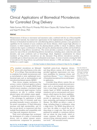
  • 1. Clinical Applications of Biomedical Microdevices for Controlled Drug Delivery Pablo Gurman, MD; Oscar R. Miranda, PhD; Kevin Clayton, BS; Yitzhak Rosen, MD; and Noel M. Elman, PhD Abstract Miniaturization of devices to micrometer and nanometer scales, combined with the use of biocompatible and functional materials, has created new opportunities for the implementation of drug delivery systems. Advances in biomedical microdevices for controlled drug delivery platforms promise a new generation of capabilities for the treatment of acute conditions and chronic illnesses, which require high adherence to treatment, in which temporal control over the pharmacokinetic profiles is critical. In addition, clinical conditions that require a combination of drugs with specific pharmacodynamic profiles and local delivery will benefit from drug delivery microdevices. This review provides a summary of various clinical appli- cations for state-of-the-art controlled drug delivery microdevices, including cancer, endocrine and ocular disorders, and acute conditions such as hemorrhagic shock. Regulatory considerations for clinical translation of drug delivery microdevices are also discussed. Drug delivery microdevices promise a remarkable gain in clinical outcomes and a substantial social impact. A review of articles covering the field of microdevices for drug delivery was performed between January 1, 1990, and January 1, 2014, using PubMed as a search engine. ª 2014 Mayo Foundation for Medical Education and Research n Mayo Clin Proc. 2014;nn(n):1-16 B iomedical microdevices are fabricated devices with critical features on the order of 1 to 100 mm. These microdevices range in complexity from simple microstructures such as microchannels to more sophisticated micro- functional parts such as microtransducers and microelectromechanical systems (MEMS).1 These devices integrate mechanisms that activate a variety of physical signals to achieve a specific function. For example, MEMS-based inertial sensors transduce a mechanical signal input to an electrical signal response. Current transducers are able to combine multiple physical inputs with multiple output signals. Biomedical microdevices present a variety of key advantages for applications in health care owing to their (1) extremely small sizes providing minimally invasive procedures, (2) low power consumption, (3) batch fabrication processes with high reproducibility, and (4) low cost per device, in conjunction with their multiple functionalities and compatibility with very large-scale integration electronics. These novel technologies have accelerated the development of a variety of micromedical devices, such as catheter pressure sensors, microelectronic components for pacemakers, hand-held point-of-care diagnostic devices, and drug delivery systems, all of which have provided significant improvement over treat- ment possibilities for numerous chronic and nonchronic illnesses.1-4 Figure 1 shows a variety of biomedical microdevices for several therapeu- tic applications. Controlled drug delivery systems that are based on microdevices contain structural micro- parts, such as microchannels and microreser- voirs, to store drugs. In addition, drug delivery systems based on MEMS incorporate micro- transducers such as microactuators and micro- sensors, which improve the device capabilities. Drug delivery devices based on MEMS provide an opportunity for improved diag- nosis, monitoring, and treatment of numerous illnesses. The MEMS can deliver a variety of drugs, including drugs in combination, using a single device. The MEMS drug delivery de- vices have the ability to control the rate of drug release to a target area. They can be pro- grammed for pulsatile or continuous delivery and can release the drug locally, which in- creases treatment efficacy using a smaller amount of drug, reducing systemic concentra- tion levels1-6 and associated toxicity. From the Institute for Soldier Nanotechnologies, Massachusetts Institute of Technology, Cambridge (P.G., O.R.M., K.C., Y.R., N.M.E.); and Department of Materials Science, University of Texas at Dallas, Richardson (P.G.). Mayo Clin Proc. n XXX 2014;nn(n):1-16 n http://dx.doi.org/10.1016/j.mayocp.2014.10.003 www.mayoclinicproceedings.org n ª 2014 Mayo Foundation for Medical Education and Research 1 REVIEW
  • 2. Finally, the scope of novel materials for biomedical devices has expanded the potential use of biocompatible platforms with high biolog- ical performance, eg, less toxic and nonreactive devices, enabling new therapeutic applications. This review provides a summary of current state-of-the-art biomedical microdevices for controlled drug delivery and their correspond- ing clinical applications. The following sections describe passive and active delivery devices based on MEMS technology. Each section pro- vides a technical description of a microdevice followed by its suggested clinical application. The review continues with a summary of the regulatory strategies for obtaining Food and Drug Administration (FDA) approval for such microdevices. Finally, a perspective on the future of these novel devices is presented. DATA SOURCES AND SEARCHES A PubMed search between January 1, 1990, and January 1, 2014, was performed. The search terms were drug delivery AND MEMS, implant- able devices AND MEMS, control release AND microchip, controlled release AND BioMEMS, neural probes AND drug delivery, vaccines AND microneedles, diabetes AND microneedles, intraoc- ular AND drug delivery devices, and inner ear AND drug delivery AND microfluidics. Papers were selected following the definition of micro- devices and MEMS. Selection also was per- formed with the aim of having examples of different types of microdevices (passive and active, actuation mechanism, and materials). Ex- amples of different clinical applications for drug delivery microdevices assisted in selecting pa- pers more close to the clinical application than those focused solely on fundamental science. Diagnostic microdevices were specifically excluded from the search. PASSIVE DEVICES Passive biomedical microdevices for drug deliv- ery do not rely on an actuation mechanism or on monitoring for feedback. These devices are reservoir based, relying on mass transfer across a permeable membrane to deliver pharmaceu- tical drugs, the biodegradation of a hermetic membrane, or a unique reservoir structure to achieve controlled release. The rate of release can be controlled by taking into account the following design parameters: (1) the effective permeability of the membranes by fine-tuning structural dimensions and materials (pore size, thickness), (2) the rate of degradation of the polymer contained on the membrane or in the reservoir, (3) the diffusivity properties of the drug, and (4) the osmotic pressure. Pas- sive delivery of drugs cannot be modified after implementation. Other passive-release devices operate based on actuation resulting from in vivo conditions inside the body, such as pH or temperature, to accelerate degradation of the materials that encapsulate the pharmaceu- tical drugs. Typically, the controlled release is achieved by considering the pharmacokinetics of the selected drug for delivery. Design and ma- terial parameters are thereafter adjusted and selected during the design process to provide a constant and superior pharmacokinetic perfor- mance, such as an improvement in treatment efficacy duration over the typical half-life of the pharmaceutical drug. Existing passive- release devices, such as the fentanyl transdermal system (DURAGESIC; Janssen Pharmaceuticals Inc) and the fluocinolone acetonide intravitreal implant (Retisert; Bausch & Lomb Inc), are used for either short-term (3 days) or ARTICLE HIGHLIGHTS n Drug delivery systems can be classified as passive and active. Passive devices do not incorporate sensors and actuators for drug delivery. n Active microdevices include microelectromechanical systems (MEMS), which comprise microparts such as microchannels and microvalves and transducers, including microsensors and microactuators, integrated into a singular microdevice. n Advantages of MEMS drug delivery systems include miniaturization, integration with microelectronics, actively controlled, low cost, multiple pharmacologic therapies in a single device, controlled over release rate, and in vivo long-term storage of drugs. n The MEMS are being used for a variety of clinical conditions, including diabetes, neurologic disorders, inner ear diseases, and cancer. n Fluzone is an example of a Food and Drug Administratione approved drug delivery microdevice for vaccine delivery. n The MEMS drug delivery devices can be considered combina- tion products. Many combination products are considered drugs, requiring a New Drug Application for Food and Drug Administration approval. MAYO CLINIC PROCEEDINGS 2 Mayo Clin Proc. n XXX 2014;nn(n):1-16 n http://dx.doi.org/10.1016/j.mayocp.2014.10.003 www.mayoclinicproceedings.org
  • 3. long-term (2.5-3 years) continuous treatment of diseases. The lack of integrated electronics re- duces the complexity of these devices.7,8 Hydrogels Implantable devices based on environmentally sensitive hydrogels were developed for controlled release (Figure 2).9 The device ar- chitecture consists of a reservoir and a 100- mm-thick silicon membrane with orifices measuring 140 mm in diameter. Each orifice contains a support post in the center and is tethered to confine the hydrogel to the mem- brane. The hydrogel is loaded around the central support post such that the entire orifice is blocked by the hydrogel in the swollen state. Under activation by chemical or physical stimuli, the hydrogel shrinks and the drug is allowed to diffuse through the resulting orifice. The response of the hydrogel opening or closing is critical in controlling the rate of drug delivery. Additional control of drug de- livery can be gained by manipulating the membrane thickness, the size of the orifices, the support posts, and the tethers. The temperature, pH, and glucose sensitivity of different hydrogels are some of the parameters that provide additional control over activation. For example, N-isopropylacrylamide hydrogels Microchannel Pt conducting wire Neural probe with microchannels for drug delivery (Figure 4) MEMS Chip Pump Into inner ear for intra cochlear delivery Cochlear implanted device with pump and electronic control (Figure 10) Stratum corneum Epidermis Dermis Subcutaneous tissue Microneedles Hypodermic needle Dendritic cell Langerhans cell Microneedle transdermal patch (Figure 8) Drug solution Drug reservoir Parylene cannula Into cannula Into eye Eye wall Electrolysis pump Pump outlet Ocular device with electrolysis pump (Figure 7) MEMS delivery for emergency (Figure 6) Microchip drug delivery for osteoporosis (Figure 5) Implantabe MEMS device for drug delivery (Figure 9) FIGURE 1. Technology map for applications of biomedical microdevices. MEMS ¼ microelectromechanical systems. CLINICAL APPLICATIONS OF BIOMEDICAL MICRODEVICES Mayo Clin Proc. n XXX 2014;nn(n):1-16 n http://dx.doi.org/10.1016/j.mayocp.2014.10.003 www.mayoclinicproceedings.org 3
  • 4. were found to rapidly contract at 34 C, resulting in a sharp increase in the flow rate of the drug from 0 to approximately 1 mL/min. This type of hydrogel exhibited a fast response to environ- mental conditions, contracting in 10 seconds at 25 C and expanding back to close the orifice in 20 seconds at 50 C. N-butan-2-ylbutan-2- amine/anodic alumina membrane hydrogel was measured to respond to a change in pH of 3.0 to 10.0 in 4 minutes, whereas for changes in glucose levels from 0 to 20 mmol/L, this hydrogel responded in 40 minutes.9 Clinical Application: Diabetes Diabetes represents a significant burden in health care as the number of people with type 2 diabetes is increasing dramatically owing to a pandemic of obesity worldwide.10 One of the key issues in diabetes is adherence with insulin administration. Adherence is limited owing to the frequent and uncomfort- able subcutaneous (SC) injections that the pa- tient needs to treat his or her diabetes. Glucose-responsive hydrogels provide an opportunity for the controlled delivery of insu- lin. Incorporating more channels with various types of hydrogels and channel sizes could improve control and treatment. Further work is needed to better understand and engineer response kinetics and the reliability of such hydrogel-based devices for clinical applications. Passive Nanochannel-Based Drug Delivery Device A novel, high-throughput nanochannel drug delivery system for the sustained delivery of chemotherapeutics was developed and tested in vitro.11 The device was developed to be implantable to improve patient adherence and quality of life by avoiding the need for repeated administrations and frequent visits to the clinic. The device passively controls the release of drugs by physical-electrostatic confinement. By manipulating the size of the nanochannels, zero-order release of chemotherapeutics was achieved. The nanochannel membrane com- prises a silicon substrate reservoir and a capping layer. An array of 161 channels, measuring 200Â200 mm and spaced by 50- mm-thick walls, makes up the membrane sur- face (Figure 3). It consists of 30-mm-wide microchannels that connect the reservoir and the capping layer. The nanochannels connect the inlet and outlet channels at the interface of the silicon substrate and the capping layer. Clinical Application: Melanoma Melanoma, a tumor originating from melano- cyte cells, represents the most aggressive form of skin cancer, with 5-year survival of 20% for advanced cases. Current pharmaco- logic therapies include the use of interferon alfa-2b as an adjuvant for stage III melanomas. Interferon alfa-2b is an immunomodulatory drug that activates the immune system against the tumor, increasing patient relapse-free sur- vival. An important issue with interferon alfa-2b is its adverse effects. Interferon alfa- 2b in high doses has been linked to hepatotox- icity and suicidal ideation.12 The use of implantable, controlled nano- channel delivery systems could potentially overcome some of the limitations associated with current therapies by decreasing the amount of drug that reaches the systemic cir- culation. This improvement could avoid adverse effects in healthy tissues while keeping high concentrations of interferon alfa-2b at the targeted site where the tumor is located. Clinical Application: Prostate Cancer Prostate cancer represents the sixth leading cause of cancer death in men, with an incidence of 233,000 new cases and 29,480 deaths in Front side tethers Central tethered post Back side tethers Flow Silicon membrane FIGURE 2. Schematic of silicon membrane with structured orifices. From Sens Actuators B Chem,9 with permission. MAYO CLINIC PROCEEDINGS 4 Mayo Clin Proc. n XXX 2014;nn(n):1-16 n http://dx.doi.org/10.1016/j.mayocp.2014.10.003 www.mayoclinicproceedings.org
  • 5. 2014.13 Leuprolide acetate is a synthetic analogue of gonadotropin-releasing hormone. Gonadotropin-releasing hormone stimulates the release of follicle-stimulating hormone and luteinizing hormone, which promote the pro- duction of estrogen and testosterone. Testos- terone is metabolized in the interior of prostate cells to dihydrotestosterone, which upregulates cell proliferation, gene expression, and protein synthesis. It is thought that leuprolide acts as a gonadotropin-releasing hormone analogue and when given continuously by the SC or intramus- cular route (leuprolide acetate could not be administered orally because is a peptide) leads to testosterone deprivation. Deprivation of pros- tate cells from testosterone would lead to apoptosis and cytoreduction of tumor volume.14 Interferon alfa-2b and leuprolide acetate were chosen to test the nanochannel micro- chip delivery device. The release of interferon alfa-2b was tested using 20-nm membranes and was measured to be a mean Æ SD of 29.7Æ1.5 mg/d for 6 days, which is in agree- ment with current maintenance doses of inter- feron alfa-2b used in patients with melanoma (10 million IU/m2 SC 3 times/wk; 10 million international units ¼ 38 mg). The release of leuprolide was tested using 5- and 15-nm channels and was measured to be zero order for the 5-nm channel at a mean Æ SD rate of 100Æ10 mg/d for 3 days, which is close in agreement with current leu- prolide doses used in prostate cancer (250 mg/d). By increasing reservoir sizes, the nanochannel-based delivery system has the potential to achieve the current dose regimens used for interferon alfa-2b and leuprolide. Multifunctional MEMS for Neural Recording and Drug Delivery Multifunctional MEMS for simultaneous recording of neural activity and drug delivery were developed (Figure 4).15,16 One such de- vice has been reported by Altuna et al16 based on flexible microprobes made of SU-8. The polymer SU-8 was used as the structural mate- rial for the probes, with platinum for the elec- trodes. Tetrode-like probes with a single microfluidic channel and linear probes with 2 microfluidic channels were tested. Electrodes for the tetrode-like probe were spaced 25 mm apart, with diameters of 20 mm to sense indi- vidual neuronal firing at the tip of the probe. The microfluidic channel measured 50Â20 mm, with 3 outlet ports also near the tip of the probe. In the linear probe, 8 electrodes were spaced 100 mm apart, allowing for sensing at different depths of the brain. The 2 microfluidic channels measured 40Â20 mm and had inde- pendent outlet ports. Both devices were 55 mm thick. The tetrode-like probe was 90 mm wide, and the linear probe was 150 mm wide. The probes were tested in vivo in anesthe- tized rats. The SU-8 linear probes were used to deliver kainate at the CA1 cell and dendritic layers at a flow rate of 3 to 6 mL/min to induce seizures. Neuronal excitability was recorded against a control delivery of saline to confirm de- livery of the drug. The tetrode-like probe deliv- ered potassium at a high flow rate of 0.6 to 1.5 mL/min to the CA1 cell layer. The probe was able to record isolated neurons together with multi-unit firing. Both probes had the ability to measure ripples and spikes common during large irregular brain activity at the CA1 cell layer. Clinical Applications: Parkinson Disease and Epilepsy Effective treatments for neurologic diseases are still lacking. Parkinson disease, the second most FIGURE 3. Schematics (A, C, and D) and optical microscopy (B) of a passive nanochannel delivery system designed for drug release with zero-order kinetics. The innovative architecture of the device includes macro- channels, microchannels, and nanochannels. M ¼ macrochannel; mO ¼ microchannel outlet; mI ¼ microchannel inlet; n¼ nanochannel; w ¼ supporting walls. From Pharm Res,11 with permission. CLINICAL APPLICATIONS OF BIOMEDICAL MICRODEVICES Mayo Clin Proc. n XXX 2014;nn(n):1-16 n http://dx.doi.org/10.1016/j.mayocp.2014.10.003 www.mayoclinicproceedings.org 5
  • 6. common neurodegenerative disorder after Alz- heimer disease, has been managed for the past few decades with L-3,4-dihydroxyphenylalanine (L-DOPA).17 The L-DOPA is a precursor to dopa- mine, which is a neurotransmitter that is absent in the brain of patients with Parkinson disease owing to the progressive loss of dopaminergic neurons. In the long-term, patients start to expe- rience L-DOPA adverse effects that deteriorate their quality of life. Epilepsy, a disorder characterized by un- controlled propagation of electrical stimuli in the brain, has been managed with drugs that reduce neuron excitability. Some types of epi- lepsy, however, remain refractory to drugs. Therefore, it is clear that current pharmaco- logic therapies alone have not reached an acceptable benefit for neurologic disorders requiring additional intervention. Implantable devices such as neural stimulators have emerged as an attractive option for patients with advanced Parkinson disease, refractory epilepsy, and other neurologic conditions.18 Despite the aforementioned benefits, these novel delivery modalities need to overcome is- sues of poor biocompatibility, such as inflamma- tory response and fibrosis around the implant, which limit overall device performance. For example, neural probes have been found to elicit glial scar formation and neuronal loss during im- plantation, impairing device performance. Hav- ing an anti-inflammatory drug in the same device could decrease the inflammatory response and, thus, the generation of fibrotic tissue (eg, glial scar formation) surrounding the implant, thus preserving functionality.19,20 Therefore, it is being realized that by combining devices with drug therapies, it is possible to maximize the benefits of both while avoiding their adverse effects. This clinical need has been met by using MEMS technologies, in which neural electrodes are being combined with microfluidic channels or microreservoirs. This combines the capability to record neural data and drug delivery. ACTIVE DEVICES Active drug delivery devices use a variety of mechanisms to release pharmaceutical drugs and provide an increased level of control. The MEMS devices have been developed using different actuation modalities, including micropumps based on gas pressure from elec- trolysis, integration of magnetic actuators, and electrochemical and electrothermal actuation systems. Active devices can be customized to treat a range of diseases requiring specific pharmacokinetic drug delivery profiles. More- over, as opposed to passive delivery systems, Bonding pads Microelectrode film Microelectrode sites Outlet Microchannel Molded PDMS film Vacuum A B C Microchannel Pt conducting wire FIGURE 4. Schematics (A and B) and optical picture (C) of a neural probe with drug delivery capabilities. A, Assembly of device components. B, Method used to incorporate the drug into the microchannels. C, Optical picture of the finished device and microchannels containing a dye solution. PDMS ¼ poly- dimethylsiloxane; Pt ¼ platinum. From Sens Actuators A Phys,15 with permission. MAYO CLINIC PROCEEDINGS 6 Mayo Clin Proc. n XXX 2014;nn(n):1-16 n http://dx.doi.org/10.1016/j.mayocp.2014.10.003 www.mayoclinicproceedings.org
  • 7. MEMS can be activated and stopped at any time after implantation. Active devices commonly require minia- turized power electronics for actuation, typi- cally increasing the overall form factor, which is a key limiting factor in implantable applications. Alternatively, telemetry systems to transfer energy for activation can be adop- ted to overcome this limiting issue. Electrothermally Actuated MEMS Drug Delivery Microchip Santini et al21 developed a device for the controlled, pulsatile release of chemicals from single or multiple reservoirs. The controlled drug release was triggered by the application of an electric potential to burst sealing gold membranes electrothermally. Drugs inside the reservoir were then free to diffuse to the tar- geted site. This original device had the func- tionality for complex release of kinetics by varying the amount or substance type placed in each reservoir and varying the timing of release. This type of device would be able to deliver drugs in a pulsatile manner. Since then, several works on active drug delivery devices based on MEMS have made substantial progress toward effectively treating various ailments. Clinical Application: Osteoporosis Osteoporosis is the progressive degradation of bone architecture and loss of mass bone den- sity that leads to bone fragility that ultimately increases the risk of fractures. Osteoporosis is more common in postmenopausal women, who are at risk for lower levels of estrogens, which are known to be involved in bone for- mation. According to the National Institute for Health and Clinical Excellence, 9 million osteoporotic fractures occur annually in the world.22 A microchip device containing parathyroid hormone (PTH) was developed for the treat- ment of osteoporosis23 ; PTH is known to stim- ulate bone formation by increasing osteoblast number and function.24 An implantable microchip device capable of releasing PTH would prevent the need for frequent injections of PTH. The first in-human testing performed in postmenopausal women evaluated the in vivo pharmacokinetic profile of a PTH-release microchip (Figure 5) against standard SC in- jections.25 The microchip was implanted sub- cutaneously in the abdomen, and the pharmacokinetic profile was measured after a fibrous capsule was formed around the implant. The rationale of the study was to deter- mine the pharmacokinetic performance of the microchip when it was surrounded by a fibrous capsule as a result of the host response to the implant. In addition, bone biomarkers were measured to determine the effect on bone formation of PTH injections vs PTH released by the microchip. A safety laboratory panel was performed to determine the safety of the microchip vs that of the SC injections. Overall, the microchip was found to be bioequivalent to the SC injections even in the presence of the fibrous capsule. The microchip was also found to be as safe as the SC injections based on a laboratory panel.25 FIGURE 5. A microchip drug delivery system for parathyroid hormone (PTH) release for osteoporosis treatment. The picture depicts the titanium packaging used for carryng the micro- chip. The device has undergone first human trials. From Sci Transl Med,25 with permission. CLINICAL APPLICATIONS OF BIOMEDICAL MICRODEVICES Mayo Clin Proc. n XXX 2014;nn(n):1-16 n http://dx.doi.org/10.1016/j.mayocp.2014.10.003 www.mayoclinicproceedings.org 7
  • 8. A Rapid-Delivery Microchip for Acute Clinical Conditions A microchip drug delivery system for rapid de- livery of vasopressin was developed by Elman et al.26 The device consists of a membrane layer, an actuation layer, and a reservoir layer. The membrane layer consists of a biocompatible sil- icon nitride film that serves as a hermetic seal for the reservoirs. The actuation layer consists of 3 microresistors. Heat is generated when a current is passed through these microresistors. The heat serves to nucleate bubbles and dramatically in- crease internal pressure inside of the reservoir. This step leads to rupturing of the silicon nitride membrane, followed by rapid release of the pharmaceutical drug, used as a bolus. A picture of the device in action is shown in Figure 6. Clinical Application: Hemorrhagic Shock Hemorrhagic shock is an acute condition that can result from severe traumatic injuries associated with massive bleeding loss, which if not treated within seconds or minutes could result in perma- nent damage or death. In most cases, critical pa- tients do not have immediate access to a health care facility where basic measures to restore he- modynamic stability are available. These mea- sures include oxygenation; restoration of intravascular volume with colloids, crystalloids, or blood products; and use of inotropic and vaso- pressor drugs. In settings with limited or no ac- cess to health care facilities, interventions to prevent massive hemorrhages include self- applied hemostatic dressings. This approach, however, does not account for internal bleeding sites, which occasionally are the main cause of death. During hemorrhagic shock, the massive loss of blood compromises vital organ activity in the brain, heart, and kid- neys, among others. The natural response of the body to avert vital organ damage is to pro- duce vasoconstriction to restore arterial blood pressure and cardiac output to the level required to maintain adequate oxygenation of vital organs while avoiding further blood loss.27 Vasopressin and inotropic agents represent an important tool in the management of hem- orrhagic shock.28-30 This biomedical microde- vice was designed to be implanted in high-risk patients to deliver vasopressin for the manage- ment of hemorrhagic shock in emergency and ambulatory settings. Finally, other potential uses of the rapid-delivery microchip include acute medical conditions that require immedi- ate intervention, such as cardiovascular and neurologic emergencies. Magnetically Controlled MEMS Drug Delivery A magnetic actuator MEMS drug delivery device was developed for the controlled release of a chemotherapeutic agent. The device was designed to avoid the use of batteries, improving form factor. The device consists of a microreser- voir sealed by a thin magnetic membrane com- posite consisting of elastic polydimethylsiloxane material integrated with iron oxide nanoparticles. An external magnetic field applied by a neodym- ium iron boron permanent magnet creates a force that allows the magnetic membrane to deflect. This process builds up pressure inside the reser- voir, enabling the drug to diffuse out through a laser-drilled micron-sized aperture. On-demand release profiles can be created for optimal treatment using this device. With no actuation, the mean Æ SD release of the drug was measured to be 0.053Æ0.014 ng/ min. With actuation of the membrane by appli- cation of a 255-mT magnetic field, the mean Æ SD release rate increased to 160Æ10.2 ng per actuation. The release rate exhibited sustained delivery for more than 35 days.31 Clinical Application: Cancer Docetaxel was selected as a test drug to study the device release profile. Docetaxel is an anti- neoplastic agent that disrupts the mitotic spin- dle, causing cell death; it is used for the FIGURE 6. Pulsatile controlled delivery profile of a microelectromechanical systems device with a thermally induced actuator releasing drug out of a reservoir for emergency applications. From Biomed Microdevices,26 with permission. MAYO CLINIC PROCEEDINGS 8 Mayo Clin Proc. n XXX 2014;nn(n):1-16 n http://dx.doi.org/10.1016/j.mayocp.2014.10.003 www.mayoclinicproceedings.org
  • 9. treatment of a variety of tumors, such as breast cancer.32 An important issue in antineoplastic drugs is to achieve maximum selectivity be- tween cancer cells and healthy cells by increasing the local concentration of the drug while decreasing systemic drug biodistribution, avoiding exposure of healthy tissues.33 This could be accomplished using a magnetically actuated MEMS device that could release the drug locally on demand. In vitro drug-release experiments using cell culture demonstrated that freshly prepared docetaxel solutions and docetaxel from the de- vice described previously herein were found to have comparable effects on target cells. Further development is still required before clinical translation. Micropump MEMS-Based Drug Delivery Devices A refillable intraocular MEMS drug delivery de- vice was developed that uses a micropump for actuation. The device was designed to deliver drugs from a 54-mL reservoir by sending the drug through a cannula and past a 1-way check valve incorporated at the end of the cannula (Figure 7).34 A dose of medication is dispensed from the device via an electrolysis micropump. The device is intended to be implanted under the conjunctiva, with the cannula pointing into the anterior chamber of the eye. Electrolysis of water is triggered by an applied voltage, producing oxygen and hydrogen gases. These gases result in an internal pressure that forces the drug out of the reservoir. For driving currents ranging from 5 mA to 1.25 mA, the flow rate of drug increased linearly from 5 to 439 mL/min. Under normal and abnormal back pressures, the device was able to release 1500 and 1300 nL/min, respectively, with a driving current of 200 mA. Silicone rubber was selected as the reservoir material and was found to be capable of resealing without leakage after repeated refills via a non-coring needle. Replenish Inc further developed a similar system called the Ophthalmic MicroPump System. Two types of micropump systems were developed: an anterior micropump and a posterior micropump. Both devices use a wireless programmer and charger for control of drug delivery. A flow sensor controls the flow rate through a feedback loop, allowing the dispensing of nanoliter volume of drugs. The final piece of the system is a separate con- sole unit to refill the implant with drug.35 Clinical Application: Ocular Disorders Traditional ocular drug treatments, such as oral drugs and eyedrops, require significant overdose because less than 5% of the drug is able to pass the physiologic barriers and reach the site of ac- tion.36 The overdose needed to achieve thera- peutic concentrations results in potential systemic adverse effects. A variety of passive im- plants were developed to overcome this issue. Current passive intraocular implants depend on polymer degradation to release the drug and have no control over the drug-release pro- file, which could lead to subtherapeutic or supratherapeutic (toxic) drug concentrations. Using the electrolysis micropump, it is possible to circumvent these limitations by providing the drug locally and controlling the pharmacokinetic profiles. Release profiles can be programmed by adjusting the current applied to the electrolysis pump. The ability of this device to be refilled makes it attractive for long-term treatment of ocular diseases of the posterior segment, such as age-related macular degeneration. The anterior micropump developed by Replenish Inc was adapted to address disor- ders of the anterior chamber (glaucoma), whereas the posterior micropump was adapt- ed to address disorders of the posterior cham- ber (retina disorders). Transdermal MEMS Microneedle Patch Array Delivery System Researchers developed a wearable patch based on a microneedle array for the transdermal Drug solution Drug reservoir Parylene cannula Into cannula Into eye Eye wall Electrolysis pump Pump outlet FIGURE 7. Cross section of the ocular device illustrating the pump and cannula. From Sens Actuators A Phys,34 with permission. CLINICAL APPLICATIONS OF BIOMEDICAL MICRODEVICES Mayo Clin Proc. n XXX 2014;nn(n):1-16 n http://dx.doi.org/10.1016/j.mayocp.2014.10.003 www.mayoclinicproceedings.org 9
  • 10. delivery of macromolecular drugs. Micronee- dles provide painless administration because they are designed to penetrate through the stratum corneum (the outer layer of the skin) without reaching the nerve terminals located deeper in the skin. Studies have reported a strong correlation between microneedle length and pain percep- tion, although other features, such as drug vol- ume and number of microneedles, have also been associated with pain development during microneedle insertion.37,38 Figure 8 compares injection depth and physiologic impact be- tween an array of microneedles and a hypo- dermic needle.39 A device was developed consisting of 400- mm-long microneedles that are inserted through the outermost layer of the skin, result- ing in pain-free drug delivery. The whole de- vice consists of an array of 25 microneedles, each with 300-mm through-holes on a 4Â4- mm cross section. A thermally expandable sil- icone composite is layered below the reservoir. A printed circuit board with heaters to expand the silicone composite layer into the reservoir layer was designed to perform controlled release of the drug through the microneedles. The amount of power applied to the electrical copper heaters controls the amount of expansion and, therefore, the flow rate of the drug. The microneedles use side openings to allow an incredibly sharp apex to avoid coring of tissue during its therapeutic application.40 Clinical Application: Diabetes The reduction of frequent SC injections of insu- lin can improve adherence to insulin therapy in diabetic patients. Moreover, emulating the physiologic release of insulin by the pancreas is a highly desirable feature. In this regard, the transdermal microneedle MEMS array pro- vides painless administration (improving pa- tient adherence) and control over the flow rate that mimics the kinetics release of insulin by the pancreas (improving efficiency while avoiding adverse effects). Furthermore, a transdermal patch is an easy-to-use device compared with current insu- lin SC injections. The device was tested in vivo on diabetic rats. With applied power of 150 to 450 mW, the device was measured to dispense 0.1 to 300 mL/h of insulin (a vial of insulin con- tains 100 IU/mL; therefore, 0.1 mL¼0.01 IU and 300 mL¼30 IU). Based on the pancreatic secretion of insulin (1 IU/h), it is likely that the operational space of the micropump is well suited to replicate the physiologic insulin production by the pancreas. Further work is needed to determine the optimal response of the thermally expandable material to allow for a precisely defined low flow rate with no leakage. In this study, an external power source was used, but a micro-sized battery for practical use could be tested in future work. Clinical Application: Vaccines Vaccines have greatly reduced the incidence of several infectious diseases and represent one of the most cost-effective interventions in health care.41 Therefore, adherence with vaccine administration has an important role in public health. Microneedle technologies for vaccines can provide painless vaccines, improving pa- tient acceptability and adherence. This is particularly relevant because most vaccines are administered to pediatric populations.42,43 Moreover, it is expected that painless vac- cines could also improve adherence in the adult population, eg, tetanus vaccine. Another important advantage of transdermal micronee- dles over intramuscular vaccines is the Stratum corneum Epidermis Dermis Subcutaneous tissue Microneedles Hypodermic needle Dendritic cell Langerhans cell FIGURE 8. Schematic comparing a traditional hypodermic needle with a microneedle array. Note how the microneedle array reaches the dermis, where Langerhans cells are found, and does not reach the subcutaneus tissue, rich in nervous terminals. Both properties make microneedles a very attractive option for vaccine delivery systems. From Clin Exp Vaccine Res,39 with permission. MAYO CLINIC PROCEEDINGS 10 Mayo Clin Proc. n XXX 2014;nn(n):1-16 n http://dx.doi.org/10.1016/j.mayocp.2014.10.003 www.mayoclinicproceedings.org
  • 11. possibility of stimulating antigen-presenting cells, located in the skin, to improve antigen transport to lymph nodes, which enhances the immune response. Microneedles also could overcome the technical problems related to intradermal vaccines (eg, poor reproduc- ibility over the injection site and the need to train health care personnel). Electrothermal MEMS Drug Delivery Device A MEMS-based intracranial drug delivery de- vice has been developed and tested for the treatment of malignant brain tumors (Figure 9).44 Passive-release implants have demonstrated some effectiveness, but incorpo- rating active MEMS to gain more control over the release kinetics could improve efficacy and decrease toxicity. The MEMS drug delivery device consisted of an injection-molded liquid crystal polymer reservoir measuring 3.7Â3.2Â2.2 mm and containing a total drug payload of 10 mg of temozolomide. A 300-mm-thick silicon micro- chip sits on top of a 200-mm lip on the interior reservoir walls. The silicon microchip contains three 300Â300-mm suspended silicon nitride membranes, which provides an effective, biocompatible barrier to diffusion. The actuation mechanism relies on using resistive heating to melt a metallic fuse that sits on top of the silicon nitride membranes. Titanium and gold layers are deposited on top of the silicon nitride membrane and are shaped into thin metallic fuses by using photo- lithography followed by wet etching. The fuse is melted using resistive heating by applying an electrical pulse. This burst results in a membrane fracture and release of the reservoir content. Each membrane can be designed to be independently opened by varying the thick- ness of the gold and titanium layers or the width of the fuse to require more or less resis- tive heating. This allows for a variable drug- release profile. Clinical Application: Glioblastoma Glioblastoma is a devastating type of human cancer with mean survival of 12 months and survival of less than 5% after 5 years.45,46 A variety of pharmacologic therapies have been explored, with very poor clinical outcomes.47 A major challenge in drug delivery to the brain is circumventing or passing the blood-brain barrier (BBB). The BBB is the separation of the vasculature system from the brain.48 The BBB maintains brain homeostasis by restricting the transport of molecules present in the circulatory system to and from the brain. This is achieved by the unique charac- teristics of the brain microvasculature that possess endothelial cells connected by very tight junctions. These tight junctions impede the passage of large macromolecules from the blood to the brain. To circumvent the BBB, local implants that release drugs directly in the brain were developed and commercialized.49 Although commercial polymeric implants already exist, survival rates are poor and new approaches are needed. By using active implantable microchips, a multitarget approach using a combination of drugs with controllable phar- macokinetics could lead to better clinical outcomes. It is important to note that active devices that require frequent drug refilling or power source exchange are not suitable alternatives for MEMS implanted in the central nervous system owing to the implicit requirement for repeated neurosurgical procedures. Repeated neurosurgical procedures may lead to a variety of serious complications in the central nervous system. Therefore, several design considerations for implantable MEMS drug delivery systems must be considered owing to the unique anatomical and physiologic features of the central ner- vous system. The electrothermal MEMS described previ- ously herein was tested in vitro and in vivo via intracranial implantation in rats. In vitro tests FIGURE 9. Microelectromechanical systems (MEMS) drug delivery device for the treatment of glioblastoma. Assembled MEMS device (A) and computer-aided design model of the reservoir (B). From Biomaterials,44 with permission. CLINICAL APPLICATIONS OF BIOMEDICAL MICRODEVICES Mayo Clin Proc. n XXX 2014;nn(n):1-16 n http://dx.doi.org/10.1016/j.mayocp.2014.10.003 www.mayoclinicproceedings.org 11
  • 12. confirmed that more membranes being opened leads to more rapid drug release. With 3 membranes activated, the release rate was measured at 0.3 mg/h, and the mean Æ SD total release was 90%Æ3.2% in 30 hours. The release rate and mean Æ SD total release decreased to 0.136 mg/h and 82%Æ1.9%, respectively, in 60 hours for 2 membranes activated; further decreases to 0.007 mg/h and 60%Æ12%, respectively, in 800 hours was observed for 1 membrane activated. Implantation and activation of the device was found to be effective in increasing survival time of 9-L glioblastoma rats. Activation of all 3 membranes in the device on the day of im- plantation was the most effective. This device showed improved efficacy via control of drug pharmacokinetics, but further studies are needed to determine optimal release rates and timing. Microfluidic Hydraulic MEMS-Based Drug Delivery Devices The MEMS devices for drug delivery to the in- ner ear were developed using microfluidics (Figure 10).50-52 A microcannula connected to a closed microfluidic circuit allows fluid to flow in and out of the cochlea. Differences in the micron-sized tubing used for the outlet and inlet loops results in discharge and recharge of fluid on the order of seconds and minutes, respectively. As the solution is continuously pumped in and out of the cochlea and mixed with peri- lymph, dilution of a dissolved compound results in net delivery. The first and second generations of devices use micropumps, and the third gener- ation uses a reciprocating delivery system to control fluid flow. Reciprocating delivery in- volves infusing and drawing the same volume of liquid, resulting in zero net volume transfer. MEMS chip Fluidic channel Drug loading port Infuse withdraw port Displacement diaphragm and chamber MEMS chip Pump Into inner ear for intracochlear delivery Fill port ~13 mm FIGURE 10. Schematic diagram describing a cochlear microfluidic delivery device to prevent sensorineural hearing loss. The miniaturized device comprises several components, including a microfluidic chip, tubing and cannula for delivery, and electronic circuitry and a battery to power the device. Device dimensions are 5.5Â4.0Â3.8 cm. The device operates under the principle of reciprocating delivery, used for drug delivery into small and sensitive regions of the body, such as the cochlea, where a volume of drug is infused while the same amount of liquid is withdrawn, keeping constant the volume in the cochlear space and allowing higher instantaneous flow rates. MEMS ¼ microelectromechanical systems. MAYO CLINIC PROCEEDINGS 12 Mayo Clin Proc. n XXX 2014;nn(n):1-16 n http://dx.doi.org/10.1016/j.mayocp.2014.10.003 www.mayoclinicproceedings.org
  • 13. This technique is suitable for small spaces where overall volume is limited, such as delivery of drugs in the cochlea. Biological back pressures in the cochlea were confirmed to have no noticeable effect on discharge. The distribution of agents in the co- chlea was tested using 6,7-dinitroquinoxaline- 2,3-dione to alter the generation of compound action potential. In vitro and in vivo studies in guinea pigs found increases in the compound action potential threshold, indicating effective drug penetration. Clinical Application: Inner Ear Disorders Inner ear disorders comprise a variety of clinical conditions affecting the inner ear structure or the auditory nerve. The inner ear anatomy in- volves the cochlea and the vestibular system. The cochlea is responsible for transducing sound waves into electrical impulses that are trans- ported through the auditory nerve to the region in the brain responsible for audition perception. Disorders that affect either the sensing (cochlear) or transducing (auditory nerve) component of the auditory system are known as sensorineural hearing loss (SNHL). It is esti- mated that SNHL affects nearly 250 million people worldwide.52 Disorders affecting the inner ear include infectious diseases (eg, congenital rubella and congenital cytomegalo- virus), genetic disorders (such as mutations on the gene for myosin VIIa, a protein found in the stereocilia), and sensing elements of the hair cells located in the cochlea. Other causes include trauma due to long- term exposure to loud sounds and drugs such as aminoglycosides.53 The physiopathology of SNHL involves damage to and death of the hair cells located in the corti organ (a region of the cochlea that contains hair cells and auditory neurons). Hair cells are a specialized type of cell that contain stereocilia, a type of organelle that in response to acoustic waves opens ionic chan- nels, resulting in depolarization of hair cell membranes. This leads to the release of neuro- transmitters, which transport action potentials along the auditory nerve to the regions of the brain responsible for auditory function. The development of the cochlear implant has been a great achievement to restore hearing to people with deafness.54 Cochlear implants aim to stimulate ganglion cells. With the contin- uous degeneration of these cells as a result of infectious, traumatic, or genetic disorders, cochlear implants lose their efficacy. Therefore, drug delivery devices such as the reciprocating micropumps described previously herein repre- sent a novel and promising modality for restoring auditory perception. These devices may allow delivery of neurotropic factors with zero net volume transfer, thus maintaining intra- cochlear pressure constant and preserving the sensing elements of the cochlea.55 REGULATORY PROCESS FOR CLINICAL TRANSLATION To date, there are a few examples of MEMS for medical applications approved by the FDA, including the CardioMEMS wireless pressure sensor (St Jude Medical, Inc), the i-STAT point-of-care blood analyzer device (Abbott Laboratories), and Fluzone (Sanofi Pasteur Inc), an influenza vaccine based on micronee- dles.56-60 Several of the MEMS drug delivery devices described previously herein have not been approved by the FDA for clinical use. It is possible, however, based on previous tech- nologies, such as prefilled syringes (a device prefilled with a drug) and the case of Fluzone (which was approved under a Biologics License Application), to describe a potential regulatory pathway for future drug delivery microdevices. First, MEMS drug delivery systems involve at least 2 components: a device and a drug. If the MEMS device incorporates the drug into the final packaged product (it is expected, owing to their small size, that the device and the drug will be copackaged in a single product), they will be considered combination products.61,62 Second, according to the FDA Office of Combination Products, because the drug incorporated into the device provides the main mechanism of ac- tion (the therapeutic effect is due to the drug; the device only releases the drug), the system is considered a drug. Drug products are subjected to premarket approval through a New Drug Application (NDA) submission or an Abbreviated NDA (ANDA) submission.61,63 As mentioned previously, Fluzone was approved under a Bio- logics License Application, which is similar to an NDA.60 An NDA requires a complete description of the manufacturing process and preclinical and clinical studies with the device to establish safety and effectiveness. When the drug being used in the device has already been approved, CLINICAL APPLICATIONS OF BIOMEDICAL MICRODEVICES Mayo Clin Proc. n XXX 2014;nn(n):1-16 n http://dx.doi.org/10.1016/j.mayocp.2014.10.003 www.mayoclinicproceedings.org 13
  • 14. an ANDA might be required. An ANDA is less stringent than an NDA, demanding only bio- equivalence studies to establish a similar phar- macokinetic profile with existing devices or formulations using the same drug.4 The future of drug delivery microdevices is promising. Their novelty, their complexity, and the fact that they are implantable, however, will make regulatory approval a challenging endeavor. PERSPECTIVE Biomedical microdevices for controlled drug de- livery represent the next generation of delivery modalities that combine miniaturization, low cost, batch manufacturability and reproduc- ibility, and integration with very large-scale inte- gration electronics, allowing programmability and active control over drug release. The current development of drug delivery microdevices is at an early stage, and most of the technologies are still in the proof-of-concept stage. There are a few examples of successful clin- ical translation of biomedical microdevices, such as the clinical use of vaccine microneedles. There are several reasons that some of the microdevices are still in the drug delivery pipeline. From a clinical standpoint, there must be a clear and identified unmet clinical need where current solutions are still lacking. Even if the clinical need exists and is identified, many appli- cations (eg, infectious diseases) demand large drug payloads that cannot be accommodated with microdevices or that would require peri- odic refilling. Moreover, bringing these devices to the market entails a very high-risk endeavor. Finally, regulatory issues could also pose a significant barrier for bringing microedrug delivery devices to the market. Some recent initiatives at the FDA, such as the Center for Devices and Radiological Health Medical Inno- vation Initiative, potentially will help ensure a faster transition of novel biomedical microde- vices into the market. CONCLUSION Recent advances in drug delivery devices that use biomedical microdevices for controlled de- livery promise improved treatment for a vari- ety of acute and chronic illnesses. Passive devices operate by releasing the pharmaceu- tical drugs from reservoirs through permeable structures, which can also be degraded by environmental triggers, such as pH and os- motic forces, to regulate the release rate. Active devices require power to actuate a part that releases the drug after the device is deployed. The release profile of the drug can be actively controlled after the device has been implanted. Passive and active devices can be used as part of minimally invasive procedures and have the ability to deliver drugs with a pre- cise pharmacokinetic profile, enhancing the effi- cacy and decreasing the toxicity of the drug being used. These devices offer a range of clinical ap- plications in which tailored pharmacokinetics, local release, and high adherence are prerequi- sites. These clinical conditions include cancer, endocrine disorders, and ocular diseases, among many others. Drug delivery devices represent a novel technology but face a variety of regulatory challenges. Further understanding of biocompatible materials, alternative techniques for drug release actuation, and closed-loop microdevices will enhance the capability of microdevices for clin- ical drug delivery. Microdevices for drug deliv- ery represent the next generation of platforms for more accurate and efficient drug delivery systems that will enable new therapeutic modal- ities. These novel platforms promise to increase patient adherence and overall significantly improve treatment outcomes. Abbreviations and Acronyms: ANDA = Abbreviated New Drug Application; BBB = blood-brain barrier; FDA = Food and Drug Administration; L-DOPA = L-3,4- dihydroxyphenylalanine; MEMS = microelectromechanical systems; NDA = New Drug Application; PTH = parathyroid hormone; SC = subcutaneous; SNHL = sensorineural hearing loss Grant Support: This work was supported by the US Army Research Office via the Institute for Soldier Nanotechnol- ogies at Massachusetts Institute of Technology (contract W911NF-07-D-0004). Correspondence: Address to Noel M. Elman, PhD, Institute for Soldier Nanotechnologies, Massachusetts Institute of Technology, 500 Technology Square, Cambridge, MA 02139 (nelman@mit.edu). REFERENCES 1. Madou MJ. Fundamentals of Microfabrication and Nanotech- nology. 3rd ed. Boca Raton, FL: CRC Press; 2011. 2. Huff MA. Medical applications of micro-electro mechanical sys- tems technology. In: Rosen Y, Elman N, eds. Biomaterials Sci- ence: An Integrated Clinical and Engineering Approach. Boca Raton, FL: CRC Press; 2012. MAYO CLINIC PROCEEDINGS 14 Mayo Clin Proc. n XXX 2014;nn(n):1-16 n http://dx.doi.org/10.1016/j.mayocp.2014.10.003 www.mayoclinicproceedings.org
  • 15. 3. Polla DL, Erdman AG, Robbins WP, et al. Microdevices in med- icine. Annu Rev Biomed Eng. 2000;2:551-576. 4. Panescu D. MEMS in medicine and biology. IEEE Eng Med Biol Mag. 2006;25(5):19-28. 5. Staples M, Daniel K, Cima MJ, Langer R. Application of micro- and nano-electromechanical devices to drug delivery. Pharm Res. 2006;23(5):847-863. 6. Jain KK. Drug delivery systems: an overview. Methods Mol Biol. 2008;437:1-50. 7. DURAGESIC website. http://www.duragesic.com. Accessed February 5, 2014. 8. Posterior uveitis/Retisert. Psivida Corp website. http://www. psivida.com/products-retisert.html. Accessed February 5, 2014. 9. Baldi A, Lei M, Gu Y, Siegel RA, Ziaie B. A microstructured silicon membrane with entrapped hydrogels for environmen- tally sensitive fluid gating. Sens Actuators B Chem. 2006; 114(1):9-18. 10. Ginter E, Simkon V. Type 2 diabetes mellitus, pandemic in 21st century. Adv Exp Med Biol. 2012;771:42-50. 11. Grattoni A, Shen H, Fine D, et al. Nanochannel technology for constant delivery of chemotherapeutics: beyond metronomic administration. Pharm Res. 2011;28(2):292-300. 12. Dieperink E, Ho SB, Tetrick L, Thuras P, Dua K, Willenbring ML. Suicidal ideation during interferon-alpha2b and ribavirin treat- ment of patients with chronic hepatitis C. Gen Hosp Psychiatry. 2004;26(3):237-240. 13. Siegel R, Ma J, Zou Z, Jemal A. Cancer statistics, 2014. CA Can- cer J Clin. 2014;64(1):9-29. 14. Sethi R, Sanfilippo N. Six-month depot formulation of leupror- elin acetate in the treatment of prostate cancer. Clin Interv Aging. 2009;4:259-267. 15. Gao K, Li G, Lia L, Cheng J, Zhao J, Xu Y. Fabrication of flexible microelectrode arrays integrated with microfluidic channels for stable neural interfaces. Sens Actuators A Phys. 2013;197(1):9-14. 16. Altuna A, Bellistri E, Cid E. SU-8 based microprobes for simul- taneous neural depth recording and drug delivery in the brain. Lab Chip. 2013;13(7):1422-1430. 17. Khatri IA, Chaudhry US. Parkinson disease: a review. J Neurol Sci. 2009;4(1):33-43. 18. Theodore WH, Fisher RS. Brain stimulation for epilepsy. Lancet Neurol. 2004;3(2):111-118. 19. Retterer ST, Smith KL, Bjornsson CS, et al. Model neural pros- theses with integrated microfluidics: a potential intervention strategy for controlling reactive cell and tissue responses. IEEE Trans Biomed Eng. 2004;51(11):2063-2073. 20. Zhong Y, Bellamkonda RV. Dexamethasone-coated neural probes elicit attenuated inflammatory response and neuronal loss compared to uncoated neural probes. Brain Res. 2007; 1148:15-27. 21. Santini JT Jr, Cima MJ, Langer R. A controlled-release microchip. Nature. 1999;397(6717):335-338. 22. National Insititute for Health and Care Excellence. Osteoporosis: Assesing the Risk of Fragility Fracture. Manchester, UK: NICE; 2012:4-23:NICE clinical guideline 146. 23. Proos ER, Prescott JH, Staples MA. Long-term stability and in vitro release of hPTH(1e34) from a multi-reservoir array. Pharm Res. 2008;25(6):1387-1395. 24. Esbrit P, Alcaraz MJ. Current perspectives on parathyroid hormone (PTH) and PTH-related protein (PTHrP) as bone anabolic therapies. Biochem Pharmacol. 2013;85(10): 1417-1423. 25. Farra R, Sheppard NF, McCabe L, et al. First-in-human testing of a wirelessly controlled drug delivery microchip. Sci Transl Med. 2012;4(122):1-12. 26. Elman NM, Ho Duc HL, Cima MJ. An implantable MEMS drug delivery device for rapid delivery in ambulatory emergency care. Biomed Microdevices. 2009;11(3):625-631. 27. Tintinally J, Kelen GD, Stapcszynsky JS. Emergency Medicine: A Comprehensive Study Guide. 6th ed. New York, NY: McGraw- Hill Education; 2003. 28. Brunton L, Lazo J, Parker K, Lazo JS. Goodman and Gilman’s The Pharmacological Basis of Therapeutics. 11th ed. New York, NY: McGraw Hill Education; 2006. 29. Liu L, Tian K, Xue M, et al. Small doses of arginine vasopressin in combination with norepinephrine “buy” time for definitive treatment for uncontrolled hemorrhagic shock in rats. Shock. 2013;40(5):398-406. 30. Beloncle F, Meziani F, Lerolle N, Radermacher P, Asfar P. Does vasopressor therapy have an indication in hemorrhagic shock? Ann Intensive Care. 2013;3(1):13. 31. Pirmoradi FN, Jackson J, Burt H, Chiao M. Delivery of an anti- cancer drug from a magnetically controlled MEMS device shows cytotoxicity in PC3 and HUVEC cells. In: Proceedings from the 16th International Solid-State Sensors, Actuators and Microsystems Conference (TRANSDUCERS); June 5-9, 2011; Beijing. 32. Alken S, Kelly MC. Benefit risk assessment and update on the use of docetaxel in the management of breast cancer. Cancer Manag Res. 2013;5:357-365. 33. Wolinsky JB, Colson YL, Grinstaff MW. Local drug delivery stra- tegies for cancer treatment: gels, nanoparticles, polymeric films, rods, and wafers. J Control Release. 2012;159(1):14-26. 34. Li P-Y, Shih J, Lo R, et al. An electrochemical intraocular drug delivery device. Sens Actuators A Phys. 2008;143:41-48. 35. Replesnish Inc website. http://www.replenishinc.com. Accesed September 1, 2014. 36. Gaudana R, Ananthula HK, Parenky A, Mitra AK. Ocular drug delivery. AAPS J. 2010;12(3):348-360. 37. Pierre MB, Rossetti FC. Microneedle-based drug delivery sys- tems for transdermal route. Curr Drug Targets. 2014;15(3): 281-291. 38. Sachdeva V, Banga AK. Microneedles and their applications. Recent Pat Drug Deliv Formul. 2011;5(2):95-132. 39. Suh H, Shin J, Kim YC. Microneedle patches for vaccine deliv- ery. Clin Exp Vaccine Res. 2014;3(1):42-49. 40. Roxhed N, Samel B, Nordquist L, Griss P, Stemme G. Painless drug delivery though Microneedle-based transdermal patches featuring active infusion. IEEE Trans Biomed Eng. 2008;55(3): 1063-1071. 41. Greenwood B, Salisbury D, Hill AV. Vaccines and global health. Philos Trans R Soc Lond B Biol Sci. 2011;366(1579):2733-2742. 42. Prausnitz MR, Mikszta JA, Cormier M, Andrianov AK. Microneedle-based vaccines. Curr Top Microbiol Immunol. 2009;333:369-393. 43. Kim YC, Park JH, Prausnitz MR. Microneedles for drug and vac- cine delivery. Adv Drug Deliv Rev. 2012;64(14):1547-1568. 44. Masi BC, Tyler BM, Bow H, et al. Intracranial MEMS based temozolomide delivery in a 9L gliosarcoma model. Biomaterials. 2012;33(23):5768-5775. 45. Davis ME, Stoiber AM. Glioblastoma multiforme: enhancing survival and quality of life. Clin J Oncol Nurs. 2011;15(3): 291-297. 46. Omuro A, DeAngelis LM. Glioblastoma and other malignant gliomas: a clinical review. JAMA. 2013;310(17):1842-1850. 47. Neyns B, D’haeseleer M, Rogiers A, et al. The role of cytotoxic drugs in the treatment of central nervous system gliomas. Acta Neurol Belg. 2010;110(1):1-14. 48. Domb A, Maniar M, Bogdansky S, Chasin M. Drug delivery to the brain using polymers. Crit Rev Ther Drug Carrier Syst. 1991; 8(1):1-17. 49. Panigrahi M, Das PK, Parikh PM. Brain tumor and Gliadel wafer treatment. Indian J Cancer. 2011;48(1):11-17. 50. Fiering J. Local drug delivery with a self-contained, programma- ble, microfluidic system. Biomed Microdevices. 2008;11(3): 571-578. 51. Pararas EE, Borkholder DA, Borenstein JT. Microsystems tech- nologies for drug delivery to the inner ear. Adv Drug Deliv Rev. 2012;64(14):1650-1660. 52. Borestein JB. Intracochlear drug delivery systems. Expert Opin Drug Deliv. 2011;8(9):1161-1174. CLINICAL APPLICATIONS OF BIOMEDICAL MICRODEVICES Mayo Clin Proc. n XXX 2014;nn(n):1-16 n http://dx.doi.org/10.1016/j.mayocp.2014.10.003 www.mayoclinicproceedings.org 15
  • 16. 53. Haddad J. The ear. In: Nelson Textbook of Pediatrics. 17th ed. Philadelphia, PA: Elsevier Health Sciences; 2003. 54. Lenarz T, Pau HW, Paasche G. Cochlear implants. Curr Pharm Biotechnol. 2013;14(1):112-123. 55. Budenz CL, Pfingst BE, Raphael Y. The use of neurotrophin therapy in the inner ear to augment cochlear implantation out- comes. Anat Rec (Hoboken). 2012;295(11):1896-1908. 56. FDA approves first implantable wireless device with remote monitoring to measure pulmonary artery pressure in certain heart failure patients. Food and Drug Administration website. http:// www.fda.gov/newsevents/newsroom/pressannouncements/ucm 399024.htm. Published May 28, 2014. Accessed June 5, 2014. 57. St. Jude Medical acquires CardioMEMS and announces FDA approval of heart failure (HF) monitoring technology. St Jude Medical website. http://www.sjm.com/cardiomems. Accessed February 5, 2014. 58. The i-STATâ system: advanced handheld and test cartridge blood analysis system that delivers lab-quality results. Abbott Laboratories website. http://www.abbottpointofcare.com/ Products-and-Services.aspx. Accessed February 5, 2014. 59. Icardi G, Orsi A, Ceravolo A, Ansaldi F. Current evidence on in- tradermal influenza vaccines administered by Soluviaä licensed micro injection system. Hum Vaccin Immunother. 2012;8(1):67-75. 60. May 15, 2012 approval letter: Fluzone, Fluzone high-dose and Fluzone intradermal. FDA website. http://www.fda.gov/ biologicsbloodvaccines/vaccines/approvedproducts/ucm305015. htm. Accessed February 5, 2014. 61. Siegel EB, ed. Development and Approval of Combination Products: A Regulatory Perspective. Hoboken, NJ: John Wiley Sons; 2008. 62. Gurman P, Chi A, Hood T, et al. Prefilled devices for parenteral applications. Expert Rev Med Devices. 2014;11(2):205-223. 63. Gurman P, Rabinovitz O, Hunter TB. Regulatory challenges on biomaterials: focus on medical devices. In: Rosen Y, Elman NM, eds. Biomaterials Science: a Clinical and Engineering Approach. Boca Raton, FL: CRC Press; 2012. MAYO CLINIC PROCEEDINGS 16 Mayo Clin Proc. n XXX 2014;nn(n):1-16 n http://dx.doi.org/10.1016/j.mayocp.2014.10.003 www.mayoclinicproceedings.org
  • 17. Recombinant Tissue Plasminogen Activators (rtPA): A Review P Gurman1,3 , OR Miranda1 , A Nathan1,2 , C Washington1 , Y Rosen1 and NM Elman1 INTRODUCTION Acute ischemic stroke (AIS), acute myocardial infarction (AMI), and pulmonary embolism (PE) represent main causes of morbidity and mortality worldwide.1 These clinical condi- tions result from an imbalance of the hemostatic system, lead- ing to thrombosis. Recombinant tissue plasminogen activators (rtPAs) are used in patients with AIS, AMI, and PE to treat thrombus. This review focuses on the pharmacology and clinical applications of rtPAs, and therapeutic strategies to improve thrombolytic therapy. PHYSIOPATHOLOGY OF HEMOSTASIS: THROMBOSIS AND FIBRINOLYSIS The hemostatic system is a combination of biochemical and cel- lular events occurring in the blood of arteries and veins designed to maintain the blood in a fluid state (fibrinolytic system) and prevent blood loss upon the injury of a blood vessel wall (coagula- tion system).2,3 Primary hemostasis results from small injuries to blood vessels that result in vasoconstriction and platelet activation, aggregation, and adhesion to the subendothelium of the damaged vessel wall, resulting in a platelet clot. Secondary hemostasis refers to the reinforcement of the platelet plug formed during primary hemo- stasis, through conversion of the soluble protein fibrinogen into an insoluble meshwork of fibrin. This process is carried out by the coagulation system in response to a larger vessel injury. The coagulation system is a complex mechanism involving coagulation factors, a number of plasma proteins, which work in a coordi- nated fashion to generate fibrin that together with the platelet clot becomes a consolidated thrombus. The interested reader is referred to the literature2–6 for a comprehensive review of the hemostatic system and mechanisms of thrombogenesis. Fibrinolysis is one of the components of the hemostatic system that functions to counteract the coagulation process and dissolve insoluble fibrin clots. The fibrinolytic system is a proteolytic enzy- matic process that consists of an inactive proenzyme, plasminogen, which has the ability to be converted to the active enzyme, plasmin, by tissue plasminogen activator (tPA). Structurally, tPA is a 70 kDa globular protein with serine proteinase activity, consist- ing of five domains including finger (F domain), epidermal growth factor (E domain), two kringle domains (K1 and K2), and the pro- tease region (P domain). While the finger domains and the second kringle domain are involved in fibrin binding, the F and E domains are involved in tPA clearance by the liver, while the prote- ase region displays plasminogen-specific proteolytic activity.7,8 tPA is synthesized primarily by endothelial cells.9 Plasminogen belongs to a class of proteins known as zymogens. These proteins are present in fibrin and remain in an inactive form until activated via hydrolysis, a kinase coupled reaction, or a change in configuration. Specifically, tPA binds to fibrin in a thrombus and converts the entrapped plasminogen to plasmin, thereby initiating local fibrinolysis. tPA has the property of fibrin- enhanced conversion of plasminogen to plasmin. It produces lim- ited conversion of plasminogen in the absence of fibrin. Plasmin is inactivated by alpha-2 antiplasmin, a serine protease inhibitor. tPA can be deactivated by a tissue plasminogen activator inhibitor known as PAI-1. In this manner, the fibrinolytic process is a tightly regulated system, designed to avoid systemic fibrinolysis, and thus excessive bleeding. Figure 1 summarizes the mechanism of action of tPA and fibrinolysis inhibitors present in the plasma.10,11 Under certain conditions, however, the fibrinolytic system can be bypassed by procoagulation states, such as alterations in blood flow or blood constituents, promoting the development of a thrombus, as shown in Figure 2.12 In these situations, external intervention with synthetic tPA agents may be necessary. These synthetic forms of tPA are known as recombinant tissue activa- tors, rtPAs, or thrombolytics. THROMBOLYTIC THERAPY General considerations Pharmacokinetics. All thrombolytic agents are administered intravenously (i.v.). Intraarterial thrombolysis (IAT) has emerged as a potential strategy for thrombolysis in patients who do not match inclusion criteria for i.v. therapy such as time window or 1 Institute for Soldier Nanotechnologies, Massachusetts Institute of Technology, Cambridge, Massachusetts, USA; 2 Sackler Faculty of Medicine, Tel-Aviv University, Ramat Aviv, Israel; 3 Department of Materials Science and Bioengineering, University of Texas at Dallas, Richardson, Texas, USA. Correspondence: N Elman (nelman@mit.edu) Received 25 August 2014; accepted 4 November 2014; advance online publication 31 January 2015. doi:10.1002/cpt.33 274 VOLUME 97 NUMBER 3 | MARCH 2015 | www.wileyonlinelibrary/cpt REVIEWS
  • 18. with large vessel occlusions. Although a number of clinical studies have been performed to determine whether IAT could offer an alternative to the i.v. thrombolysis, further large, prospective, randomized clinical trials comparing IAT with standard i.v. ther- apy will be needed to demonstrate a clinical advantage of IAT over i.v. thrombolysis.13 Indications for thrombolytic therapy. Thrombolytics have been approved by the US Food and Drug Administration (FDA) for clinical use in the treatment of AIS, AMI, and PE, as shown in Figure 3.14 Contraindications for thrombolytic therapy. Contraindications in the use of thrombolytics include: serious gastrointestinal bleeding during the last 3 months; surgery within 10 days including organ biopsy, puncture of noncompressible vessels, serious trauma, and cardiopulmonary resuscitation; history of hypertension (diastolic pressure 110 mmHg); active bleeding; previous cerebrovascular accident or active intracranial process; aortic dissection and acute pericarditis.15 Side effects of thrombolytic therapy. Bleeding is the major risk of thrombolytic therapy, particularly intracranial hemorrhage. The causes of bleeding result from systemic activation of plasmin out- side the thrombus that leads to systemic fibrinolysis. This might be attributed to the fact that under physiological conditions the concentration of tPA around the fibrin clot (5–10 ng/mL) makes the systemic conversion of plasminogen to plasmin unlikely. When external administration of rtPAs becomes necessary, how- ever, the plasma concentration of rtPAs could rise to 300–3,000 ng/mL, increasing the chances of a hyper fibrinolytic state result- ing in hemorrhage.15 Risk factors associated with intracranial hemorrhage during thrombolytic therapy include patients age Figure 1 Schematic representation of the mechanism of action of tPA. Plasminogen is converted to the proteolytic enzyme plasmin by tissue-type plas- minogen activator (tPA). tPA can be inhibited by tissue plasminogen activator inhibitor or PAI-1. Free plasmin in the blood is rapidly inactivated by a2- antiplasmin, but plasmin generated at the fibrin surface is partially protected from inactivation. [Color figure can be viewed in the online issue, which is available at wileyonlinelibrary.com.] Figure 2 Schematic depicting the evolution of a thrombus in the vascula- ture system. The thrombogenic process involves activation, aggregation, and adhesion of platelets to the subendothelium, precipitation of fibrino- gen into a fibrin meshwork, and subsequent trapping of red blood cells. [Color figure can be viewed in the online issue, which is available at wileyonlinelibrary.com.] REVIEWS CLINICAL PHARMACOLOGY THERAPEUTICS | VOLUME 97 NUMBER 3 | MARCH 2015 275
  • 19. 70 years old and those patients who had taken aspirin before starting thrombolytic therapy.16 Thrombolytic agents Significant advances in thrombolytic therapies have been made since the 1980s. Since 2010 several thrombolytics have been developed. Currently, there are five principal thrombolytic agents approved for clinical use: 1) recombinant tissue plasminogen acti- vators (rtPAs) including alteplase, reteplase, and tenecteplase; 2) streptokinase (SK); and 3) urokinase (UK).17–26 SK is a bacterial product and thus antigenic, resulting in the production of antibodies that preclude repeat doses of SK. In addition, SK is nonfibrin-selective.27 UK has been shown to be more expensive than alteplase and has suffered from manufactur- ing shortfalls.28 For these reasons, rtPAs are among the most widely adopted thrombolytic drugs in the clinical setting for the management of thrombolytic diseases. Therefore, this review will focus on the pharmacology and clin- ical applications of rtPAs. Table 1 summarizes key pharmacologi- cal and nonpharmacological thrombolytic therapies. Table 1 Summary of pharmacological and nonpharmacological approaches in thrombolytic therapies Drug name Advantages Limitations Stage of development Streptokinase First thrombolytic discovered Allergenic FDA-approved (streptase) Urokinase Second thrombolytic discovered Expensive Manufacturing issues Withdrawn from the market in 1999. Reintroduced in 2002 (abbokinase) Alteplase, Reteplase, Tenecteplase Current standards for Stroke, AMI, and PE Poor selectivity towards fibrin Long infusion time (alteplase) Neurotoxicity FDA-approved (activase, retavase, TNKase) Desmoteplase Potential use after 6 hours stroke onset Long half-life allowing single bolus administration High fibrin selectivity Lack of neurotoxicity Under clinical development the DIAS-2 clinical trial has demonstrated higher mortality rates with higher doses, without clinical improvement Clinical trials: DIAS-3, DIAS-4 studies (ongoing) Mechanical thrombectomy Can be performed in patients where rtPAs are contraindicated More clinical trials needed to assess clinical outcome as endpoints MERCI retriever FDA-approved Mechanical thrombectomy Successful recanalization demonstrated after 8 hours of the onset of stroke Cost (requires interventional neurologist and angiography team) Equipment is expensive Careful selection of patients is needed Trevo stent retriever FDA-approved AMI, acute myocardial infarction; PE, pulmonary embolism; FDA, Food and Drug Administration; DIAS, Desmoteplase in Acute Ischemic Stroke Trial. Figure 3 FDA-approved uses of rtPAs. (A) Acute ischemic stroke. (B) Pulmonary embolism (PE). (C) Acute myocardial infarction (AMI). [Color figure can be viewed in the online issue, which is available at wileyonlinelibrary.com.] REVIEWS 276 VOLUME 97 NUMBER 3 | MARCH 2015 | www.wileyonlinelibrary/cpt
  • 20. Recombinant tissue plasminogen activators (rtPAs) rtPAs are produced using genetic engineering techniques through mutations in the DNA sequence of native tPA. These new thera- peutic agents exhibit longer half-lives than native tPA, allowing convenient bolus dosing, enhanced fibrin specificity, and higher resistance to inactivation by PAI-1. Three tPA analogs are approved in the United States for use as therapeutic agents in thrombotic disorders including: 1) alteplase, 2) tenecteplase, and 3) reteplase. Desmoteplase, a fourth recombinant form of tPA, is currently being tested in clinical trials.29,30 In this section, key features of tPA analogs are summarized. Alteplase (Activase) is synthesized using the complementary natural cDNA sequence of native tPA. Alteplase is administered i.v. in patients experiencing AIS, AMI, or PE. Alteplase is admin- istrated in a single i.v. bolus and then in a 3-hour or 90-minute (accelerated) infusion regime. Circulating fibrinogen levels decrease about 16% to 36% when 100 mg of alteplase is administered. Alteplase has a half-life of 4–8 minutes, requiring long infu- sion times to achieve recanalization of occluded arteries. The liver mediates clearance of alteplase from the plasma. The most frequent adverse reaction to alteplase in all approved indications is bleeding. Alteplase has been associated with neurotoxic properties.31 This is because alteplase has been shown to activate matrix metal- loproteinases (MMP), resulting in breakdown of the blood–brain barrier (BBB) with an increased risk of cerebral hemorrhage and edema. In addition, alteplase has been shown to interact with N- methyl-D-aspartate (NMDA) receptors and elicit calcium excito- toxicty and cell death. The failure of alteplase to achieve rapid reperfusion, the increased risk of cerebral hemorrhage, and its potential neurotoxicity has led to the development of newer thrombolytic agents, as described below. Reteplase is another recombinant form of tissue plasminogen activator. Reteplase is composed of the second kringle domain and protease domain of native tPA and is normally used for patients who experience AMI. It has a longer half-life than alte- plase (13–16 minutes), which makes reteplase easier to adminis- ter than alteplase, allowing a double bolus injection (second injection given 30 minutes after the first injection) and thus avoiding the longer infusion times needed for alteplase. Reteplase has been shown to possess similar specificity towards fibrin but with lower binding affinity than alteplase. This property allows reteplase to penetrate the thrombus more efficiently and improve the reperfusion time in occluded arteries compared to alteplase. Clinical trials comparing the efficacy and safety of both thrombolytics in AMI, however, did not find a significant difference in mortality rates between either agent. The liver and kidneys mediate reteplase clearance from plasma, another difference with alteplase, which is cleared mainly by the liver. Similarly, the most common adverse effect of reteplase is excessive bleeding. Tenecteplase was designed by multiple point mutations of the native tPA DNA sequence resulting in a molecule with longer half-life (20–24 minutes compared to 5–10 minutes), enhanced fibrin specificity, and increased resistance to PAI-1 when com- pared to alteplase. Tenecteplase is approved for the treatment of AMI. Tenecteplase can be administered in a single i.v. bolus over 5 seconds, which was demonstrated to provide similar efficacy to a 90-minute infusion of alteplase. A recent phase IIb randomized controlled trial comparing tenec- teplase vs. alteplase has shown better reperfusion rates as measured by magnetic resonance imaging (MRI), as well as better clinical outcomes after 24 hours of administration of the drugs, without a significant difference in intracranial hemorrhage between the groups. Tenecteplase is cleared from the plasma by the liver. Desmoteplase is a recombinant form of native tPA derived from a chemical found in the saliva of vampire bats with similar structure to native tPA. Desmoteplase has a half-life of 4 hours and higher selectivity for fibrin than alteplase. Due to its high fibrin specificity that avoids systemic activation of plasminogen, and the lack of neurotoxic effects, researchers have sought to replace alteplase by desmoteplase. Desmoteplase alpha I (DSPA a1) exhibits the most favorable profile based on preliminary biochemical and pharmacological analysis and therefore has been chosen for most clinical studies. DSPA a1 shares 70% structural homology with native tPA, but they differ in their proteolytic activities. Currently, a phase III clinical trial, the Desmoteplase in Acute Ischemic Stroke Trial (DIAS-4), is being conducted to study the efficacy and safety of a single i.v. bolus of 90 lg/kg dose of desmoteplase given between 3–9 hours after the onset of AIS. Table 2 summarizes the key pharmacological features of rtPAs CURRENT CLINICAL USES OF rtPAs Acute ischemic stroke (AIS) Stroke remains as the main cause of disability and the third cause of mortality in industrialized nations. Stroke can be divided into ischemic and hemorrhagic. Ischemic stroke accounts for 85% of cases of stroke while hemorrhagic stroke accounts for 15% of the cases of stroke.32 Ischemic stroke results from a region of the brain that becomes hypoperfused as a consequence of the obstruction of a vessel with a thrombus or embolus. Diagnosis of ischemic stroke requires computed tomography (CT) that must be performed as soon as a stroke is suspected, as shown in Figure 4A. While management of hemorrhagic stroke remains elusive, ischemic stroke can be managed pharmacologically or mechanically.33 One of the drugs that has demonstrated more suc- cess in the management of acute ischemic stroke is alteplase. Alte- plase has been shown to be effective for the treatment of acute ischemic stroke and was approved for this use in the US in 1996. Clinical trials to assess the optimal therapeutic window for rtPAs in AIS. Traditionally, thrombolytic therapies have been shown to be effective within the first 3 hours after the onset of stroke symp- toms.34,35 Recent studies, however, have suggested that some rtPAs could also be effective up to 4.5 hours after the onset of the symptoms.36,37 Furthermore, ongoing clinical trials are study- ing the effectiveness of desmoteplase within 3–9 hours of the onset of the symptoms of AIS. Determining the optimal therapeutic window for adminis- tration of rtPAs after the onset of AIS results is critical for an REVIEWS CLINICAL PHARMACOLOGY THERAPEUTICS | VOLUME 97 NUMBER 3 | MARCH 2015 277
  • 21. effective treatment and will provide an opportunity to broaden the inclusion criteria for rtPAs therapies and thus benefit more patients previously excluded from rtPAs treatments. A brief description of some of the clinical trials designed to study the best therapeutic window for rtPAs therapy is detailed below. The National Institute for Neurological Disorders and Stroke Study (NIDDS): rtPAs within 3 hours of symptoms onset One of the key clinical studies assessing the effectiveness of tPA within 3 hours of the onset of stroke was the National Institute for Neurological Disorders and Stroke study (NINDS). This study demonstrated early improvement of neurological symptoms Table 2 Summary of key pharmacological features of rtPAs Drug name Pharmacokinetics Fibrin selectivity Clinical use/FDA status Study Alteplase Single bolus followed by 90 minutes to 3 hours infusion Half-life: 4–8 minutes Clearance mediated by liver 11 Stroke, AMI PE (FDA-approved) (activase) NIDDS, ECASS-3, IST-3 Wardlaw et al. Goldhaber et al. Yamasawa et al. Dong et al. GUSTO Reteplase Double bolus injection (2nd injection given 30 minutes after 1st injection) Half-life: 13–16 minutes Clearance mediated by liver and kidneys 1 AMI (FDA-approved) (retavase) GUSTO-3 Tenecteplase Single bolus injection given in 5 seconds Half-life: 20–24 minutes Clearance mediated by liver 111 AMI (FDA-approved) (TNKase) ASENT-2 Desmoteplase Single bolus Half-life: 4 hours 111111 Stroke (phase III clinical trial) DIAS-4 AMI, acute myocardial infarction; FDA, Food and Drug Administration; PE, pulmonary embolism; NIDDS, National Institute of Neurological Disorders and Stroke Trial; ECASS, European Cooperative Acute Stroke Study; IST, International Stroke Trial 3; GUSTO, Global Utilization of Streptokinase and Tissue plasminogen activator for occluded coronary arteries. Figure 4 (A) CT images of the brain depicting a thrombus formation in the brain vasculature system. The hyperdense image shown at the right repre- sents a clot formation in the M1 segment of the middle cerebral artery (MCA), while the loss of gray-white differentiation in the insular cortex represents an infarct secondary to the thrombus (courtesy of Dr Eble, Dept. of Medical Imaging, University of Arizona). (B) Axial image from a contrast-enhanced CT of the chest demonstrating a saddle embolus in the main pulmonary artery with extension into the lobar arterial branches bilaterally (courtesy of Dr Oliva, Dept. of Medical Imaging, University of Arizona). REVIEWS 278 VOLUME 97 NUMBER 3 | MARCH 2015 | www.wileyonlinelibrary/cpt
  • 22. after 24 hours of symptoms onset and improved clinical out- comes after 3 months. The study also showed an increased inci- dence of intracerebral hemorrhage in the tPA group compared to the placebo group. European Cooperative Acute Stroke Study III (ECASS-3 trial): evaluation of alteplase within 3–4.5 hours of the onset of AIS symptoms In the ECASS-3 study, the safety and efficacy of alteplase admin- istered between 3–4.5 hours after the onset of stroke symptoms was evaluated. ECASS III demonstrated an improvement in clini- cal outcomes in a 90-day period in the groups treated with tPA compared with the placebo group. Safety analysis showed that, in spite of the fact that intracranial hemorrhage was more fre- quently found in patients receiving alteplase compared to patients receiving placebo, the overall mortality did not change signifi- cantly between the groups. A meta-analysis by Lansberg et al.36 involving the clinical studies ECASS-1, ECASS-2, and ECASS-3 and the Alteplase Thrombolysis for Acute Noninterventional Therapy in Ischemic Stroke (ATLANTIS), concluded that rtPAs therapy between 3–4.5 hours after the onset of AIS was associ- ated with increased chances of favorable outcomes without adversely affecting mortality. The International Stroke Trial (IST-3): alteplase within 6 hours of the onset of AIS symptoms onset The International Stroke Trial (IST-3) was a multicenter, randomized, open-label study designed to study whether more patients could benefit from thrombolytic therapy by extending the therapeutic window for rtPAs administration beyond 6 hours after the onset of the AIS. Patients were allocated to either rtPA or placebo. The primary endpoint of the study was the propor- tion of patients alive and independent after 6 months. The study confirmed the need for an early intervention of thrombolytic therapy after the onset of stroke symptoms, although it also provided data that some patients might benefit from rtPAs administration even 6 hours after the onset of the symptoms of stroke. The study also provided data that justify the use of rtPA in patients older than 80 years and did not support any restriction in the use of rtPA according to stroke severity. The study also encouraged that more controlled clinical trials are needed to evaluate the efficacy of rtPAs beyond 4.5 hours of the onset of stroke symptoms. DIAS-4 trial: desmoteplase within 3–9 hours of the onset of AIS symptoms DIAS-4 is an ongoing randomized, double-blind, parallel group placebo-controlled phase III study to determine the efficacy and safety of desmoteplase vs. placebo within 3–9 hours of the onset of AIS symptoms. The study started in 2009 and is expected to be completed by 2015. SYSTEMATIC REVIEW AND META-ANALYSIS OF rtPAs IN AIS A recent systematic review and meta-analysis of clinical trials involving i.v. rtPAs vs. placebo was conducted by Wardlaw et al.38 The meta-analysis included 12 randomized controlled tri- als totaling 7,012 patients with i.v. rtPAs vs. placebo (including NIDDS, ECASS III, and IST-3) administrated up to 6 hours of the development of stroke symptoms. In terms of the optimal therapeutic window for rtPAs therapy, the meta-analysis con- cluded that rtPAs should be initiated as early as possible, but did not rule out the possibility that some patients might benefit from rtPAs therapy even 6 hours after the onset of ischemic stroke. The study reported that interindividual variability could explain why some individuals might benefit even after 6 hours of the onset of stroke while others do not. In addition, the meta- analysis also concluded that the reason that later treatment (after 6 hours) could not benefit more patients than early treatment (between 3–6 hours) might be attributed to less benefit in tissue to salvage rather than increased odds of intracranial hemorrhage. Acute myocardial infarction Acute myocardial infraction (AMI) is a life-threatening condi- tion that results from oxygen deprivation to the heart tissue (ischemia) that occurs when a coronary artery becomes occluded. AMI produces the death of myocardial tissue affecting cardiac output with a drop in blood pressure. The drop in blood pressure increases sympathetic reflexes leading to vasoconstriction, which decreases coronary flow hindering even more cardiac contractil- ity, ultimately leading to cardiogenic shock and death. Therefore, AMI must be treated immediately to save as much tissue as possi- ble before cardiogenic shock can occur. Current approaches in the management of AMI aim at restor- ing the blood flow to myocardial cells in the shortest time possi- ble. These include minimally invasive procedures such as percutaneous coronary interventions (PCI), and pharmacological therapies such as rtPAs.39 Due to the increasing importance of prehospital management of AMI in terms of mortality gain with decreasing delay time to reperfusion after the onset of the symp- toms, prehospital thrombolysis became a very important thera- peutic tool. Prehospital thrombolysis offers several advantages, such as immediate access (only 25% of US hospitals provide primary angioplasty and the delay time to receive PCI treatment cannot exceed 90 minutes), and the possibility to add adjunctive pharma- cotherapy.40 Alteplase, reteplase, and tenecteplase are the throm- bolytics approved for the management of AMI in the US. Tenecteplase has found a place in the management of AMI during prehospital management, before PCI is available.41 The ASSENT-2 clinical trial demonstrated that tenecteplase was simi- lar to alteplase in terms of mortality rate, with an additional advantage in terms of major bleeding and reduced rate of conges- tive failure.41 Additional advantages of tenecteplase over alteplase include its ease of administration (single bolus), its higher affinity to fibrin than alteplase, its 80 times less inhibition by PAI-1 than alteplase, and that it is not affected by nitrates, as seems to occur with alteplase. It is important to note that, although PCI has been demon- strated to be superior to fibrinolysis therapy in reducing mortality in patients with ST elevation, there seems to be a synergistic effect when PCI is performed after thrombolytic therapy. REVIEWS CLINICAL PHARMACOLOGY THERAPEUTICS | VOLUME 97 NUMBER 3 | MARCH 2015 279
  • 23. Goodman and Cantor reviewed the clinical studies comparing early fibrinolytic therapy with PCI in patients entering the emer- gency room with AMI with ST elevation.42 These studies, which include the “TRANSFER”AMI trial (Trial of Routine Angioplasty and Stenting after Fibrinolysis to Enhance Reperfusion in Acute Myocardial Infarction), have shown a benefit in administering early fibrinolytic therapy fol- lowed by PCI.42 Pulmonary embolism Pulmonary embolism (PE) is one of the manifestations of venous thromboembolism together with thrombophlebitis and deep vein thrombosis. PE can range from asymptomatic findings to life- threatening clinical entities (massive pulmonary embolism) with mortality rates of up to 65%. PE refers to the migration of emboli to the lung capillaries leading to pulmonary complications includ- ing a decrease in gas exchange from the lungs to the systemic cir- culation and pulmonary edema, leading to a fall in oxygen saturation in the blood, difficulty breathing, and increase in heart rate. PE increases pulmonary artery pressure affecting the right ven- tricle. In severe cases, PE leads to a failure of the left ventricle with cardiogenic shock. Figure 4B shows a CT image of PE of the main pulmonary artery and lobar arterial branches. Fibrinolytics have an important role in pulmonary embolism by dissolving the thrombus and releasing the pressure on the pul- monary arteries, improving cardiac output. Pioneer studies con- ducted by Buneameux and Goldhaber and coworkers demonstrated the usefulness of intravenous tPA for the manage- ment of this serious condition.43–50 Yamasawa et al.47 compared the effect on oxygen pressure and lung perfusion of tPA vs. heparin alone in 45 patients, demon- strating a clinical benefit of tPA vs. heparin in these parameters. Meneveau49 highlighted the need for thrombolytic therapy as a first-line treatment in patients presenting pulmonary embolism with signs of cardiogenic shock. Wan et al.50 reported a meta-analysis of randomized controlled trials comparing heparin alone vs. thrombolytic therapy. The meta-analysis showed that no clear benefit of using thrombolytic therapy was observed. However, subgroup analysis showed that those patients with massive pulmonary embolism at risk of death have a benefit from thrombolytics compared with heparin alone. NOVEL USES OF rtPAs Frostbite Frostbite has been defined as the freezing of tissues resulting from exposure of intact skin to temperatures below the freezing point. This traumatic freezing leads to devastating ischemic-based damage to distal extremities, which in severe cases leads to ampu- tation of devitalized tissue.51 The damage caused by frostbite can be divided into two sepa- rate categories: mechanical and ischemic. Mechanical damage refers to the injury caused by the formation of ice crystals, whereas ischemic damage refers to injury caused by thrombosis. Intravenous rtPAs administration, together with heparin and aspirin, has been suggested as a pharmacological approach in the management of frostbite. tPA has been recommended to be administered as fast as possible after exposure to freezing temper- ature. It is thought that tPA would limit the formation of micro- vascular thrombus, avoiding reperfusion injuries.52 The Wilderness Medical Society (WMS)53 developed one of the most detailed guidelines on frostbite management. This is because frostbite tends to occur in wild environments and reports of cases in urban settings is very limited. The strongest level of clinical evidence provided to date in the use of tPA for frostbite is detailed below. Twomey et al.54 performed an open label study to assess the safety and efficacy of rtPA for treating severe frostbite. Patients included in the study had suffered severe frostbite, exhibited no improvement with rewarming, lacked Doppler pulses in distal limb or digits, and had no perfusion indicated by the use of a technetium (Tc) 99m three-phase bone scan. Six patients received intra-arterial rtPA, while 13 patients were treated with intravenous tPA. In addition, all patients of the study received i.v. heparin. Patients who did not respond to thrombolytic therapy had more than 24 hours exposure to cold, warm ischemia greater than 6 hours, or multiple freeze-thaw cycles. Twomey et al. saw a reduction in digits requiring amputa- tions from a predicted, at-risk number of 174 to only 33 digits amputated (in 18 patients) after treatment with i.v. rtPA. The authors indicated that i.v. rtPA with heparin after rapid rewarm- ing was safe and reduced significantly the amount of predicted digit amputations. In another open-label study conducted by Cauchy et al.,55 three treatment regimens were randomly assigned to 47 patients with severe frostbite. The regimens assigned randomly included either of the following therapies for 8 days: aspirin plus buflo- medil, aspirin plus prostacyclin, or aspirin plus iloprost along with recombinant rtPA for the first day as the additive therapy. Cauchy et al. indicated that adding rtPA should be based on a case-by-case basis, depending on severity level (at least stage 4), presence of trauma such as head trauma, related contraindica- tions, and amount of time since rewarming. Cauchy et al. did not rule out the possible additive effects of rtPA in selected patients. Submacular hemorrhage A randomized clinical trial to treat submacular hemorrhage caused by wet macular degeneration, a degenerative disorder of the retina, is currently being performed using rtPAs and perfluor- opropane (C3F8). The rationale of using tPA is to help to dis- solve the clot formed during the hemorrhage, while C3F8 is used to shift the clot away from the central region of the retina (mac- ula) where high resolution vision is achieved.56 Pediatric empyema Empyema represents a complication of pneumonia where liquid and pus are accumulated in the pleural cavity. In children, the incidence of this condition has been increasing in the last years, requiring prolonged hospitalization for recovery. In order to shorten the hospitalization times, a new therapeutic modality involving rtPAs is being explored. REVIEWS 280 VOLUME 97 NUMBER 3 | MARCH 2015 | www.wileyonlinelibrary/cpt