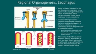
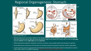
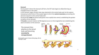
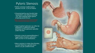
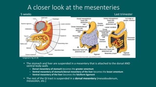
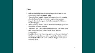
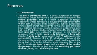
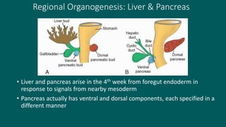
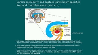
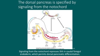
Foregut
• Foregut gives rise to:
• Esophagus
• Stomach
• Liver
• Gallbladder and bile ducts
• Pancreas
• Upper Duodenum
Regional Organogenesis: Esophagus
• Region of foregut just caudal to lung
bud develops into esophagus –errors
in forming the esophagotracheal septa
and/or re-canalization lead to
tracheoesophageal fistulas and/or
esophageal atresia, respectively.
• Endodermal lining is stratified
columnar and proliferates such that
the lumen is obliterated; patency of
the lumen established by re-
canalization –errors in this process lead
to esophageal stenosis
• NOTE: this process of recanalization occurs
throughout the gut tube, so occlusion can
occur anywhere along the GI tract (e.g.
duodenal stenosis)
• Tube initially short and must grow in
length to “keep up” with descent of
heart and lungs –failure of growth in
length leads to congenital hiatal hernia
in which the cranial portion of the
stomach is pulled into the hiatus.
Langman’s fig 14-06
Larsen’s fig 14-22
5th week 7th week
9th week
Regional Organogenesis: Stomach
• Stomach appears first as a fusiform dilation of the foregut endoderm which undergoes a 90° rotation such that the left
side moves ventrally and the right side moves dorsally (the vagus nerves follow this rotation which is how the left vagus
becomes anterior and the right vagus becomes posterior).
• Differential growth establishes the greater and lesser curvatures; cranio-caudal rotation tips the pylorus superiorly
• Dorsal AND ventral mesenteries of the stomach are retained to become the greater and lesser omenta, respectively
• Caudal end of the stomach separated from the duodenum by formation of the pyloric sphincter (dependent on factors
such as SOX-9, NKX-2.5, and BMP-4 signaling) –errors in this process lead to pyloric stenosis.
Greater omentum
Langman’s figs 14-08, 11, 12
3rd month
3rd month
4th week
3rd month
5th week
6th week
Gastric Features
• Mixes and Emulsifies
• Reservoir
• Secretes
• Acid  Host Defense and Digestion
• Digestive Enzymes Pepsin, Lipase
• Intrinsic Factor Vitamin B12 absorption
• Regulates release of gastric chyme
• Prevents acid reflux - (LES)
http://www.acm.uiuc.edu/sigbio/project/digestive/middle/stomach.html
Gastric Anatomy
• Neuromuscular
http://www.britannica.com/EBchecked/media/68634/
Gastric pits
GASTROENTEROLOGY 2008;134:1842–1860
Gastric Glands
• Cells line the gastric pits with differing products
• Acid secretion
• Mucus secretion
• Bicarbonate secretion
• Hormone secretion
Endocrine Cells of Stomach
(Located in the stomach body)
GASTROENTEROLOGY 2008;134:1842–1860
Pyloric Stenosis
• Rather common malformation:
present in 0.5% - 0.1% of infants
• Characterized by very forceful (aka
“projectile”), non-bilious vomiting
~1hr. after feeding (when pyloric
emptying would occur).
NOTE: the presence of bile would indicate
POST-duodenal blockage of some sort.
• Hypertrophied sphincter can often be
palpated as a spherical nodule;
peristalsis of the sphincter seen/felt
under the skin.
• Stenosis is due to overproliferation /
hypertrophy of pyloric sphincter…
NOT an error in re-canalization.
• More common in males than females,
so most likely has a genetic basis
which is as yet undetermined.
A closer look at the mesenteries
• The stomach and liver are suspended in a mesentery that is attached to the dorsal AND
ventral body walls
– Dorsal mesentery of stomach becomes the greater omentum
– Ventral mesentery of stomach/dorsal mesentery of the liver becomes the lesser omentum
– Ventral mesentery of the liver becomes the falciform ligament
• The rest of the GI tract is suspended in a dorsal mesentery (mesodoudenum,
mesocolon, etc.)
Langman’s fig 14-26
Langman’s fig 14-30
5 weeks Last trimester
Pancreas
• 1. Development.
• The dorsal pancreatic bud is a direct outgrowth of foregut
endoderm, whose formation is induced by the notochord. The
ventral pancreatic bud is a direct outgrowth of foregut
endoderm, whose formation is induced by hepatic mesoderm.
Within both pancreatic buds, endodermal tubules surrounded
by mesoderm branch repeatedly to form acinar cells and ducts
(i.e., exocrine pancreas). Isolated clumps of endodermal cells
bud from the tubules and accumulate within the mesoderm to
form islet cells (i.e., endocrine pancreas) in the following
sequence (first S last): alpha cells (glucagon) S beta cells
(insulin) S delta cells (somatostatin) and PP cells (pancreatic
polypeptide). Because of the 90_ clockwise rotation of the
duodenum, the ventral bud rotates dorsally and fuses with the
dorsal bud to form the definitive adult pancreas. The ventral
bud forms the uncinate process and a portion of the head of
the pancreas. The dorsal bud forms the remaining portion of
the head, body, and tail of the pancreas.
Regional Organogenesis: Liver & Pancreas
• Liver and pancreas arise in the 4th week from foregut endoderm in
response to signals from nearby mesoderm
• Pancreas actually has ventral and dorsal components, each specified in a
different manner
Langman’s fig 14-19
Cardiac mesoderm and septum transversum specifies
liver and ventral pancreas (sort of…)
• ALL foregut endoderm has the potential to develop into either liver or pancreas but local signals
repress these fates (mesodermal Wnts ↓ Liver; mesodermal Wnts + endodermal Shh ↓ Pancreas)
• FGFs and BMPs from cardiac mesoderm and septum transversum inhibit Wnt signaling, but the
endoderm still expresses Shh and thus develops into liver.
• Meanwhile, endoderm just caudal to liver bud is out of reach from the FGFs and BMPs but still sees
Wnts, so the tissue does NOT become liver. Unlike the rest of the endoderm, it does NOT express
Shh and so develops into pancreas.
ventral pancreas
Langman’s fig 14-22
The dorsal pancreas is specified by
signaling from the notochord
Signaling from the notochord represses Shh in caudal foregut
endoderm, which permits dorsal pancreatic differentiation.
Larsen’s fig 14-05
• The main pancreatic duct is formed by the anatomosis of the distal
two thirds of the dorsal pancreatic duct (the proximal one third
regresses) and the entire ventral pancreatic duct (48% incidence).
The main pancreatic duct and common bile duct form a single
opening (hepatopancreatic ampulla of Vater) into the duodenum at
the tip of a major papillae (hepatopancreatic papillae).
• 2. Sources. Acinar cells, islet cells, and simple columnar or cuboidal
epithelium lining the pancreatic ducts of the definitive pancreas are
derived from endoderm. Surrounding connective tissue and vascular
components of the definitive pancreas are derived from visceral
mesoderm.
• 3. Clinical considerations
• a. Accessory pancreatic duct: develops when the proximal one third
of the dorsal pancreatic duct persists and opens into the duodenum
through minor papillae at a site proximal to the ampulla of Vater (33%
incidence).
• b. Pancreas divisum: occurs when the distal two thirds of the dorsal
pancreatic duct and the entire ventral pancreatic duct fail to anastomose
and the proximal one third of the dorsal pancreatic duct persists, thereby
forming two separate duct systems (4% incidence). The dorsal pancreatic
duct drains a portion of the head, body, and tail of the pancreas by
opening into the duodenum through minor papillae. The ventral pancreatic
duct drains the uncinate process and a portion of the head of the
pancreas by opening into the duodenum through the major papillae.
Patients with pancreas divisum are prone to pancreatitis, especially if the
opening of the dorsal pancreatic duct at the minor papillae is small. Note
that the distal two thirds of the dorsal pancreatic duct and the ventral
pancreatic bud fail to anastomose, thereby forming two separate duct
systems.
• c. Annular pancreas: occurs when the ventral pancreatic bud fuses
with the dorsal bud both dorsally and ventrally, thereby forming a
ring of pancreatic tissue around the duodenum and causing severe
duodenal obstruction. Newborns and infants are intolerant of oral
feeding and often have bilious vomiting. Radiographic evidence of an
annular pancreas is indicated by a duodenal obstruction, where a
“double bubble” sign is often seen due to dilation of the stomach and
distal duodenum (also associated with Down syndrome).
Rotation of the duodenum also causes it and the
pancreas to become SECONDARILY retroperitoneal
Secondarily retroperitoneal = a structure that was originally in the body coelom but
then got pushed into the body wall during development
Langman’s fig 14-11
5th week 3rd month
Rotation of the
duodenum brings
the ventral and
dorsal pancreas
together
Aberrations in this process may
result in an annular pancreas,
which can constrict the duodenum.
Also, since the dorsal and ventral
pancreas arise by different
mechanisms, it’s possible that one
or the other may be absent in the
adult.
Larsen’s fig 14-09
Larsen’s fig 14-10
Intraperitoneal vs. retroperitoneal vs.
secondarily retroperitoneal
Larsen’s fig 14-16

Foregut

  • 1.
    Foregut • Foregut givesrise to: • Esophagus • Stomach • Liver • Gallbladder and bile ducts • Pancreas • Upper Duodenum
  • 2.
    Regional Organogenesis: Esophagus •Region of foregut just caudal to lung bud develops into esophagus –errors in forming the esophagotracheal septa and/or re-canalization lead to tracheoesophageal fistulas and/or esophageal atresia, respectively. • Endodermal lining is stratified columnar and proliferates such that the lumen is obliterated; patency of the lumen established by re- canalization –errors in this process lead to esophageal stenosis • NOTE: this process of recanalization occurs throughout the gut tube, so occlusion can occur anywhere along the GI tract (e.g. duodenal stenosis) • Tube initially short and must grow in length to “keep up” with descent of heart and lungs –failure of growth in length leads to congenital hiatal hernia in which the cranial portion of the stomach is pulled into the hiatus. Langman’s fig 14-06 Larsen’s fig 14-22 5th week 7th week 9th week
  • 3.
    Regional Organogenesis: Stomach •Stomach appears first as a fusiform dilation of the foregut endoderm which undergoes a 90° rotation such that the left side moves ventrally and the right side moves dorsally (the vagus nerves follow this rotation which is how the left vagus becomes anterior and the right vagus becomes posterior). • Differential growth establishes the greater and lesser curvatures; cranio-caudal rotation tips the pylorus superiorly • Dorsal AND ventral mesenteries of the stomach are retained to become the greater and lesser omenta, respectively • Caudal end of the stomach separated from the duodenum by formation of the pyloric sphincter (dependent on factors such as SOX-9, NKX-2.5, and BMP-4 signaling) –errors in this process lead to pyloric stenosis. Greater omentum Langman’s figs 14-08, 11, 12 3rd month 3rd month 4th week 3rd month 5th week 6th week
  • 6.
    Gastric Features • Mixesand Emulsifies • Reservoir • Secretes • Acid  Host Defense and Digestion • Digestive Enzymes Pepsin, Lipase • Intrinsic Factor Vitamin B12 absorption • Regulates release of gastric chyme • Prevents acid reflux - (LES) http://www.acm.uiuc.edu/sigbio/project/digestive/middle/stomach.html
  • 7.
  • 8.
    Gastric pits GASTROENTEROLOGY 2008;134:1842–1860 GastricGlands • Cells line the gastric pits with differing products • Acid secretion • Mucus secretion • Bicarbonate secretion • Hormone secretion
  • 9.
    Endocrine Cells ofStomach (Located in the stomach body) GASTROENTEROLOGY 2008;134:1842–1860
  • 10.
    Pyloric Stenosis • Rathercommon malformation: present in 0.5% - 0.1% of infants • Characterized by very forceful (aka “projectile”), non-bilious vomiting ~1hr. after feeding (when pyloric emptying would occur). NOTE: the presence of bile would indicate POST-duodenal blockage of some sort. • Hypertrophied sphincter can often be palpated as a spherical nodule; peristalsis of the sphincter seen/felt under the skin. • Stenosis is due to overproliferation / hypertrophy of pyloric sphincter… NOT an error in re-canalization. • More common in males than females, so most likely has a genetic basis which is as yet undetermined.
  • 11.
    A closer lookat the mesenteries • The stomach and liver are suspended in a mesentery that is attached to the dorsal AND ventral body walls – Dorsal mesentery of stomach becomes the greater omentum – Ventral mesentery of stomach/dorsal mesentery of the liver becomes the lesser omentum – Ventral mesentery of the liver becomes the falciform ligament • The rest of the GI tract is suspended in a dorsal mesentery (mesodoudenum, mesocolon, etc.) Langman’s fig 14-26 Langman’s fig 14-30 5 weeks Last trimester
  • 13.
    Pancreas • 1. Development. •The dorsal pancreatic bud is a direct outgrowth of foregut endoderm, whose formation is induced by the notochord. The ventral pancreatic bud is a direct outgrowth of foregut endoderm, whose formation is induced by hepatic mesoderm. Within both pancreatic buds, endodermal tubules surrounded by mesoderm branch repeatedly to form acinar cells and ducts (i.e., exocrine pancreas). Isolated clumps of endodermal cells bud from the tubules and accumulate within the mesoderm to form islet cells (i.e., endocrine pancreas) in the following sequence (first S last): alpha cells (glucagon) S beta cells (insulin) S delta cells (somatostatin) and PP cells (pancreatic polypeptide). Because of the 90_ clockwise rotation of the duodenum, the ventral bud rotates dorsally and fuses with the dorsal bud to form the definitive adult pancreas. The ventral bud forms the uncinate process and a portion of the head of the pancreas. The dorsal bud forms the remaining portion of the head, body, and tail of the pancreas.
  • 14.
    Regional Organogenesis: Liver& Pancreas • Liver and pancreas arise in the 4th week from foregut endoderm in response to signals from nearby mesoderm • Pancreas actually has ventral and dorsal components, each specified in a different manner Langman’s fig 14-19
  • 15.
    Cardiac mesoderm andseptum transversum specifies liver and ventral pancreas (sort of…) • ALL foregut endoderm has the potential to develop into either liver or pancreas but local signals repress these fates (mesodermal Wnts ↓ Liver; mesodermal Wnts + endodermal Shh ↓ Pancreas) • FGFs and BMPs from cardiac mesoderm and septum transversum inhibit Wnt signaling, but the endoderm still expresses Shh and thus develops into liver. • Meanwhile, endoderm just caudal to liver bud is out of reach from the FGFs and BMPs but still sees Wnts, so the tissue does NOT become liver. Unlike the rest of the endoderm, it does NOT express Shh and so develops into pancreas. ventral pancreas Langman’s fig 14-22
  • 16.
    The dorsal pancreasis specified by signaling from the notochord Signaling from the notochord represses Shh in caudal foregut endoderm, which permits dorsal pancreatic differentiation. Larsen’s fig 14-05
  • 17.
    • The mainpancreatic duct is formed by the anatomosis of the distal two thirds of the dorsal pancreatic duct (the proximal one third regresses) and the entire ventral pancreatic duct (48% incidence). The main pancreatic duct and common bile duct form a single opening (hepatopancreatic ampulla of Vater) into the duodenum at the tip of a major papillae (hepatopancreatic papillae).
  • 20.
    • 2. Sources.Acinar cells, islet cells, and simple columnar or cuboidal epithelium lining the pancreatic ducts of the definitive pancreas are derived from endoderm. Surrounding connective tissue and vascular components of the definitive pancreas are derived from visceral mesoderm. • 3. Clinical considerations • a. Accessory pancreatic duct: develops when the proximal one third of the dorsal pancreatic duct persists and opens into the duodenum through minor papillae at a site proximal to the ampulla of Vater (33% incidence).
  • 22.
    • b. Pancreasdivisum: occurs when the distal two thirds of the dorsal pancreatic duct and the entire ventral pancreatic duct fail to anastomose and the proximal one third of the dorsal pancreatic duct persists, thereby forming two separate duct systems (4% incidence). The dorsal pancreatic duct drains a portion of the head, body, and tail of the pancreas by opening into the duodenum through minor papillae. The ventral pancreatic duct drains the uncinate process and a portion of the head of the pancreas by opening into the duodenum through the major papillae. Patients with pancreas divisum are prone to pancreatitis, especially if the opening of the dorsal pancreatic duct at the minor papillae is small. Note that the distal two thirds of the dorsal pancreatic duct and the ventral pancreatic bud fail to anastomose, thereby forming two separate duct systems.
  • 24.
    • c. Annularpancreas: occurs when the ventral pancreatic bud fuses with the dorsal bud both dorsally and ventrally, thereby forming a ring of pancreatic tissue around the duodenum and causing severe duodenal obstruction. Newborns and infants are intolerant of oral feeding and often have bilious vomiting. Radiographic evidence of an annular pancreas is indicated by a duodenal obstruction, where a “double bubble” sign is often seen due to dilation of the stomach and distal duodenum (also associated with Down syndrome).
  • 25.
    Rotation of theduodenum also causes it and the pancreas to become SECONDARILY retroperitoneal Secondarily retroperitoneal = a structure that was originally in the body coelom but then got pushed into the body wall during development Langman’s fig 14-11 5th week 3rd month
  • 26.
    Rotation of the duodenumbrings the ventral and dorsal pancreas together Aberrations in this process may result in an annular pancreas, which can constrict the duodenum. Also, since the dorsal and ventral pancreas arise by different mechanisms, it’s possible that one or the other may be absent in the adult. Larsen’s fig 14-09 Larsen’s fig 14-10
  • 28.
    Intraperitoneal vs. retroperitonealvs. secondarily retroperitoneal Larsen’s fig 14-16