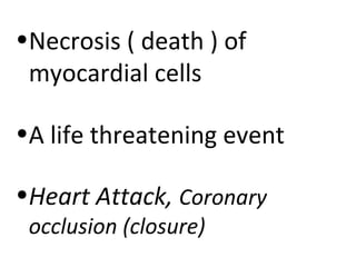
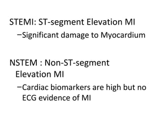
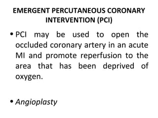
•Necrosis ( death ) of
 myocardial cells

•A life threatening event

•Heart Attack, Coronary
 occlusion (closure)
•   MI is usually caused by reduced blood flow/
    oxygen supply in a coronary artery
•   vasospasm (sudden constriction/ narrowing) of a
    coronary artery
•   and increased demand for oxygen
Nonmodifiable Risk Factors
• Family history of coronary heart disease
• Increasing age
• Gender (heart disease occurs more often in
  men and in premenopausal women) *men and
 premenopausal women have low estrogen = helps in ↑HDL and ↓LDL

• Race : higher incidence of heart disease in
  African Americans than in Caucasians
  (NHLBI, 2002)
Modifiable Risk Factors
• High blood cholesterol level (high blood lipids)
• Cigarette smoking, tobacco use (nicotine causes
  vasoconstriction)
• Hypertension (high blood pressure causes vasoconstriction)
• Diabetes mellitus (impaired insulin production- excess glucose-
  liver uses glucose to make fatty acids)

• Lack of estrogen in women
• Physical inactivity (fats are not burned, lodges in blood
  vessels)
• Obesity (high fat deposits in the body)
• Cardiovascular
  – Chest pain or discomfort.
    • Palliative: not relieved by rest or sublingual
      vasodilator therapy, requires opioids.
    • Quality: variable, but often diffuse, steady substernal
      chest pain. Other sensations include a crushing and
      squeezing feeling in the chest.
    • Radiation: pain may radiate to the arms (commonly
      the left), and to the shoulders, neck, back, or jaw.
    • Time: pain continues for more than 15 minutes.


  – Increased jugular venous distention may be
    seen if the MI has caused ® heart failure.
• Respiratory
   – Shortness of breath, dyspnea, tachypnea,
     and crackles (decreased oxygen supply)
• Genitourinary
   – Decreased urinary output (*when there is decreased
     blood flow RAAS is activated, aldosterone = water retention)
• Skin
   – Cool, clammy, diaphoretic, (compensatory
    mechanism = Pumping blood through clogged arteries takes more
    effort from your heart, so your body will sweat more to try o keep
    your body temperature down during the extra exertion. )
                          and Pale appearance


• Neurologic
  – Anxiety, restlessness, light-headedness
     (there is a decrease in contractility and cerebral oxygenation.)
ECG


•Ischemia causes inversion of T wave because of altered repolarization.
•Cardiac muscle injury causes elevation of the ST segment and tall,
symmetrical T waves.
•With Q-wave infarction, Q or QS waves develop because of the absence of
depolarization current from the necrotic tissue and opposing currents from
other parts of the heart.
STEMI: ST-segment Elevation MI
 – Significant damage to Myocardium


NSTEM : Non-ST-segment
 Elevation MI
 – Cardiac biomarkers are high but no
   ECG evidence of MI
LABORATORY TESTS
• Creatine Kinase and Its Isoenzymes.
• There are three CK isoenzymes:
   – CK-MM (skeletal muscle),
   – CK-MB (heart muscle), N = 0-3% of total CK
   – CK-BB (brain tissue).
• CK-MB is the cardiac-specific isoenzyme;
• CK-MB is found mainly in cardiac cells and therefore
  rises only when there has been damage to these cells.
• the most specific index for the diagnosis of acute MI
  (Braunwald et al., 2001).
• The level starts to increase within a few hours and peaks
  within 24 hours of an MI. If the area is reperfused (eg,
  due to thrombolytic therapy or PTCA), it peaks earlier.
• Nonspecific enzyme myoglobin may be
  elevated
  – Myoglobin: a heme protein that helps to
    transport oxygen. Like CK-MB enzyme,
    myoglobin is found in cardiac and skeletal
    muscle.
  – The normal ("negative") range is 0 - 85
    nanograms per milliliter (ng/mL).
• Triponin T and I are myocardial proteins that
  increase in the serum about 3 to 4 hours after
  an MI, peak in 4 to 24 hours, and are
  detectable for up to 2 weeks;

• the test can help diagnose an MI up to 2
  weeks earlier, and only unstable angina
  causes a false positive.
• N = < 0.2 mcg/L
• N = < 3.1 mcg/L
Pharmacologic Intervention:

•Analgesics: Morphine
•Vasodilator : Nitrogylcerine
•ACE Inhibitors
•Thrombolytics: to dissolve
 thrombus formation
• Anticoagulants or other anti-platelet
  medications : Aspirin / Heparin
• Beta-adrenergic blockers, to improve
  oxygen supply and demand, decrease
  sympathetic stimulation to the heart,
  promote blood flow in the small vessels
  of the heart, and provide antiarrhythmic
  effects.
EMERGENT PERCUTANEOUS CORONARY
       INTERVENTION (PCI)
• PCI may be used to open the
  occluded coronary artery in an acute
  MI and promote reperfusion to the
  area that has been deprived of
  oxygen.

• Angioplasty
Nursing Interventions:
• Monitor baseline vital signs before and 10 to 15
  minutes after administering drugs. Also monitor
  blood pressure continuously when giving
  nitroglycerin I.V. (causes vasodilation =
  improving blood flow)

• Monitor continuous ECG to watch for life
  threatening arrhythmias (common within 24
  hours after infarctions) and evolution of the MI
  (changes in ST segments and T waves).
• Administer O2 at 2-5 L per cannula : Oxygen
  therapy may increase the oxygen supply to
  the myocardium and other tissues

• pain relief : administer analgesics promptly. Place
  the patient in supine position during administration
  (to minimize hypotension)

• Promote rest (to decrease cardiac workload) with
  early gradual increase in mobilization (to prevent
  deconditioning)
Cardiac Rehabilitation
• After the MI patient is free of symptoms, an active
  rehabilitation program is initiated.
• Targets risk reduction by means of education,
  individual and group support, and physical activity.
• Goals are extend and improve the quality of life.
• The immediate objectives
   – to limit the effects and progression of atherosclerosis,
   – return the patient to work and a pre-illness lifestyle,
   – enhance the psychosocial and vocational status of the
     patient,
   – and prevent another cardiac event.
Phase I
• may begin with the diagnosis of atherosclerosis,
  which may occur when the patient is admitted to the
  hospital for ACS (unstable angina, acute MI).

• It consists of low-level activities and initial education
  for the patient and family.

• The amount of activity recommended at discharge
  depends on the age of the patient, his or her
  condition before the cardiac event, the extent of the
  disease, the course of the hospital stay, and the
  development of any complications.
Phase II
• occurs after the patient has been discharged.
• It usually lasts for 4 to 6 weeks but may last up to 6
  months.
• This outpatient program consists of supervised, often
  ECG-monitored, exercise training that is
  individualized based on the results of an exercise
  stress test.
• Support and guidance related to the treatment of
  the disease and education and counseling related to
  lifestyle modification for risk factor reduction are a
  significant part of this phase.
Phase III
• focuses on maintaining cardiovascular
  stability and long-term conditioning.
• The patient is usually self-directed
  during this phase and does not require a
  supervised program, although it may be
  offered.
• The goals of each phase build on the
  accomplishments of the previous phase.

Myocardial infarction

  • 1.
    •Necrosis ( death) of myocardial cells •A life threatening event •Heart Attack, Coronary occlusion (closure)
  • 2.
    MI is usually caused by reduced blood flow/ oxygen supply in a coronary artery • vasospasm (sudden constriction/ narrowing) of a coronary artery • and increased demand for oxygen
  • 4.
    Nonmodifiable Risk Factors •Family history of coronary heart disease • Increasing age • Gender (heart disease occurs more often in men and in premenopausal women) *men and premenopausal women have low estrogen = helps in ↑HDL and ↓LDL • Race : higher incidence of heart disease in African Americans than in Caucasians (NHLBI, 2002)
  • 5.
    Modifiable Risk Factors •High blood cholesterol level (high blood lipids) • Cigarette smoking, tobacco use (nicotine causes vasoconstriction) • Hypertension (high blood pressure causes vasoconstriction) • Diabetes mellitus (impaired insulin production- excess glucose- liver uses glucose to make fatty acids) • Lack of estrogen in women • Physical inactivity (fats are not burned, lodges in blood vessels) • Obesity (high fat deposits in the body)
  • 6.
    • Cardiovascular – Chest pain or discomfort. • Palliative: not relieved by rest or sublingual vasodilator therapy, requires opioids. • Quality: variable, but often diffuse, steady substernal chest pain. Other sensations include a crushing and squeezing feeling in the chest. • Radiation: pain may radiate to the arms (commonly the left), and to the shoulders, neck, back, or jaw. • Time: pain continues for more than 15 minutes. – Increased jugular venous distention may be seen if the MI has caused ® heart failure.
  • 7.
    • Respiratory – Shortness of breath, dyspnea, tachypnea, and crackles (decreased oxygen supply) • Genitourinary – Decreased urinary output (*when there is decreased blood flow RAAS is activated, aldosterone = water retention)
  • 8.
    • Skin – Cool, clammy, diaphoretic, (compensatory mechanism = Pumping blood through clogged arteries takes more effort from your heart, so your body will sweat more to try o keep your body temperature down during the extra exertion. ) and Pale appearance • Neurologic – Anxiety, restlessness, light-headedness (there is a decrease in contractility and cerebral oxygenation.)
  • 9.
    ECG •Ischemia causes inversionof T wave because of altered repolarization. •Cardiac muscle injury causes elevation of the ST segment and tall, symmetrical T waves. •With Q-wave infarction, Q or QS waves develop because of the absence of depolarization current from the necrotic tissue and opposing currents from other parts of the heart.
  • 10.
    STEMI: ST-segment ElevationMI – Significant damage to Myocardium NSTEM : Non-ST-segment Elevation MI – Cardiac biomarkers are high but no ECG evidence of MI
  • 11.
    LABORATORY TESTS • CreatineKinase and Its Isoenzymes. • There are three CK isoenzymes: – CK-MM (skeletal muscle), – CK-MB (heart muscle), N = 0-3% of total CK – CK-BB (brain tissue). • CK-MB is the cardiac-specific isoenzyme; • CK-MB is found mainly in cardiac cells and therefore rises only when there has been damage to these cells. • the most specific index for the diagnosis of acute MI (Braunwald et al., 2001). • The level starts to increase within a few hours and peaks within 24 hours of an MI. If the area is reperfused (eg, due to thrombolytic therapy or PTCA), it peaks earlier.
  • 12.
    • Nonspecific enzymemyoglobin may be elevated – Myoglobin: a heme protein that helps to transport oxygen. Like CK-MB enzyme, myoglobin is found in cardiac and skeletal muscle. – The normal ("negative") range is 0 - 85 nanograms per milliliter (ng/mL).
  • 13.
    • Triponin Tand I are myocardial proteins that increase in the serum about 3 to 4 hours after an MI, peak in 4 to 24 hours, and are detectable for up to 2 weeks; • the test can help diagnose an MI up to 2 weeks earlier, and only unstable angina causes a false positive. • N = < 0.2 mcg/L • N = < 3.1 mcg/L
  • 15.
    Pharmacologic Intervention: •Analgesics: Morphine •Vasodilator: Nitrogylcerine •ACE Inhibitors •Thrombolytics: to dissolve thrombus formation
  • 16.
    • Anticoagulants orother anti-platelet medications : Aspirin / Heparin • Beta-adrenergic blockers, to improve oxygen supply and demand, decrease sympathetic stimulation to the heart, promote blood flow in the small vessels of the heart, and provide antiarrhythmic effects.
  • 17.
    EMERGENT PERCUTANEOUS CORONARY INTERVENTION (PCI) • PCI may be used to open the occluded coronary artery in an acute MI and promote reperfusion to the area that has been deprived of oxygen. • Angioplasty
  • 18.
    Nursing Interventions: • Monitorbaseline vital signs before and 10 to 15 minutes after administering drugs. Also monitor blood pressure continuously when giving nitroglycerin I.V. (causes vasodilation = improving blood flow) • Monitor continuous ECG to watch for life threatening arrhythmias (common within 24 hours after infarctions) and evolution of the MI (changes in ST segments and T waves).
  • 19.
    • Administer O2at 2-5 L per cannula : Oxygen therapy may increase the oxygen supply to the myocardium and other tissues • pain relief : administer analgesics promptly. Place the patient in supine position during administration (to minimize hypotension) • Promote rest (to decrease cardiac workload) with early gradual increase in mobilization (to prevent deconditioning)
  • 20.
    Cardiac Rehabilitation • Afterthe MI patient is free of symptoms, an active rehabilitation program is initiated. • Targets risk reduction by means of education, individual and group support, and physical activity. • Goals are extend and improve the quality of life. • The immediate objectives – to limit the effects and progression of atherosclerosis, – return the patient to work and a pre-illness lifestyle, – enhance the psychosocial and vocational status of the patient, – and prevent another cardiac event.
  • 21.
    Phase I • maybegin with the diagnosis of atherosclerosis, which may occur when the patient is admitted to the hospital for ACS (unstable angina, acute MI). • It consists of low-level activities and initial education for the patient and family. • The amount of activity recommended at discharge depends on the age of the patient, his or her condition before the cardiac event, the extent of the disease, the course of the hospital stay, and the development of any complications.
  • 22.
    Phase II • occursafter the patient has been discharged. • It usually lasts for 4 to 6 weeks but may last up to 6 months. • This outpatient program consists of supervised, often ECG-monitored, exercise training that is individualized based on the results of an exercise stress test. • Support and guidance related to the treatment of the disease and education and counseling related to lifestyle modification for risk factor reduction are a significant part of this phase.
  • 23.
    Phase III • focuseson maintaining cardiovascular stability and long-term conditioning. • The patient is usually self-directed during this phase and does not require a supervised program, although it may be offered. • The goals of each phase build on the accomplishments of the previous phase.