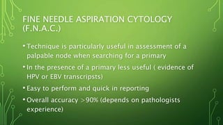
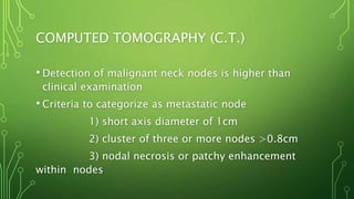
METASTATIC NECK DISEASE
AND MANAGEMENT
DR SANKALPA GAMAGE
REGISTRAR IN OMF SURGERY
TEACHING HOSPITAL KARAPITIYA
GALLE, SRI LANKA.
INTRODUCTION
•Most important prognostic factor in head and
neck cancer is presence or absence, level and
size of metastatic neck disease
•Controversies exist about management of neck
disease (Choice of treatment, timing and
combination of modalities)
TUMOUR BIOLOGY RELATED TO
METASTASIS
• Tumours do not have primary lymphatics
• Solid tumours can induce lymphangiogenesis
• VEGF-C and VEGF-D show active role in
lymphangiogenesis ( New research pathway for anti
lymphangiogenetic therapeutics)
• Cellular adhesion molecules as E cadherin downdrading
happens therefore cancer cells allowed to freely migrate
• MMPs produced by tumour cells act on ECM to enhance
migration
• Active production of autocrine paracrine cytokynes
mediate the active migration of CA cells in to lymphatics
NECK LEVELS
NECK DISSECTION (HISTORY)
• In 1906 George Washington Crile introduced Radical
Neck Dissection
• In 1951 Hays Martin popularized RND with step wise
descriptions
• 1963 Osvaldo Suarez introduced Functional Neck
Dissection
NECK DISSECTION TERMINOLOGY
• First introduces by the American academy of
Otolaryngology and updated by committee for neck
dissection classification of the American head and
neck society in 2002
1) Comprehensive neck dissection (Removes level
I-V) subdivided in to RND, MRND, ERND
2) Selective neck dissection (Preserves one or
more LN levels)
NECK DISSECTION (HISTORY) CONTD..
• Earlier division of SND in to subdivisions (SOBD,
ESOBD) is now superseded due to confusion
• Therefore levels and sublevels removed in neck
dissection should be clearly mentioned in operative
notes
• Term elective neck dissection (END) is used to any
type of neck dissection done in a clinically and
radiologicaly negative neck
CLASSIFICATION
• Radical Neck Dissection - Removal of I to V neck node
levels and Accessory nerve, Internal jugular vein and
Sternocleidomastoid
• Modified Radical Neck Dissection – removal of level I-V
preservation of one or more non lymphatic structures
• Extended Radical Neck Dissection – Removal of additional
lymphatic and/ or non lymphatic structures
• Selective Neck Dissection – Preservation of one or more
levels of LN
OCCULT NODAL DISEASE
-Presence of neck metastasis in the neck nodes that
cannot be clinically or radiologically identified-
•2 categories
1) Occult metastasis that can be
identified by the light microscopy
2) Micrometastases less than 2mm that
need special histological techniques
• Lymph node negative in histology means
CANCER NEVER REACHED THE NODE
Or else
IT REACHED BUT DESTROYED IN THE NODE
•This make several controversies in the Management
of occult neck
•Observational data suggests conversion of N0 to
N+ (30%) is similar to the incidence of pathological
positive nodes In END specimens
PROGNOSTIC NODAL FEATURES
•Site Size and number
•Low neck nodes
•Extra capsular spread
•Morphology
•Bilateral or contralateral spread
CLINICAL STAGING
•Nx – Regional lymph nodes cannot be
asscessed
•N0 - No regional lymph node metastasis
•N1 – Single ipsilateral node <3mm
•N2a – single ipsilateral node 3-6mm
•N2b – Multiple ipsilateral nodes 3-6mm
•N2c – Bilateral or contralateral nodes 3-6mm
•N3- Node/ Nodes >6mm
FACTORS IMPLICATED IN PATTERN OF
METASTATIC NODAL DISEASE
•Tumour size
•Tumour site
•Tumour thickness
•Previous treatment
•Tumour recurrence
ASSESSMENT OF CERVICAL
LYMPHADENOPATHY
• Clinical assessment
• Fine needle aspiration cytology
• Ultra sound scan
• Computed Tomography
• Magnetic Resonance Imaging
• Positron Emission Tomography
• Open Biopsy
CLINICAL EXAMINATION
• Remains the most important initial method of
assessment
• Variable in accuracy ( Clinicians experience)
• Sensitivity of 74% specificity of 81% overall accuracy
of 77% (Merrit et al)
• Difficult in short stocky necks
• Drawback is nodes may go unnoticed until they reach
a considerable size
FINE NEEDLE ASPIRATION CYTOLOGY
(F.N.A.C.)
• Technique is particularly useful in assessment of a
palpable node when searching for a primary
• In the presence of a primary less useful ( evidence of
HPV or EBV transcripsts)
• Easy to perform and quick in reporting
• Overall accuracy >90% (depends on pathologists
experience)
ULTRA SOUND SCAN (U.S.S.)
•Can detect the presence malignant cervical
lymph nodes with sensitivity of 70% to 90%
•When combined with FNAC it increases to 90%
•No absolute criteria to differentiate between
malignant vs benign nodes
COMPUTED TOMOGRAPHY (C.T.)
• Detection of malignant neck nodes is higher than
clinical examination
• Criteria to categorize as metastatic node
1) short axis diameter of 1cm
2) cluster of three or more nodes >0.8cm
3) nodal necrosis or patchy enhancement
within nodes
MAGNETIC RESONANCE IMAGING (M.R.I.)
•Meta analysis suggests MRI can detect
metastatic neck disease similar to CT
•MRI is better in assessing N0 neck
POSITRON EMISSION TOMOGRAPHY (P.E.T.)
• Uses F-2-fluoro-2-deoxy-D-glucose (FDG) as a
radioactive tracer
• Can identify the areas od higher glucose uptake
• Anatomical localization difficult
• Coregistered anatomic and functional imaging
systems CT-PET lead to accurate image fusion by
harvesting advantages of both techniques
• Role in CUP, recurrent and residual disease
OPEN BIOPSY
• In general best avoided as initial diagnostic modality
• When FNAC not available, non diagnostic, results
suggesting a lymphoma, or anaplastic Carcinoma or
primary site is occult may be necessary
• Prognosis will not be different if the definitive
treatment is done within 6 weeks
• Any incision should made to facilitate to remove the
scar via subsequent neck dissection and vital
structures like sternocleidomastoid may have to be
SENTINEL NODE BIOPSY
•Used to assess the presence of occult disease
in N0 neck
•Assumption is ‘‘If these echelon nodes are
negative for tumour deposits rest of the neck is
free of tumour metastasis’’
•Technique uses radioactive probes and/or blue
dye around tumour then first third echelon
TREATMENT FOR METASTATIC NECK
DISEASE
• Decided by the stage of neck disease
FEW POINTS TO REMEMBER
1)Untreated neck patterns of spread is predictable
2) In occult neck metastasis usually found at first
echelon LN basin
3) Previous RX alters the pattern of spread
4) PTs with palpable neck nodes likely to have non
palpable spread in other levels
N0 NECK
• Historically evaluation and treatment for No is great dilemma
and still controversial
• Weather to treat or not to treat
• Number of Rx options
Elective surgery
Elective radiotherapy
Wait and watch
Elective neck investigations
INDICATIONS FOR ELECTIVE NECK
TREATMENT
•15% to 20% chance of subclinical neck disease
•Vigilant follow up not possible
•Clinical evaluation of the neck is difficult
•Access to the neck for reconstruction
CHOICE OF TREATMENT MODALITY FOR N0
• NO prospective data available
• Elective neck dissection and irradiation are equally
effective
• Choice dictated by many factors
Patient and surgeons choices
quality of life issues and how the primary is
treated
ARGUMENTS FOR ELECTIVE NECK SURGERY
• High incidence of occult metastatic disease
• SND got low morbidity and mortality
• Routine follow up will not detect the earliest
conversion from No to N+
• Untreated neck masses can induce distant metastasis
• Elective neck surgery can perform at primary surgery
• Patient compliance for regular follow ups cannot be
assured
ARGUMENTS AGAINST ELECTIVE NECK
SURGERY
•careful clinical follow up combined with
imaging has the potential to detect early
conversion
•Control rates no lower than when wait and
watch policy is followed
•Elective surgery result in inevitable morbidity
•END removes the natural barrier to the spread
•END creates a scarred hypoxic field that can
ELECTIVE SURGERY
• Therapeutic equivalence of SND and CND is widely
accepted
• Levels II to IV need to be cleared in laryngeal and
hypolaryngeal tumours
• Level I to III for oral cavity tumours addition of level IV
for tongue cancers
• Appears to have Little advantage of adding level V for
any mucosal primaries electively
• END will serve as a biopsy to indicate the risk of
THE N+ NECK (N1 NECK)
•Treatment modality is usually dictates by
treatment to primary site
•In palpable neck disease all 5 LN levels should be
dissected
•Majority of the cases accessory nerve can be
preserved (MRND)
•Many patients subsequently need RT
INDICATIONS OF POST OPERATIVE
RADIOTHERAPY
•Extra capsular spread
•Two or more positive nodes
•N2 N3 Stages
•Residual disease
N2A AND N2B NECK
•MRND sparing spinal accessory nerve where
possible
•PORT is indicated
N2C NECK
• Common primary site is tongue base, supraglottic
larynx and hypopharynx
• Earlier it was considered as grave prognosis
• Now evidence shows the size ,number and the ECS
determines the prognosis rather than the laterality
• Conservational neck surgery on less affected side and
radical surgery on more affected side
• PORT
N3 NECK (MASSIVE NODES)
• Commonly fixed to skin or underlying structures
• Decision to operate or not decided by
stage and primary site
presence of absence of fixation
what the node is fixed to
experience of the surgeon
needs of the patient
• Long term control is poor and the benefit of treatment should
be carefully weighted against morbidity
• Radical treatment is warranted in patients who have less
advanced primary
• MANY PATIENTS PALLIATION WILL BE THE BEST OPTION
THANK YOU FOR LISTENING…

Metastatic neck disease and management

  • 1.
    METASTATIC NECK DISEASE ANDMANAGEMENT DR SANKALPA GAMAGE REGISTRAR IN OMF SURGERY TEACHING HOSPITAL KARAPITIYA GALLE, SRI LANKA.
  • 2.
    INTRODUCTION •Most important prognosticfactor in head and neck cancer is presence or absence, level and size of metastatic neck disease •Controversies exist about management of neck disease (Choice of treatment, timing and combination of modalities)
  • 3.
    TUMOUR BIOLOGY RELATEDTO METASTASIS • Tumours do not have primary lymphatics • Solid tumours can induce lymphangiogenesis • VEGF-C and VEGF-D show active role in lymphangiogenesis ( New research pathway for anti lymphangiogenetic therapeutics) • Cellular adhesion molecules as E cadherin downdrading happens therefore cancer cells allowed to freely migrate • MMPs produced by tumour cells act on ECM to enhance migration • Active production of autocrine paracrine cytokynes mediate the active migration of CA cells in to lymphatics
  • 4.
  • 5.
    NECK DISSECTION (HISTORY) •In 1906 George Washington Crile introduced Radical Neck Dissection • In 1951 Hays Martin popularized RND with step wise descriptions • 1963 Osvaldo Suarez introduced Functional Neck Dissection
  • 6.
    NECK DISSECTION TERMINOLOGY •First introduces by the American academy of Otolaryngology and updated by committee for neck dissection classification of the American head and neck society in 2002 1) Comprehensive neck dissection (Removes level I-V) subdivided in to RND, MRND, ERND 2) Selective neck dissection (Preserves one or more LN levels)
  • 7.
    NECK DISSECTION (HISTORY)CONTD.. • Earlier division of SND in to subdivisions (SOBD, ESOBD) is now superseded due to confusion • Therefore levels and sublevels removed in neck dissection should be clearly mentioned in operative notes • Term elective neck dissection (END) is used to any type of neck dissection done in a clinically and radiologicaly negative neck
  • 8.
    CLASSIFICATION • Radical NeckDissection - Removal of I to V neck node levels and Accessory nerve, Internal jugular vein and Sternocleidomastoid • Modified Radical Neck Dissection – removal of level I-V preservation of one or more non lymphatic structures • Extended Radical Neck Dissection – Removal of additional lymphatic and/ or non lymphatic structures • Selective Neck Dissection – Preservation of one or more levels of LN
  • 9.
    OCCULT NODAL DISEASE -Presenceof neck metastasis in the neck nodes that cannot be clinically or radiologically identified- •2 categories 1) Occult metastasis that can be identified by the light microscopy 2) Micrometastases less than 2mm that need special histological techniques
  • 10.
    • Lymph nodenegative in histology means CANCER NEVER REACHED THE NODE Or else IT REACHED BUT DESTROYED IN THE NODE •This make several controversies in the Management of occult neck •Observational data suggests conversion of N0 to N+ (30%) is similar to the incidence of pathological positive nodes In END specimens
  • 11.
    PROGNOSTIC NODAL FEATURES •SiteSize and number •Low neck nodes •Extra capsular spread •Morphology •Bilateral or contralateral spread
  • 12.
    CLINICAL STAGING •Nx –Regional lymph nodes cannot be asscessed •N0 - No regional lymph node metastasis •N1 – Single ipsilateral node <3mm •N2a – single ipsilateral node 3-6mm •N2b – Multiple ipsilateral nodes 3-6mm •N2c – Bilateral or contralateral nodes 3-6mm •N3- Node/ Nodes >6mm
  • 13.
    FACTORS IMPLICATED INPATTERN OF METASTATIC NODAL DISEASE •Tumour size •Tumour site •Tumour thickness •Previous treatment •Tumour recurrence
  • 14.
    ASSESSMENT OF CERVICAL LYMPHADENOPATHY •Clinical assessment • Fine needle aspiration cytology • Ultra sound scan • Computed Tomography • Magnetic Resonance Imaging • Positron Emission Tomography • Open Biopsy
  • 15.
    CLINICAL EXAMINATION • Remainsthe most important initial method of assessment • Variable in accuracy ( Clinicians experience) • Sensitivity of 74% specificity of 81% overall accuracy of 77% (Merrit et al) • Difficult in short stocky necks • Drawback is nodes may go unnoticed until they reach a considerable size
  • 16.
    FINE NEEDLE ASPIRATIONCYTOLOGY (F.N.A.C.) • Technique is particularly useful in assessment of a palpable node when searching for a primary • In the presence of a primary less useful ( evidence of HPV or EBV transcripsts) • Easy to perform and quick in reporting • Overall accuracy >90% (depends on pathologists experience)
  • 17.
    ULTRA SOUND SCAN(U.S.S.) •Can detect the presence malignant cervical lymph nodes with sensitivity of 70% to 90% •When combined with FNAC it increases to 90% •No absolute criteria to differentiate between malignant vs benign nodes
  • 18.
    COMPUTED TOMOGRAPHY (C.T.) •Detection of malignant neck nodes is higher than clinical examination • Criteria to categorize as metastatic node 1) short axis diameter of 1cm 2) cluster of three or more nodes >0.8cm 3) nodal necrosis or patchy enhancement within nodes
  • 19.
    MAGNETIC RESONANCE IMAGING(M.R.I.) •Meta analysis suggests MRI can detect metastatic neck disease similar to CT •MRI is better in assessing N0 neck
  • 20.
    POSITRON EMISSION TOMOGRAPHY(P.E.T.) • Uses F-2-fluoro-2-deoxy-D-glucose (FDG) as a radioactive tracer • Can identify the areas od higher glucose uptake • Anatomical localization difficult • Coregistered anatomic and functional imaging systems CT-PET lead to accurate image fusion by harvesting advantages of both techniques • Role in CUP, recurrent and residual disease
  • 21.
    OPEN BIOPSY • Ingeneral best avoided as initial diagnostic modality • When FNAC not available, non diagnostic, results suggesting a lymphoma, or anaplastic Carcinoma or primary site is occult may be necessary • Prognosis will not be different if the definitive treatment is done within 6 weeks • Any incision should made to facilitate to remove the scar via subsequent neck dissection and vital structures like sternocleidomastoid may have to be
  • 22.
    SENTINEL NODE BIOPSY •Usedto assess the presence of occult disease in N0 neck •Assumption is ‘‘If these echelon nodes are negative for tumour deposits rest of the neck is free of tumour metastasis’’ •Technique uses radioactive probes and/or blue dye around tumour then first third echelon
  • 23.
    TREATMENT FOR METASTATICNECK DISEASE • Decided by the stage of neck disease FEW POINTS TO REMEMBER 1)Untreated neck patterns of spread is predictable 2) In occult neck metastasis usually found at first echelon LN basin 3) Previous RX alters the pattern of spread 4) PTs with palpable neck nodes likely to have non palpable spread in other levels
  • 24.
    N0 NECK • Historicallyevaluation and treatment for No is great dilemma and still controversial • Weather to treat or not to treat • Number of Rx options Elective surgery Elective radiotherapy Wait and watch Elective neck investigations
  • 25.
    INDICATIONS FOR ELECTIVENECK TREATMENT •15% to 20% chance of subclinical neck disease •Vigilant follow up not possible •Clinical evaluation of the neck is difficult •Access to the neck for reconstruction
  • 26.
    CHOICE OF TREATMENTMODALITY FOR N0 • NO prospective data available • Elective neck dissection and irradiation are equally effective • Choice dictated by many factors Patient and surgeons choices quality of life issues and how the primary is treated
  • 27.
    ARGUMENTS FOR ELECTIVENECK SURGERY • High incidence of occult metastatic disease • SND got low morbidity and mortality • Routine follow up will not detect the earliest conversion from No to N+ • Untreated neck masses can induce distant metastasis • Elective neck surgery can perform at primary surgery • Patient compliance for regular follow ups cannot be assured
  • 28.
    ARGUMENTS AGAINST ELECTIVENECK SURGERY •careful clinical follow up combined with imaging has the potential to detect early conversion •Control rates no lower than when wait and watch policy is followed •Elective surgery result in inevitable morbidity •END removes the natural barrier to the spread •END creates a scarred hypoxic field that can
  • 29.
    ELECTIVE SURGERY • Therapeuticequivalence of SND and CND is widely accepted • Levels II to IV need to be cleared in laryngeal and hypolaryngeal tumours • Level I to III for oral cavity tumours addition of level IV for tongue cancers • Appears to have Little advantage of adding level V for any mucosal primaries electively • END will serve as a biopsy to indicate the risk of
  • 30.
    THE N+ NECK(N1 NECK) •Treatment modality is usually dictates by treatment to primary site •In palpable neck disease all 5 LN levels should be dissected •Majority of the cases accessory nerve can be preserved (MRND) •Many patients subsequently need RT
  • 31.
    INDICATIONS OF POSTOPERATIVE RADIOTHERAPY •Extra capsular spread •Two or more positive nodes •N2 N3 Stages •Residual disease
  • 32.
    N2A AND N2BNECK •MRND sparing spinal accessory nerve where possible •PORT is indicated
  • 33.
    N2C NECK • Commonprimary site is tongue base, supraglottic larynx and hypopharynx • Earlier it was considered as grave prognosis • Now evidence shows the size ,number and the ECS determines the prognosis rather than the laterality • Conservational neck surgery on less affected side and radical surgery on more affected side • PORT
  • 34.
    N3 NECK (MASSIVENODES) • Commonly fixed to skin or underlying structures • Decision to operate or not decided by stage and primary site presence of absence of fixation what the node is fixed to experience of the surgeon needs of the patient
  • 35.
    • Long termcontrol is poor and the benefit of treatment should be carefully weighted against morbidity • Radical treatment is warranted in patients who have less advanced primary • MANY PATIENTS PALLIATION WILL BE THE BEST OPTION
  • 36.
    THANK YOU FORLISTENING…